View Article

Abstract

Atherosclerotic cardiovascular disease (CVD) is one of the most significant global health problems today. Current standard therapies for cardiovascular disease and revascularization do not eliminate plaque locally nor alleviate residual risk of developing future cardiovascular events. Implantable cell-based therapeutic approaches provide a method for delivering therapeutic cells/cell-derived products directly to the plaque, increasing local retention and limiting systemic side effects from these therapies. This review discusses advances in bioengineered vascular interfaces and delivery technologies and immune-metabolically-mediated modulation of atherosclerotic plaque to establish a framework of precision implantable therapies. The approaches outlined in this review regulate macrophage polarization, smooth muscle cell phenotype, and endothelial repair to stabilize and reduce plaque size. Engineered exosomes and biomimetic nanovesicles are two approaches enhancing the targeting and effectiveness of precision implantable therapies. Clinical trials have demonstrated good safety and moderate effectiveness for implantable cell-based therapies used for peripheral and coronary artery disease, highlighting the need to establish biomarkers that can guide identification and selection of appropriate patients for treatment and to optimize delivery of these therapies. Several systemic factors, including but not limited to age, diabetes, and clonal haematopoiesis may affect integration of implanted cells within the atherosclerotic plaque and patient outcomes and demonstrate the value of considering both systemic elements as well as implant design in future development of implantable cell-based therapeutic modalities. Future directions emphasize, there is a need to establish standardized methods for evaluation, scalable manufacturing processes, and regulatory pathways, to provide a continued and long-term clinical benefit beyond current systemic therapies

Keywords

Atherosclerosis, Implantable Cell Therapy, Plaque Reprogramming, Bioengineered Vascular Interfaces, Precision Medicine

Introduction

1.1. Mechanistic Insights and Rationale

The burden of atherosclerotic cardiovascular disease (CVD) worldwide continues to grow significantly; however, the decline in the age-standardized rates makes it difficult to determine how much of this increase is due to improvements in medical care versus an increase in population age or size. In fact, the global prevalence of CVD has increased by approximately two-fold (271 million cases in 1990 vs. 523 million cases in 2019). The two largest contributors of CVD mortality are ischemic heart disease (IHD) (49.2% of total deaths due to CVD) and stroke (approximately 25% of total deaths due to CVD), with approximately half of the strokes due to ischemic causes. Incidence trends are heterogeneous, with the number of incident CVD cases increasing by 77% globally from 1990 to 2019, but age-standardized incidence only declining by 1.6% in the most recent decade.  Peripheral artery disease (PAD) incidence has also declined globally, although the rate of decline has slowed in the recent decade. The implications of this increasing absolute burden of atherosclerotic CVDs are significant for health systems and resource availability globally.[1]

1.2 Limitations of Current Therapies for Local Plaque Control

The key limitations of current therapies for local plaque control are that systemic administration of drugs often leads to off-target effects and low local drug concentrations at the plaque site, post-stenting restenosis induced by vascular injury response and non-specific therapeutic drugs limits the effectiveness of stent-based approaches. Mechanical trauma from balloon angioplasty can cause endothelial damage, platelet deposition, and smooth muscle cell hyperplasia, leading to restenosis, and surgical approaches like bypass grafting, while effective, are highly invasive and carry risks of complications.[2]

Administration of pharmacological agents in the clinical context, statins are central to atherosclerosis treatment but often fail to sufficiently lower LDL or are poorly tolerated due to side effects.  Alternative therapies such as fibrates, ezetimibe, bile acid Sequestrants and niacin provide only limited benefit, while PCI and drug-eluting stent (DESs) re-establish blood flow but expose the endothelium to injury due to the DESs obstructing re-endothelialization. This creates a greater risk for stenting and neo atherosclerosis. These treatments leave a significant residual risk of disease and surgery has done nothing to prevent disease from progressing. Current treatments do not adequately treat the local plaque pathology creating an urgent need for new and improved therapies. [3, 4]

1.3 Rationale for Plaque-Directed Implantable Cell Therapies

Cell therapy is designed to implant therapeutic cells or derived products directly into their intended delivery site to enhance the concentration, retention, and management of the cells in this specific area while reducing the risk of systemic toxicity. While the use of implantable cell therapies has significant potential for improving the treatment of systemic diseases or conditions, it may also provide unique advantages for treating focal pathologic conditions such as atherosclerotic plaques, tumours, fractures, infarcts, periodontitis, etc. [5, 6, 7].


Table 1: Local versus Systemic Delivery of Therapeutic Cells: Mechanistic and Clinical Considerations

Sr. No.

Dimension

Plaque directed/Local delivery

Systemic delivery

Citations

1

Primary goal

Targeted retention and tissue regeneration

Natural homing and minimally invasive

[5,6]

2

Typical routes

Intraplaque, intracoronary,

intramyocardial

Peripheral IV, as systemic approach

[5,6]

3

Cell retention

at target

High retention

Low retention

[5,6]

4

Regenerative efficacy

Local delivery and better retention

Systemic therapy and less effective repair

[5,6]

5

Targeting

Local,site-specific(plaque)

Whole body distribution

[5,7]

6

Dependence

on homing signals

Less dependent

Strongly dependent

[6,7]

7

Off-target distribution

Lowersystemic exposure

High off-target trapping

[5,6]

8

Invasiveness/ procedural risk

More invasive

(procedural/implant)

Less invasive

(IV/Injection)

[6,7]

9

Technical complexity

Specialized procedure, operator skills required

Technically simple

[6,7]

10

When preferred

Accessible lesions and local targeting

Inaccessible lesions and non-invasive approach

[5,6]


Implantable cell therapies—whether delivered as live cells, cell-seeded scaffolds, or cell-derived products like exosomes—offer a strategic advantage for localized, plaque-directed therapy by providing sustained, targeted, and controlled therapeutic effects while minimizing systemic exposure [5, 6, 7].

1.4 Objectives and Novel Contributions of this review

This purpose of this review is to give a focused assessment of therapeutic devices that can be used for atherosclerosis treatment. The review has been structured in such a way that it combines information on the many disciplines relevant to the study; including engineering, plaque biology, and clinical data acquisition. Thus, the true value of this review lies in the explanation of how bioengineered vascular devices (bioengineered Vascular Interfacing Systems - BVI) and implantable cell therapies can provide a precisely targeted and sustained stabilization and regeneration of atherosclerotic plaques, which relate directly to the methodology for developing a clinical translation pathway. This review then proposes design principles for therapeutic devices based upon biomechanical and biological limitations, methods of delivering drugs via a therapeutic device and techniques of using imaging to evaluate a therapeutic device. Furthermore, this review provides a roadmap for conducting clinical trials in which the use of a biomarker will guide the selection of patients for participation and will be used to stratify patients as either a responder or non-responder. This roadmap will maximize both the safety and efficacy of precision implantable therapeutic devices for patients with atherosclerotic cardiovascular disease. [1,2,3,4,5,6,7]

2. Cellular and Molecular Landscape of Atherosclerotic Plaques

2.1 Cellular Composition of the Atherosclerotic Plaque

Atherosclerotic plaques are made up of a heterogeneous population of cells, including ECs, VSMCs, macrophages and immune cells. Endothelial dysfunction, as seen in patients with hypercholesterolemia, hypertension, and/or diabetes, stimulates lipid deposition and results in inflammation within the plaque. VSMCs undergo phenotypic transitions, including through endothelial mesenchymal transition and converting into foam cells; this process increases the size of the plaque and contributes to the calcification of the plaque. Macrophages, deriving both from resident sources as well as monocyte sources, can take on pro-inflammatory (M1) or pro-resolution (M2) phenotypes, which influence the stability and progression of the plaque. The recruitment of pro-atherogenic T cells through the release of chemokines increases the overall inflammatory process within the plaque and contributes significantly to atherosclerotic progression [3, 8].

2.2 Targetable Pathobiological Processes in Atherosclerosis

The principal target areas for therapeutic intervention in atherosclerosis include: (i) Endothelial dysfunction, (ii) oxidative stress (iii) chronic inflammation, (iv) impaired efferocytosis and (v) plaque instability/erosion. Endothelial cell injury is responsible for increasing low-density lipoprotein (LDL) penetration into the vessel wall and leukocyte adherence to endothelial cells. Oxidation of LDL and the presence of proinflammatory cytokines are both driving forces behind subsequent plaque development. Impaired efferocytosis leads to the formation of necrotic cores, which further destabilise the plaque, rendering it susceptible to rupture and resulting in acute coronary syndrome. Continued plaque expansion will also impede blood flow and facilitate thrombosis. Therefore, nanoparticle-based therapies aim to modulate these pathways through improving endothelial function, reducing oxidative stress, controlling inflammation, enhancing efferocytosis and stabilising plaques. [4, 8].

2.3 Plaque Reprogramming as an Emerging Therapeutic Paradigm

The concept of plaque reprogramming in atherosclerosis involves the therapeutic modification of the following three important cell types associated with plaque development: macrophages, vascular smooth muscle cells (VSMCs) and endothelial cells (ECs). This concept has the following three therapeutic implications to inhibit disease progression: 1) shift macrophages from pro-inflammatory (M1) to anti-inflammatory (M2) phenotypes to increase cholesterol efflux and efferocytosis, 2) shift VSMCs towards a contractile/functionally quiescent phenotype to stabilize the fibrous cap, and 3) restore the balance of ECs to their homeostatic state. The mechanisms used to achieve these responses in macrophages, VSMCs and ECs will utilize cell- and exosome-based therapies, such as engineered exosomes from macrophages exhibiting anti-inflammatory and pro-efferocytosis properties, mesenchymal stem cell therapy to enhance EC function and increase VSMC quiescence, and nanoparticle-based delivery of miRNAs (e.g., miRNA-146a for macrophages and miR-145 for VSMCs) to reprogram cellular functioning. Finally, the use of biomimetic nanoparticles and extracellular vesicles derived from endothelial progenitor cells to promote vascular repair/angiogenesis and the use of immunomodulatory therapies, such as engineered regulatory T cells and senolytic CAR-T cells, to target inflammation and senescence of plaque, will expand the therapeutic approaches for plaque reprogramming. [9, 10, 11]

There are multiple ways in which stem cell-derived exosomes can positively impact the treatment of atherosclerosis. They can influence macrophage polarization to the anti-inflammatory M2 phenotype by altering cellular signaling through the following miRNAs: miR-let7, miR-182 and HMGA2/NF-κB. They can also inhibit VSMC mineralization by inhibiting the expression of genes that drive VSMC mineralization (e.g., RUNX2, BGLAP, BMP2) and through the inhibition of the NFATc3-osteocalcin pathway.

Additionally, stem-cell-derived exosomes from multiple sources (e.g., MSCs, CPCs, and EPCs) can restore endothelial cell function by delivering miRNAs (e.g., miR-342-5p, miR-512-3p, and miR-10a) that protect against oxidative stress and ox-LDL-induced endothelial cell dysfunction. They may further stimulate angiogenesis. As demonstrated by their diverse actions, stem cell-derived exosomes are a potentially comprehensive therapeutic option for treating atherosclerosis. [8]


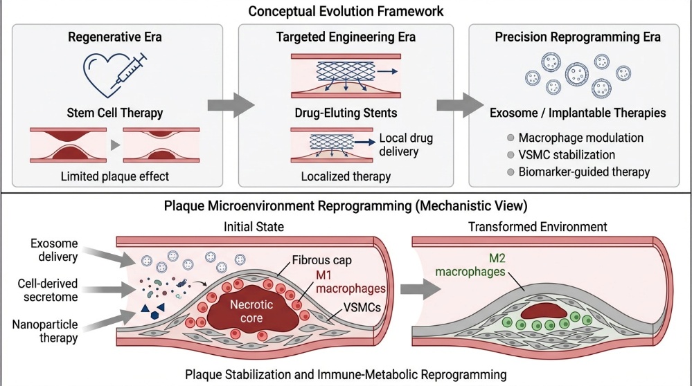
Figure 1. The Advancement of Implantable Therapies for Precision Reprogramming of the Plaque Microenvironment in Atherosclerosis


3. Clinical Status of Cell-Based Therapies in Atherosclerotic CVD

3.1 Clinical Evidence in Peripheral Atherosclerotic Disease

Several meta-analyses have evaluated the clinical evidence for autologous cell therapy in patients with peripheral artery disease (PAD) and chronic limb-threatening ischemia (CLTI):


Table 2: Key clinical trial findings [12]

Author(s) of trial/

meta-analysis

Key findings

Rigato et al. [13]

A meta-analysis of 19 randomized clinical trials (involving 837 patients) found that autologous cell therapy may

improve amputation-free survival, limb perfusion, and functional capacity. However, the studies had significant

heterogeneity and were underpowered.

Gao et al. [14]

An analysis of 27 randomized trials (with 1186 patients) revealed that autologous stem cell therapy improved ulcer

healing compared to standard treatments, but limb salvage outcomes were similar. The authors noted that many

studies had small sample sizes and there was considerable variation among trials.

Pu et al. [15]

A meta-analysis of 12 randomized controlled trials (involving 630 patients with CLTI) showed significant

improvements in amputation rates, ankle-brachial index, transcutaneous oxygen tension, and rest pain scores when

compared to placebo or standard care. The findings suggest autologous stem cell therapy is a promising treatment

for “no-option” candidates.

Arango-

Rodríguez et al. [16]

A randomized, double-blind, controlled study in diabetic patients with CLTI compared auto-BM-MNC and allo-WJ-

MSCs. Both cell types demonstrated potential therapeutic benefits, indicating stem cell therapy could be a promising

alternative treatment.


Meta-analyses reveal that studies on autologous cell therapy s for PAD/CLTI, mostly have small sample sizes and considerable heterogeneity with respect to study design, characteristics of the disease, types and doses of cells, and length of follow-up, resulting in limited statistical power and clarity of results. Primary outcomes evaluated were amputation-free survival (AFS), perfusion of the limb (e.g., ABIs and TcPO?), and functional ability (e.g., rest pain and pain-free walking distance). All of the above outcomes had considerable variability; however, autologous cell therapy has generally demonstrated some potential for improving these outcomes, particularly in patient’s ineligible for revascularization. Thus, these data suggest a promising avenue for limb salvage and an improved quality of life; although, more well-designed studies are needed to determine their safety and effectiveness. [12]

3.2 Coronary and cardiac cell?therapy trials

Over 200 clinical trials have examined stem cell therapies—including MSCs, EPCs, and BM-MNCs—for acute MI, chronic ischemic cardiomyopathy, CAD, and IHD. The results of these trials confirm safety but show modest, inconsistent efficacy, with some improvements in LVEF, perfusion, and function lacking statistical significance. Key insights highlight optimizing cell dose, delivery route, timing, and patient selection, alongside advanced imaging to track cell fate and assess outcomes. These findings can guide the development of targeted, plaque-directed cell therapies addressing CAD pathophysiology more precisely. [108, 109, 110, 110]

3.3 Therapeutic Potential of Adipose-Derived MSC-Derived Exosomes

Adipose-derived mesenchymal stem cells (ADSCs) and their exosomes show promise for stabilization in atherosclerosis plaque by modulating lipids, reducing inflammation, and promoting repair via paracrine factors. ADSC exosomes assist the process of cholesterol exit from cells through the upregulation of the cholesterol transporters ABCA1 and ABCG1. A challenge to clinical translation of ADSC exosomes is variability among donors, such as type 2 diabetes and/or obesity. Ongoing trials will assess the safety and efficacy of ADSC exosomes from patients with diabetes and/or obesity; however, additional research is warranted with regard to donor variables influencing ADSC exosome product quality. [8]

4. Shifting from Regeneration to Plaque Reprogramming

4.1 Limitations of Regenerative Strategies in Atherosclerosis

Current evidence suggests that fully “regenerating” normal arterial wall in advanced atherosclerosis is unlikely, but reprogramming plaque cells and microenvironment toward a stable, regressive state is increasingly plausible. [19, 20]

There are several significant limitations in the "regenerative" paradigm of the treatment of atherosclerosis and heart disease; even with 20 years of research into this area, stem cell therapies have yet to achieve clinical success; one factor contributing to the failure of stem cell therapies is a disconnect between the mechanisms of tissue repair and the mechanisms of disease. The clinical trial results obtained so far have demonstrated that stem cells can exhibit dysfunction and have genetic defects which underlie the conditions associated with atherosclerosis and heart disease; therefore, the authors of this article recommend an increased understanding of mechanisms through enhanced computational data analysis, through a common data repository, and through collaboration at a scientific/clinical level (clinical trial/longitudinal studies and integration of basic research with clinical trials and long-term). The existence of cardiovascular risk factors such as hypertension, diabetes, hyperlipidaemia, and smoking has a significant detrimental effect on the number and functionality of EPCs; therefore, there is a loss of therapeutic potential for autologous EPC therapies because of this factor, and the use of allogeneic EPCs will continue to be limited due to immunological incompatibility. Currently, no effective interventions exist to overcome EPC dysfunction caused by these risk factors. [19, 20]

Complete vascular regeneration in established atherosclerosis is limited, especially in patients with multiple risk factors. However, reprogramming plaque cells and their microenvironment—targeting macrophages, endothelium, innate immunity, and surrounding tissue—provides a mechanistic, scalable approach for plaque regression and stabilization, now the leading therapeutic framework. [19, 20]

4.2 Immunometabolic Modulation by Implanted Cellular Products

Novel findings from recent advances confirm the therapeutic potential of exosomes produced from stem cells to change the polarization of macrophages leading to alleviation of several inflammatory diseases.

Exosomes derived from mesenchymal stem cells (MSCs) can reduce the development of atherosclerosis through the induction of M2-type polarization of macrophages and decrease the presence of M1-type macrophages present within plaque by upregulating M2 candidate markers and downregulating M1 candidate markers via miR-21a-5p. This miRNA enhances expression of the M2 marker, Arg-1, while reducing expression of the M1 marker, iNOS, and decreasing expression of the transcription factor KLF6 resulting in a greater concentration of M2 macrophages [21]. Metabolically engineered stem cell-derived exosomes (DS-EXOs) can also affect macrophage heterogeneity within rheumatoid arthritis by promoting the ability of macrophages to convert from an M1-type to an M2-type macrophage. In-vitro, DS-EXOs will decrease expression of iNOS and increase expression of CD206 via miRNA let-7b-5p and miRNA-24-3p through modulation of the JAK-STAT pathway. Systemic DS-EXO treatment shifts joint macrophages toward M2, reducing proinflammatory M1 cells and synovial fibroblast activity, reprogramming the synovial microenvironment early in disease [22].

Implanted cells and engineered exosomes act as immune-metabolic modulators across models, coordinating macrophage polarization, autophagy, endothelial repair, and VSMC phenotype/calcification. This frames therapies as programmable tools that rewire plaque and vascular microenvironments along interconnected pathways rather than simple regenerators.[22]

4.3 Representative Engineered Cell-Derived Therapeutic Platforms


Table 3: Engineered Stem Cell–Derived Therapeutic Platforms

Engineered Stem Cell-derived Product

Modification / Feature

Mechanism/ Effect

Therapeutic outcome

Citation

MSCs overexpressing Akt

Akt protein overexpression

Promotes angiogenesis

Improved cardiac function

[8]

Platelet membrane -coated exosome-mimetic nanovesicles

Platelet membrane coating

Upregulates cholesterol transporters ABCA1and ABCG1

Reduces intracellular cholesterol accumulation

[8]

MSC-ExoP

Platelet- membrane fusion

Enhances plaque targeting, activates VSMC autophagy

Reduces VSMC proliferation, migration, foam-cell formation

[23]

Platelet- mimetic exosomes for restenosis (PM-Exo)

Platelet- membrane fusion

Improves injured artery homing, promotes endothelial repair

Limits neointimal hyperplasia

[24]

Platelet- MSC hybrid exosomes for myocardial infarction

Platelet- membrane fusion

Boosts uptake by endothelial cells and cardiomyocytes

Enhances cardiac targeting and post-MI functional recovery

[25]


These case studies show that platelet?membrane–engineered exosomes/nanovesicles greatly improve vascular and myocardial targeting, enhancing uptake by endothelial cells, cardiomyocytes, and plaque regions. Across models of atherosclerosis, restenosis, and myocardial infarction, they reprogram plaque and vascular cells [8, 8, 23, 24, 25].

4.4 Relevance of Plaque Reprogramming for Clinical Evaluation

Atherosclerosis therapy is shifting from a limited “regeneration” view to plaque reprogramming, where implanted cells and exosomes are designed as immune?metabolic modulators that tune macrophage polarization, efferocytosis, autophagy, endothelial repair, VSMC phenotypic switching and anti?calcific pathways to shrink necrotic core and thicken fibrous cap, thereby stabilizing plaques and reducing events [26,27,28,29].

By engineering mesenchymal stem cells (MSCs) and their exosomes to provide mechanistic therapy tailored to individual patients—rather than simply a broad regenerative response—targeted therapy can be achieved. For example, through use of exosomes released from MSCs that overexpress Akt, angiogenesis is enhanced and cardiac function is improved post-myocardial infarction by acting through the PDGF-D/Akt pathway.

Exosome-mimetic nanovesicles (P-ENVs) derived from platelets are directed toward atherosclerotic plaque sites, activate ABCA1/ABCG1, enhance cholesterol efflux, and decrease both lipid- and necrotic-core volume. Exosomes derived from MSCs that fuse with platelet membranes (MSC-ExoP) concentrate at the site of plaques, activate autophagy in vascular smooth muscle cells (VSMCs), and inhibit VSMC proliferation, migration, and foam-cell formation, thereby promoting stabilization of these lesions. Platelet-mimetic exosomes for restenosis (PM-EXOs) show fourfold higher uptake in injured arteries, promote endothelial repair, shift macrophages to anti-inflammatory phenotypes, and prevent VSMC phenotypic switching, reducing neointimal hyperplasia without systemic toxicity [23,24]. Platelet–MSC hybrid exosomes for MI exploit platelet homing to enhance uptake by endothelial cells and cardiomyocytes, improving exosome accumulation in injured myocardium and post?MI functional recovery [25]

5. Bioengineered Vascular Interfaces for Plaque-Targeted Therapy

Bioengineered vascular interfaces have evolved from bare-metal and drug-eluting stents to bio functional, cell-interactive devices designed to stimulate endothelialisation and site-specific repair. Injectable/implantable scaffolds of PBMNC for CLTI and plaques are intended to improve cell retention, survival, local therapeutic?genic effects with safety. New biomimetic approaches such as membrane-guided nanoparticles and exosome-loaded devices also offer the potential for specific cutting-edge of plaque targeting and signal-oriented vascular regeneration. Together, these approaches straddle the line between promise and delivery in terms of translation, scaling, regulation and established long-term safety [4, 23, 24, 25, 30, 31, 32, 33, 34].


Table 4: Mapping bioengineered vascular interface examples

Bioengineered vascular interface

Examples

Citations

Cell-interactive stents

Platelet?membrane–coated rapamycin DES; exosome?eluting stents; exosome?functionalized stent surfaces

[30,31,32]

Resorbable scaffolds (ICS-001)

PBMNC?loaded ICS?001 intramuscular scaffold for CLTI; exploratory trial data

[33]

Biomimetic nano/exosome hybrids

Platelet?decorated exosomes for restenosis; platelet?exosome hybrids for plaques and MI; hybrid membrane NPs for CVD

[24,4,34,

23,25]


 6. Delivery Science and Design Principles for Arterial Implants

Therapeutic arterial implants face distinct mechanical and biological constraints in coronary vs peripheral beds, which shape feasible delivery routes, drug/cell dosing, and imaging readouts. Existing stent, liquid-drug, and graft technologies provide a practical “design window” for new cell or gene–based arterial implants [48, 49, 50].

6.1 Physical constraints of coronary and peripheral delivery

Shear stress & geometry: Coronary and peripheral stenting produce localized low and high shear stress forces at the edges of struts and in relation to the anastomotic connection. Variables that significantly influence the recirculation zones and risk of restenosis include curvature; the anastomotic angle; the material stiffness; and ratio of graft diameter to recipient artery diameter. [35,36,37]

Access routes: Contemporary practice spans femoral, radial/ulnar, axillary, carotid, and transcaval access, chosen by sheath size, tortuosity, and comorbidities. These limit device profile and support for bulky cell/gene systems. [38]

Microvasculature: CABG and peripheral graft modelling show downstream resistance and uncertain peripheral hemodynamic substantially affect wall stress and strain predictions, underscoring the need for robust design margins. [39,37]

The major mechanical constraint and an associated design consideration for each of the four procedures were collectively described as follows: For coronary percutaneous coronary interventions (PCIs) employing drug-eluting stents, shear stress disturbances due to the curvature of blood vessels and the forces acting on the edges of the stent are primarily localized. Therefore, the design must have thin, flexible, conformable struts that preserve radial strength while minimizing poor positioning to reduce both the risk of restenosis and stent thrombosis. [40,41, 42]

The presence of repetitive flexion, compression, and torsion combined with complicated plaque in superficial femoral and femoropopliteal interventions with low shear or oscillatory shear mean that fatigue-resistant structural supports as well as optimizing drug delivery are required to maintain patency during these procedures and prevent fracture or an excessive amount of neointimal growth following these interventions. [41,43,44]

Designs for femoral bypass grafts, including peripheral vascular bypass grafts and tissue-engineered femoral grafts, focus on mechanical matching, long-term durability when loaded with physiologic loads, and promotion of arteriogenesis rather than intimal hyperplasia in order to drive failure due to a lack of compliance and disturbed shear at the anastomoses [45, 41, 46, 47]

6.2 Insights from Existing Trials to Define Design Windows

Endovascular drug devices: Drug?eluting stents, balloons, and local liquid?drug delivery (LLD) catheters (Bullfrog, ClearWay, OPC, TAPAS) reliably deliver high local doses over 10–150 mm segments with favourable safety, supporting “leave?nothing?behind” concepts. [48, 49, 50]

Imaging?guided PCI: IVUS/OCT guidance reduces target lesion failure, MACE, restenosis, and stent thrombosis vs angiography alone across 15,000–18,000 patients. This defines a practical imaging backbone for arterial implant trials. [51,52,53,54,55,36]

Peripheral imaging: In femoropopliteal disease, IVUS/OCT detect more dissections, calcification, and under expansion than angiography and are associated with lower restenosis when used to guide therapy. [56, 57]

Imaging Endpoints for Plaque Stabilisation / Implant Performance

Intravascular imaging (core for “design rules”):

IVUS: Vessel size, plaque burden, stent expansion, edge dissections; prognostic for TLR, TVR, MACE. [51, 52, 53, 54, 55]

OCT: High?resolution neointimal thickness, malposition, tissue protrusion, microcalcification, and healing; useful for mechanistic readouts of endothelialisation and drug/cell distribution. [51, 54, 55, 50]

For peripheral implants: IVUS/OCT in femoropopliteal arteries refine sizing and correlate with incidence of restenosis and reintervention. [56, 57]

Non?invasive / systemic: MRI/CT angiography for lumen and remodelling, PET for inflammatory activity, and clinical endpoints (TVF, limb salvage, CLTI improvement) are established as logical higher?order outcomes variables extrapolated from endovascular drug?device literature. [58, 49, 50, 59]

6.3 Design Principles for Optimizing Retention, Homing, and Viability

From drug?eluting and liquid?delivery platforms, key transferable principles:

6.3.1 Matching of the mechanical characteristics of devices to local haemodynamic should attempt to avoid extremes of shear stress and oscillation index value, both of which stimulate neointima formation and thrombosis. [35, 55, 37]

6.3.2 Lesion?specific delivery: Drug/cell type and formulation (polymeric capsules, reservoirs); Coating method will be determined by the plaque morphology (fibrotic vs lipid rich and calcific), as determined by IVUS/OCT/VH. [55, 50, 57]

6.3.3 Uniform intramural distribution: LLD catheters and microneedle/adventitial devices show that penetrating beyond the lumen (adventitial, intramural) can improve homogeneity and reduction in systemic spillover. [48, 49, 50]

6.4.4 Chronic support vs leave?nothing?behind: Reservoir implants and intravascular pumps maintain stable therapeutic windows for years but at the cost of device complexity; “leave?nothing?behind” LLD and balloons prioritize vessel natural history and future re?intervention options. [58, 48, 49, 59]

7. Systemic Modifiers and Precision Context for Implantable Therapies

Implant integration and cardiovascular cell therapies are strongly shaped by systemic factors such as ageing, obesity, diabetes, clonal haematopoiesis, and baseline inflammatory tone. Emerging work also links anti?inflammatory biologics and colchicine with outcomes, suggesting shared pathways that could guide precision patient selection [60, 66, 67, 68].

7.1 Impact of Systemic Risk Factors on Implant Response

Systemic factors critically shape both cell product quality and host responses to implants. Aging and immunosenescence skew macrophage polarization, delay resolution, and prolong mesh?induced inflammation in an organ?specific manner, indicating that “biocompatibility” is age? and context?dependent [60, 61, 62]. Obesity and diabetes sustain peri?implant inflammation and impair tissue and bone integration (e.g., titanium), characterized by excess neutrophils/M1 macrophages, reduced MSC recruitment, and dysfunctional regenerative cells [63,64,65]. Obesity?driven inflammation also accelerates clonal expansion of CHIP?mutant hematopoietic stem/progenitor cells [66]. Clonal haematopoiesis amplifies inflammatory signalling and vascular risk, while IL?1 pathway blockade (e.g., anakinra) and other agents can suppress mutant clone growth in preclinical models [67, 68, 69, 70].

7.2 Responder and Non-Responder Phenotypes in Cell-Therapy Trials

Baseline clinical factors predict response to intracoronary CD34+ therapy in end?stage CAD: prior smoking (positive), female sex, lower angina class, and milder diastolic dysfunction (negative predictors: male sex, severe angina, higher diastolic grade) [71]. After MI, responders to BM?MNC therapy have broadly higher baseline cytokine/growth factor levels (e.g., stem cell factor, PDGF?BB, IL?15), suggesting a “primed” reparative inflammatory milieu [72]. In NIDCM, genotype strongly stratifies response to MSCs: patients without pathogenic variants gain EF and survival benefit, whereas those with pathogenic variants may not and have worse outcomes [73]. Reviews emphasize that age, diabetes, heart failure, and overall risk profile reduce autologous bone marrow cell quality and may underlie non?response [74, 75].

7.3 Integration with Anti-Inflammatory Therapeutic Strategies

Inhibition of IL-1β in the CANTOS study and low-dose colchicine in the COLCOT study demonstrate that targeting the NLRP3-IL-1-IL-6 axis reduces recurrent events in atherosclerotic cardiovascular disease (CVD) and establishes residual inflammatory risk as an exploitable modifiable risk factor [76, 77 78, 3]. High sensitivity CRP and IL-6 can serve to identify patients who may benefit from anti-inflammatory therapies to better allocate precision medicine resources in the treatment of atherosclerosis [76, 79, 77, 78, 3]. The purpose of the upcoming CADENCE trial will be to determine if FDG-PET imaging of arterial inflammation and biomarker levels (hs-CRP, IL-6, IL-1β, TNF-α, MCP-1) can identify patients who responded to colchicine treatment after recent vascular events and with diabetes/prediabetes [80].

 7.4 Framework for precision implantable therapy in atherosclerosis

An Implantable Therapy Precision Framework for the treatment of Atherosclerosis can be achieved through a comprehensive profile for the patient, which includes their systemic inflammatory state, their metabolic state, their presence of Clonal Haematopoiesis, and their immune phenotype. As such, this will enable the clinician to select potential candidates for implants in a tailored manner and also optimise the design of those implants by, if necessary, combining cell therapy with targeted, anti-inflammatory agents. Using biomarkers to monitor the patient prior to, and after, implanting their device, the clinician will be able to dynamically adjust the patient's therapy to ensure maximum efficacy and minimise adverse effects. Ultimately, integrating systemic context with implant design and patient management will provide the basis for precision medicine in atherosclerotic treatment [81, 82, 83, 84].

8. Unmet gaps in Current Reviews and Proposed Clinical Roadmap for Implantable Therapies in Atherosclerosis

Most regenerative reviews emphasize systemic stem/cell therapy and generic tissue regeneration, which are often in bone or dental models, with limited attention to vascular implants or ischemic limb salvage [85, 86, 87, 88, 89, 90].

8.1 Key gaps:

8.1.1. Implantable/localised platforms (drug?eluting or cell?loaded scaffolds, nano?engineered surfaces) although there is an increasing amount of information on implantable/localised platforms are under?represented despite emerging data in CLTI and dental implants [33, 91, 92, 93, 94].

8.1.2. Engineering–plaque biology–trial design integration is weak: biomaterial advances (scaffolds, nanocarriers, antimicrobial/therapeutic coatings) frequently exceed the development of in?vivo plaque?relevant models, sterility/packaging standards, and clinical translation frameworks [91, 92, 9, 94].

8.1.3. Responder biology and systemic modifiers (age, diabetes, inflammation, immune milieu) are rarely built into inclusion criteria or analysis, despite having significant effect on cell function and outcomes in PAD/diabetic foot and PBMNC?based CLTI therapy [13, 86, 87, 95, 15, 96].

8.2 Current Clinical Signal; across ASO/CLI/diabetic foot, autologous cell therapy provides moderate, realistic, and achievable benefits in limb salvage, perfusion (ABI, TcPO?), ulcer healing, pain and walking distance, with no mortality benefit and effect sizes attenuating in higher?quality RCTs. Safety is generally favourable; serious cell?related adverse events are rare, but there is still insufficient long-term monitoring of cellular therapies [13, 86, 95, 15, 96].

8.3 Proposed Clinical Roadmap

(I)CLTI scaffold?assisted cell implants: adequately powered RCTs using injectable or resorbable scaffolds plus PBMNCs/stem cells, with primary endpoints of major amputation/limb salvage and perfusion [33, 13, 86, 91, 92, 93].(II) Previous plaque?stabilisation trials: localized, implant or nanocarrier?based therapies with imaging and patency endpoints, modelled on BTK scaffold studies and targeted delivery platforms [91, 9, 92, 93].(III) Biomarker/immunophenotype?guided inclusion: enrich for likely responders using inflammatory, perfusion, and immune markers (e.g., PBMNC signatures, TcPO?, diabetes status) and stratify analyses accordingly [86, 87, 9, 95, 15, 96].

9. Future Directions and Translational Perspectives

Cell based implants are progressing towards engineered, mechanism driven localized implants that provide prolonged localized therapy and are synergistic with systemic treatments and not to replace systemic treatments. Future success will depend on a rational association of cell type with device, indication, and standardized end points to each regulatory pathways' definition. Currently implantable strategies designs emphasize: (i) type/source of implanted cells (primary, iPSC-derived, immune or stroma) selected for targeted type of paracrine, immune, or replacement function; [97, 98, 99]. (ii) Engineering of allergenic cells or substrates for survival and immune evasion via encapsulation, scaffold design, large devices or modifying membranes / or local substrates for controllable delivery [97, 100, 101, 102, 103, 84]. (iii) Delivery of cell-based therapies via pre-existing scaffold(s), hydrogels, or large encapsulating devices, locally applied to the disease site or resection site(s); [97, 101, 102, 84]. (iv) Target mechanisms expansion shift from engraftment to create paracrine, immunomodulatory, or drug carrier function; [97, 104, 105, 99].  (v) Ideal patient for implantation has focal, high risk, or refractory disease where sustained local therapy complements systemic therapy; [97, 101, 84, 106]. (vi) Key Endpoints for determination will include safety (fibrosis, immune response), device performance (release of drug / biomaterial or continuing percutaneous or per se), and clinical outcomes (organ function, reduction of clinical events) and standardized measures to determine these endpoints [107, 102, 104, 103].

Strategic 5–10?year priorities include harmonizing development/assessment; analysing mechanisms which underpin the paracrine & immune interactions; developing scalable GMP production facilities; and providing bespoke, modular biomaterials [107, 101, 98, 102, 104, 103, 99]. For cardiometabolic disorders, implantable cellular deposits can supplement the use of systemic lipid lowering & anti-inflammatory medicines by providing local plaque or myocardial conditioning; sustained anti-inflammatory/paracrine signals; and reduced levels of systemically active products [97, 104, 103, 99]. Ultimately, the successful clinical implementation & regulatory acceptance of these complex products will depend on demonstrating robust mechanism-related efficacy; reproducible manufacturing; long-term safety (tumorigenicity, device failure, immune responses); and establishing that these complicated implants are superior to optimum systemic pharmacotherapy in terms of long-term & consistent overall benefit [107, 101, 102, 104, 103, 99, 106].

Bibliometric Analysis


Figure 2. Bibliometric Network Visualization Depicting the Evolution Over Time of Research on Implantable Therapies for Atherosclerosis. Nodes in This Network Represent Key Authors or Documents, and Edges Represent Co-Citation Relationships That Are Weighted by the Year of Publication. The transition from early, foundational work on delivery of cells and stenting technologies, to contemporary science on plaque reprogramming, exosome engineering, and precision bio interfaces is evidenced by clusters indicating the increasing multidisciplinary integration of bioengineering, immunology, and clinical translation. These clusters also highlight the new rise in targeted implantable systems as primary therapeutic leads in the treatment of cardiovascular disease.


CONCLUSION

Cell-based implant therapies provide an innovative localized strategy for modifying the progression of atherosclerosis through sustained targeted interventions to overcome the limitations of systemic approaches. In addition, bioengineered vascular interfaces and exosome delivery systems enhance an aggregate of plaque biology through immune/exocytic metabolic reprogramming (i.e., repopulation of endothelial cells along with plaque stabilization). Maximizing therapeutic benefit will depend on integrating delivery science, imaging, and systemic modifiers (e.g., inflammation, age, and metabolic comorbidities). Although clinical studies in PAD and limb ischemia have shown promise, large, rigorously designed biomarker-based trials with standardized endpoints must be performed to assess long-term safety and efficacy. Future advances toward establishing implantable therapies as adjunctive agents to systemic medications for the management of chronic atherosclerotic disease will require ongoing collaboration between scientists, engineers, and regulators to create scalable manufacturing, refine the designs of devices, and develop appropriate regulatory pathways.

Declaration of Conflict of interest

The authors confirm that there is no conflict of interest related to this manuscript. No funding was received for this study.

Abbreviation list

  • ADSC: Adipose-Derived Mesenchymal Stem Cells
  • ABI: Ankle-Brachial Index
  • BM-MNC: Bone Marrow Mononuclear Cells
  • CAD: Coronary Artery Disease
  • CANTOS: Canakinumab Anti-inflammatory Thrombosis Outcomes Study
  • CD34+: Cluster of Differentiation 34 Positive Cells
  • CHIP: Clonal Haematopoiesis of Indeterminate Potential
  • CLTI: Chronic Limb-Threatening Ischemia
  • CVD: Cardiovascular Disease
  • EC: Endothelial Cells
  • EF: Ejection Fraction
  • EPC: Endothelial Progenitor Cells
  • HDL: High-Density Lipoprotein
  • HMGA2: High Mobility Group AT-Hook 2
  • IL-1β: Interleukin-1 Beta
  • IVUS: Intravascular Ultrasound
  • LLD: Local Liquid-Drug Delivery
  • LVEF: Left Ventricular Ejection Fraction
  • M1: Pro-inflammatory Macrophage Phenotype
  • M2: Anti-inflammatory Macrophage Phenotype
  • MSC: Mesenchymal Stem Cells
  • MSC-ExoP: Platelet-Membrane Fused MSC Exosomes
  • NIDCM: Non-Ischemic Dilated Cardiomyopathy
  • OCT: Optical Coherence Tomography
  • PAD: Peripheral Artery Disease
  • PBMNC: Peripheral Blood Mononuclear Cells
  • PCI: Percutaneous Coronary Intervention
  • PDGF: Platelet-Derived Growth Factor
  • PM-Exo: Platelet-Mimetic Exosomes
  • RUNX2: Runt-Related Transcription Factor 2
  • TcPO?: Transcutaneous Oxygen Tension
  • TLR: Target Lesion Revascularization
  • VSMC: Vascular Smooth Muscle Cells

REFERENCES

  1. Nedkoff L, Briffa T, Zemedikun D, Herrington S, Wright FL. Global trends in atherosclerotic cardiovascular disease. Clinical therapeutics. 2023 Nov 1;45(11):1087-91. https://doi.org/10.1016/j.clinthera.2023.09.020
  2. Li L, Liu S, Tan J, Wei L, Wu D, Gao S, Weng Y, Chen J. Recent advance in treatment of atherosclerosis: Key targets and plaque-positioned delivery strategies. Journal of tissue engineering. 2022 Mar; 13:20417314221088509. https://doi.org/10.1177/20417314221088509
  3. Hetherington I, Totary-Jain H. Anti-atherosclerotic therapies: milestones, challenges, and emerging innovations. Molecular Therapy. 2022 Oct 5;30(10):3106-17. https://doi.org/10.1016/j.ymthe.2022.08.024
  4. Bartusik-Aebisher D, Podgórski R, Serafin I, Aebisher D. Targeted and biomimetic nanoparticles for atherosclerosis therapy: a review of emerging strategies. Biomedicines. 2025 Jul 14;13(7):1720. https://doi.org/10.3390/biomedicines13071720
  5. Elhaieg A, Farag A, Koung Ngeun S, Kaneda M, Yokoi A, Mandour AS, Tanaka R. Therapeutic Potential of Local and Systemic Adipose-Derived Mesenchymal Stem Cell Injections in a Rat Model of Experimental Periodontitis: Implications for Cardiac Function. International Journal of Molecular Sciences. 2025 Apr 23;26(9):3984. https://doi.org/10.3390/ijms26093984
  6. Dib N, Khawaja H, Varner S, McCarthy M, Campbell A. Cell therapy for cardiovascular disease: a comparison of methods of delivery. Journal of cardiovascular translational research. 2011 Apr;4(2):177-81. https://doi.org/10.1007/s12265-010-9253-z
  7. Li J, Hu S, Zhu D, Huang K, Mei X, López de Juan Abad B, Cheng K. All roads lead to Rome (the heart): cell retention and outcomes from various delivery routes of cell therapy products to the heart. Journal of the American Heart Association. 2021 Apr 20;10(8):e020402. https://doi.org/10.1161/JAHA.120.020402
  8. Tariq H, Bukhari SZ, An R, Dong J, Ihsan A, Younis MR. Stem cell-derived exosome delivery systems for treating atherosclerosis: the new frontier of stem cell therapy. Materials Today Bio. 2025 Feb 1;30:101440. https://doi.org/10.1016/j.mtbio.2024.101440
  9. Karakasis P, Theofilis P, Vlachakis PK, Grigoriou K, Patoulias D, Antoniadis AP, Fragakis N. Reprogramming atherosclerosis: Precision drug delivery, nanomedicine, and immune-targeted therapies for cardiovascular risk reduction. Pharmaceutics. 2025 Aug 7;17(8):1028. https://doi.org/10.3390/pharmaceutics17081028
  10. Mondal AR, Misra A. Emerging cell-specific therapies in cardiovascular disease. Vascular Pharmacology. 2025 Jun 20:107516. https://doi.org/10.1016/j.vph.2025.107516
  11. Yang L, Zang G, Li J, Li X, Li Y, Zhao Y. Cell-derived biomimetic nanoparticles as a novel drug delivery system for atherosclerosis: predecessors and perspectives. Regenerative Biomaterials. 2020 Aug;7(4):349-58. https://doi.org/10.1093/rb/rbaa019
  12. Caradonna E, Setacci F, Lanza J, Ferrara F, Setacci C, Danese M, Lanza G. Autologous Cell Therapy for Chronic Limb Threatening Ischemia: Towards Personalized Medicine. Angiology. 2025 Oct 17:00033197251376093.
  13. Rigato M, Monami M, Fadini GP. Autologous cell therapy for peripheral arterial disease: systematic review and meta-analysis of randomized, nonrandomized, and noncontrolled studies. Circulation research. 2017 Apr 14;120(8):1326-40. https://doi.org/10.1161/CIRCRESAHA.116.309045
  14. Gao W, Chen D, Liu G, Ran X. Autologous stem cell therapy for peripheral arterial disease: a systematic review and meta-analysis of randomized controlled trials. Stem cell research & therapy. 2019 May 21;10(1):140.
  1. https://doi.org/10.1186/s13287-019-1254-5
  1. Pu H, Huang Q, Zhang X, Wu Z, Qiu P, Jiang Y, Wang R, Zhao Z, Xu Z, Qin J, Lu X. A meta-analysis of randomized controlled trials on therapeutic efficacy and safety of autologous cell therapy for atherosclerosis obliterans. Journal of Vascular Surgery. 2022 Apr 1;75(4):1440-9. https://doi.org/10.1016/j.jvs.2021.10.051
  2. Arango-Rodríguez ML, Mateus LC, Sossa CL, Becerra-Bayona SM, Solarte-David VA, Ochoa Vera ME, Viviescas LT, Berrio AM, Serrano SE, Vargas O, Isla AC. A novel therapeutic management for diabetes patients with chronic limb-threatening ischemia: comparison of autologous bone marrow mononuclear cells versus allogenic Wharton jelly-derived mesenchymal stem cells. Stem Cell Research & Therapy. 2023 Aug 25;14(1):221. https://doi.org/10.1186/s13287-023-03427-z
  3. Chepeleva EV. Cell therapy in the treatment of coronary heart disease. International Journal of Molecular Sciences. 2023 Nov 28;24(23):16844. https://doi.org/10.3390/ijms242316844
  4. Patel T, Meši? J, Meretzki S, Bronshtein T, Brlek P, Kivity V, Pancholy SB, Petrovi? M, Primorac D. Therapeutic Potential and Mechanisms of Mesenchymal Stem Cells in Coronary Artery Disease: Narrative Review. International Journal of Molecular Sciences. 2025 Jun 5;26(11):5414. https://doi.org/10.3390/ijms26115414
  5. Steinhoff G, Nesteruk J, Wolfien M, Große J, Ruch U, Vasudevan P, Mueller P. Stem cells and heart disease-Brake or accelerator? Advanced drug delivery reviews. 2017 Oct 1;120:2-4. https://doi.org/10.1016/j.addr.2017.10.007
  6. Werner N, Nickenig G. Influence of cardiovascular risk factors on endothelial progenitor cells: limitations for therapy? Arteriosclerosis, thrombosis, and vascular biology. 2006 Feb 1;26(2):257-66. https://doi.org/10.1161/01.ATV.0000198239.41189.5d
  7. Ma J, Chen L, Zhu X, Li Q, Hu L, Li H. Mesenchymal stem cell-derived exosomal miR-21a-5p promotes M2 macrophage polarization and reduces macrophage infiltration to attenuate atherosclerosis. Acta biochimica et biophysica Sinica. 2021 Sep 9;53(9):1227-36. https://doi.org/10.1093/abbs/gmab102
  8. You DG, Lim GT, Kwon S, Um W, Oh BH, Song SH, Lee J, Jo DG, Cho YW, Park JH. Metabolically engineered stem cell–derived exosomes to regulate macrophage heterogeneity in rheumatoid arthritis. Science advances. 2021 Jun 2;7(23):eabe0083. https://doi.org/10.1126/sciadv.abe0083
  9. Jiang Y, Wei ZY, Song ZF, Yu M, Huang J, Qian HY. Platelet membrane-modified exosomes targeting plaques to activate autophagy in vascular smooth muscle cells for atherosclerotic therapy. Drug Delivery and Translational Research. 2025 Jan 28:1-21. https://doi.org/10.1007/s13346-025-01792-1
  10. Lu S, Wang R, Cai M, Yuan C, Gao B, Guo D, Xu Y, Fu W, Yu X, Si Y. Platelet membrane decorated exosomes enhance targeting efficacy and therapeutic index to alleviate arterial restenosis. Theranostics. 2025 Jan 1;15(2):408. https://doi.org/10.7150/thno.103747
  11. Hu S, Wang X, Li Z, Zhu D, Cores J, Wang Z, Li J, Mei X, Cheng X, Su T, Cheng K. Platelet membrane and stem cell exosome hybrids enhance cellular uptake and targeting to heart injury. Nano today. 2021 Aug 1; 39:101210. https://doi.org/10.1016/j.nantod.2021.101210
  12. Wang S, Wang X, Lv Y, Zhang Z, He T, Hao X, et al. M2 Macrophage-Derived Exosomes Inhibit Atherosclerosis Progression by Regulating the Proliferation, Migration, and Phenotypic Transformation of Smooth Muscle Cells. Front Biosci (Landmark Ed). 2024 Aug 19;29(8).
  13. Wang H, Chen K, Xiao J, Salvador AM, Xie Y, Zhang Z, et al. Exosomes: Multifaceted Messengers in Atherosclerosis. Curr Atheroscler Rep. 2020 Aug 9;22(10).
  14. Hou P, Fang J, Liu Z, Shi Y, Agostini M, Bernassola F, Bove P, Candi E, Rovella V, Sica G, Sun Q. Macrophage polarization and metabolism in atherosclerosis. Cell death & disease. 2023 Oct 20;14(10):691.
  1. https://doi.org/10.1038/s41419-023-06206-z
  1. Grootaert MO, Bennett MR. Vascular smooth muscle cells in atherosclerosis: time for a re-assessment. Cardiovascular research. 2021 Oct 1;117(11):2326-39. https://doi.org/10.1093/cvr/cvab046
  2. Hu S, Li Z, Shen D, Zhu D, Huang K, Su T, Dinh PU, Cores J, Cheng K. Exosome-eluting stents for vascular healing after ischaemic injury. Nature biomedical engineering. 2021 Oct;5(10):1174-88. https://doi.org/10.1038/s41551-021-00705-0
  3. Hou YC, Li JA, Zhu SJ, Cao C, Tang JN, Zhang JY, Guan SK. Tailoring of cardiovascular stent material surface by immobilizing exosomes for better pro-endothelialisation function. Colloids and Surfaces B: Biointerfaces. 2020 May 1; 189:110831. https://doi.org/10.1016/j.colsurfb.2020.110831
  4. Li Y, Zhang B, Liu X, Wan H, Qin Y, Yan H, Wang Y, An Y, Yang Y, Dai Y, Yang L. A bio-inspired nanoparticle coating for vascular healing and immunomodulatory by cGMP-PKG and NF-kappa B signaling pathways. Biomaterials. 2023 Nov 1; 302:122288. https://doi.org/10.1016/j.biomaterials.2023.122288
  5. Yamahara K, Akahori H, Kawai K, Suna S, Yoshihara S, Ishihara M, Kakibuchi M, Furukawa M, Kogai Y, Sato H, Fukumoto S. Autologous peripheral blood mononuclear cell-loaded injectable cell scaffold (ICS-001) for revolutionising angiogenic therapy: a study protocol for an exploratory clinical trial. BMJ open. 2025 Oct 1;15(10):e107486. https://doi.org/10.1136/bmjopen-2025-107486
  6. Lu H, Jiang Y, Luo R, Zhou D, Zheng F, Shi L, Zhang H, Wang Y, Xu X, Zou R, Zhou Y. Engineered hybrid cell membrane nanosystems for treating cardiovascular diseases. Materials Today Bio. 2025 Jun 17:101992. https://doi.org/10.1016/j.mtbio.2025.101992
  7. Wentzel JJ, Whelan DM, van der Giessen WJ, van Beusekom HM, Andhyiswara I, Serruys PW, Slager CJ, Krams R. Coronary stent implantation changes 3-D vessel geometry and 3-D shear stress distribution. Journal of biomechanics. 2000 Oct 1;33(10):1287-95. https://doi.org/10.1016/S0021-9290(00)00066-X
  8. LaDisa Jr JF, Ghorbannia A, Marks DS, Mason P, Otake H. Advancements and opportunities in characterizing patient-specific wall shear stress imposed by coronary artery stenting. Fluids. 2022 Oct 11;7(10): 325.https://doi.org/10.3390/fluids7100325
  9. Schoenborn S, Lloyd T, Sivakumaran Y, Woodruff MA, Fletcher DF, Pirola S, Allenby MC. Haemodynamic impact of implant materials and anastomotic angle in peripheral vascular grafts. bioRxiv. 2024 Dec 30:202412.
  1. https://doi.org/10.1101/2024.12.18.629298
  1. Darbha K, Long VJ, Lowe CM, Abotsi EJ, Anandaraj A, Seto AH. Vascular access: Femoral, radial and large-bore alternative access. Progress in Cardiovascular Diseases. 2025 Jan 1; 88:2-19. https://doi.org/10.1016/j.pcad.2024.12.009
  2. Tran JS, Schiavazzi DE, Kahn AM, Marsden AL. Uncertainty quantification of simulated biomechanical stimuli in coronary artery bypass grafts. Computer methods in applied mechanics and engineering. 2019 Mar 1; 345:402-28. https://doi.org/10.1016/j.cma.2018.10.024
  3. Watson T, Webster MW, Ormiston JA, Ruygrok PN, Stewart JT. Long and short of optimal stent design. Open Heart. 2017 Oct 30;4(2).
  1. doi:10.1136/openhrt-2017-000680
  1. Pan C, Han Y, Lu J. Structural design of vascular stents: A review. Micromachines. 2021 Jun 29;12(7):770. https://doi.org/10.3390/mi12070770
  2. Conway C, Nezami FR, Rogers C, Groothuis A, Squire JC, Edelman ER. Acute stent-induced endothelial denudation: biomechanical predictors of vascular injury. Frontiers in Cardiovascular Medicine. 2021 Oct 15; 8:733605. https://doi.org/10.3389/fcvm.2021.733605
  3. Li J, Tzafriri R, Patel SM, Parikh SA. Mechanisms underlying drug delivery to peripheral arteries. Interventional Cardiology Clinics. 2017 Apr 1;6(2):197-216.
  1. https://doi.org/10.1016/j.iccl.2016.12.004
  1. Tzafriri AR, Parikh SA, Edelman ER. Taking paclitaxel coated balloons to a higher level: Predicting coating dissolution kinetics, tissue retention and dosing dynamics. Journal of Controlled Release. 2019 Sep 28; 310:94102. https://doi.org/10.1016/j.jconrel.2019.08.019
  2. Carrabba M, De Maria C, Oikawa A, Reni C, Rodriguez-Arabaolaza I, Spencer H, Slater S, Avolio E, Dang Z, Spinetti G, Madeddu P. Design, fabrication and perivascular implantation of bioactive scaffolds engineered with human adventitial progenitor cells for stimulation of arteriogenesis in peripheral ischemia. Biofabrication. 2016 Mar 24;8(1):015020. DOI 10.1088/1758-5090/8/1/015020
  3. Gupta P, Lorentz KL, Haskett DG, Cunnane EM, Ramaswamy AK, Weinbaum JS, Vorp DA, Mandal BB. Bioresorbable silk grafts for small diameter vascular tissue engineering applications: In vitro and in vivo functional analysis. Acta biomaterialia. 2020 Mar 15; 105:146-58. https://doi.org/10.1016/j.actbio.2020.01.020
  4. Shumal M, Saghafian M, Shirani E, Nili-AhmadAbadi M. Association of Murray's law with atherosclerosis risk: Numerical validation of a general scaling law of arterial tree. Computers in Biology and Medicine. 2025 Mar 1; 186:109741.
  1. https://doi.org/10.1016/j.compbiomed.2025.109741
  1. Todd M, Nair PK, Ohayon J, Pettigrew RI, Yazdani SK. Liquid drug delivery approaches for the treatment of occlusive arterial disease: a systematic review. Journal of Endovascular Therapy. 2024 Apr;31(2):203-13. https://doi.org/10.1177/15266028221120755
  2. Quarterman JC, Geary SM, Salem AK. Evolution of drug-eluting biomedical implants for sustained drug delivery. European journal of pharmaceutics and biopharmaceutics. 2021 Feb 1; 159:21-35. https://doi.org/10.1016/j.ejpb.2020.12.005
  3. Lungu CN, Creteanu A, Mehedinti MC. Endovascular drug delivery. Life. 2024 Mar 28;14(4):451. https://doi.org/10.3390/life14040451
  4. Koros R, Karanasos A, Papafaklis MI, Xygka G, Vasilagkos G, Apostolos A, Kallinikos F, Papageorgiou M, Tampaki NM, Fotopoulou CM, Lolou E. Latest Evidence on Intravascular Imaging: A Literature Review. Journal of Clinical Medicine. 2025 Jul 3;14(13):4714. https://doi.org/10.3390/jcm14134714
  5. Niu Y, Bai N, Ma Y, Zhong PY, Shang YS, Wang ZL. Efficacy of intravascular imaging-guided drug-eluting stent implantation: a systematic review and meta-analysis of randomized clinical trials. BMC Cardiovascular Disorders. 2022 Jul 23;22(1):327. https://doi.org/10.1186/s12872-022-02772-w
  6. Giacoppo D, Laudani C, Occhipinti G, Spagnolo M, Greco A, Rochira C, Agnello F, Landolina D, Mauro MS, Finocchiaro S, Mazzone PM. Coronary angiography, intravascular ultrasound, and optical coherence tomography for guiding of percutaneous coronary intervention: a systematic review and network meta-analysis. Circulation. 2024 Apr 2;149(14):1065-86.
  1. DOI: 10.1161/CIRCULATIONAHA.123.067583
  1. Stone GW, Christiansen EH, Ali ZA, Andreasen LN, Maehara A, Ahmad Y, Landmesser U, Holm NR. Intravascular imaging-guided coronary drug-eluting stent implantation: an updated network meta-analysis. The Lancet. 2024 Mar 2;403(10429):824-37. https://doi.org/10.1016/S0140-6736(23)02454-6
  2. Lingamsetty SS, Doma M, Kritya M, Thyagaturu H, Ubaid M, Jitta SR, Prajapati K, Ramadan A, Al-Shammari AS, Martignoni FV, Seto A. Mechanical outcomes of coronary stenting guided by intravascular ultrasound versus optical coherence tomography: A systematic review and meta-analysis with trial sequential analysis of randomized trials. International Journal of Cardiology. 2025 May 13:133387. https://doi.org/10.1016/j.ijcard.2025.133387
  3. Rutten L, Reijnen SE, van de Velde L, Versluis M, Reijnen MM. A Systematic Review on the Use of Intravascular Ultrasound and Optical Coherence Tomography During Femoropopliteal Endovascular Intervention. Catheterization and Cardiovascular Interventions. 2025 May 29. https://doi.org/10.1002/ccd.31636
  4. Xhepa G, Inzerillo A, Constantinescu I, Faerber P, Gleyzolle A, Biondetti P, Del Grande F, Xhepa E, Mortellaro S, Carrafiello G, Pellegrino G. Limus Devices for the Treatment of SFA: Latest Outcomes and Future Perspectives. Journal of Clinical Medicine. 2025 May 21;14(10):3594. https://doi.org/10.3390/jcm14103594
  5. Pons-Faudoa FP, Ballerini A, Sakamoto J, Grattoni A. Advanced implantable drug delivery technologies: transforming the clinical landscape of therapeutics for chronic diseases. Biomedical microdevices. 2019 Jun;21(2):47.
  1. doi:10.1007/s10544-019-0389-6
  1. Gomberg-Maitland M, Bourge RC, Shapiro SM, Tarver III JH, Zwicke DL, Feldman JP, Chakinala MM, Frantz RP, Torres F, Bag R, Murphy JA. Long-term results of the DelIVery for Pulmonary Arterial Hypertension trial. Pulmonary Circulation. 2019 Nov;9(4):2045894019878615. DOI: 10.1177/2045894019878615
  2. Hachim D, Wang N, Lopresti ST, Stahl EC, Umeda YU, Rege RD, Carey ST, Mani D, Brown BN. Effects of aging upon the host response to implants. Journal of Biomedical Materials Research Part A. 2017 May;105(5):1281-92. https://doi.org/10.1002/jbm.a.36013
  3. Kulkarni MM, Popovic B, Nolfi AL, Skillen CD, Brown BN. Distinct impacts of aging on the immune responses to extracellular matrix-based versus synthetic biomaterials. Biomaterials. 2025 Sep 1; 320:123204. https://doi.org/10.1016/j.biomaterials.2025.123204
  4. Brown BN, Haschak MJ, Lopresti ST, Stahl EC. Effects of age-related shifts in cellular function and local microenvironment upon the innate immune response to implants. In Seminars in immunology 2017 Feb 1 (Vol. 29, pp. 24-32). Academic Press. https://doi.org/10.1016/j.smim.2017.05.001
  5. Avery D, Morandini L, Sheakley L, Alajmi A, Bergey L, Donahue HJ, Martin RK, Olivares-Navarrete R. Obesity prolongs the pro-inflammatory response and attenuates bone healing on titanium implants. Acta Biomaterialia. 2025 Jan 15; 192:473-86. https://doi.org/10.1016/j.actbio.2024.11.040
  6. De Oliveira PG, Bonfante EA, Bergamo ET, de Souza SL, Riella L, Torroni A, Jalkh EB, Witek L, Lopez CD, Zambuzzi WF, Coelho PG. Obesity/metabolic syndrome and diabetes mellitus on peri-implantitis. Trends in Endocrinology & Metabolism. 2020 Aug 1;31(8):596-610. https://doi.org/10.1016/j.tem.2020.05.005
  7. Ma B, Chen F, Liu X, Zhang Y, Gou S, Meng Q, Liu P, Cai K. Modified titanium implants satisfy the demands of diabetic osseointegration via sequential regulation of macrophages and mesenchymal stem cells. Advanced Healthcare Materials. 2024 Dec;13(30):2401556. https://doi.org/10.1002/adhm.202401556
  8. Pasupuleti SK, Ramdas B, Burns SS, Palam LR, Kanumuri R, Kumar R, Pandhiri TR, Dave UP, Yellapu NK, Zhou X, Zhang C. Obesity-induced inflammation exacerbates clonal hematopoiesis. The Journal of clinical investigation. 2023 Jun 1;133(11). https://doi.org/10.1172/JCI163968
  9. Bick AG, Pirruccello JP, Griffin GK, Gupta N, Gabriel S, Saleheen D, Libby P, Kathiresan S, Natarajan P. Genetic interleukin 6 signaling deficiency attenuates cardiovascular risk in clonal hematopoiesis. Circulation. 2020 Jan 14;141(2):124-31.
  1. DOI: 10.1161/CIRCULATIONAHA.119.044362
  1. Jaiswal S, Libby P. Clonal haematopoiesis: connecting ageing and inflammation in cardiovascular disease. Nature Reviews Cardiology. 2020 Mar 15;17(3):137-44.
  1. https://doi.org/10.1038/s41569-019-0247-5
  1. Pasupuleti SK, Ramdas B, Burns SS, Palam LR, Kanumuri R, Kumar R, Pandhiri TR, Dave UP, Yellapu NK, Zhou X, Zhang C. Obesity-induced inflammation exacerbates clonal hematopoiesis. The Journal of clinical investigation. 2023 Jun 1;133(11). https://doi.org/10.1172/JCI163968
  2. Amancherla K, Wells IV JA, Bick AG. Clonal hematopoiesis and vascular disease. InSeminars in immunopathology 2022 May (Vol. 44, No. 3, pp. 303-308). Berlin/Heidelberg: Springer Berlin Heidelberg. https://doi.org/10.1007/s00281-022-00913-z
  3. Sung PH, Chiang HJ, Li YC, Chiang JY, Chu CH, Shao PL, Lee FY, Lee MS, Yip HK. Baseline factors identified for the prediction of good responders in patients with end-stage diffuse coronary artery disease undergoing intracoronary CD34+ cell therapy. Stem cell research & therapy. 2020 Jul 29;11(1):324. https://doi.org/10.1186/s13287-020-01835-z
  4. Jokerst JV, Cauwenberghs N, Kuznetsova T, Haddad F, Sweeney T, Hou J, Rosenberg-Hasson Y, Zhao E, Schutt R, Bolli R, Traverse JH. Circulating biomarkers to identify responders in cardiac cell therapy. Scientific reports. 2017 Jun 30;7(1):4419. DOI:10.1038/s41598-017-04801-7
  5. Rieger AC, Myerburg RJ, Florea V, Tompkins BA, Natsumeda M, Premer C, Khan A, Schulman IH, Vidro-Casiano M, DiFede DL, Heldman AW. Genetic determinants of responsiveness to mesenchymal stem cell injections in non-ischemic dilated cardiomyopathy. EBioMedicine. 2019 Oct 1; 48:377-85. https://doi.org/10.1016/j.ebiom.2019.09.043
  6. Zwetsloot PP, Gremmels H, Assmus B, Koudstaal S, Sluijter JP, Zeiher AM, Chamuleau SA. Responder definition in clinical stem cell trials in cardiology: will the real responder please stand up? Circulation research. 2016 Aug 5;119(4):514-8. DOI: 10.1161/CIRCRESAHA.116.308733
  7. Salybekov AA, Wolfien M, Kobayashi S, Steinhoff G, Asahara T. Personalized cell therapy for patients with peripheral arterial diseases in the context of genetic alterations: artificial intelligence-based responder and non-responder prediction. Cells. 2021 Nov 23;10(12):3266. https://doi.org/10.3390/cells10123266
  8. Libby P. Targeting inflammatory pathways in cardiovascular disease: the inflammasome, interleukin-1, interleukin-6 and beyond. Cells. 2021 Apr 20;10(4):951. https://doi.org/10.3390/cells10040951
  9. Libby P. Inflammation in atherosclerosis—no longer a theory. Clinical chemistry. 2021 Jan;67(1):131-42. https://doi.org/10.1093/clinchem/hvaa275
  10. Liberale L, Montecucco F, Schwarz L, Lüscher TF, Camici GG. Inflammation and cardiovascular diseases: lessons from seminal clinical trials. Cardiovascular research. 2021 Feb 1;117(2):411-22. https://doi.org/10.1093/cvr/cvaa211
  11. Wang Q, Sui YX, Zhang LT, Meng N, Chen HY. Systemic review of inflammatory pathways and immune modulation in atherosclerotic cardiovascular disease. The American Journal of Cardiology. 2025 Aug 14. https://doi.org/10.1016/j.amjcard.2025.08.013
  12. Boczar KE, Shin S, Robert AD, Dowlatshahi D, Tavoosi A, Wiefels C, Liu P, Lochnan H, MacPherson PA, Chong AY, Torres C. The Canadian Study of Arterial Inflammation in Patients with Diabetes and Recent Vascular Events, Evaluation of Colchicine Effectiveness (CADENCE): protocol for a randomised, double-blind, placebo-controlled trial. BMJ open. 2023 Nov 1;13(11): e074463. doi:10.1136/
  1. bmjopen-2023-074463
  1. Sherafat NS, Keshavarz A, Mardi A, Mohammadiara A, Aghaei M, Aghebati-Maleki L, et al. Rationale of using immune checkpoint inhibitors (ICIs) and anti-angiogenic agents in cancer treatment from a molecular perspective. Clin Exp Med. 2025 Jan 1;25(1). https://doi.org/10.1007/s10238-025-01751-7
  2. Li P, Lee GH, Kim SY, Kwon SY, Kim HR, Park S. From Diagnosis to Treatment: Recent Advances in Patient-Friendly Biosensors and Implantable Devices. ACS Nano. 2021 Feb 3;15(2):1960–2004. 10.1021/acsnano.0c06688.
  3. Weber C, Habenicht AJR, Von Hundelshausen P. Novel mechanisms and therapeutic targets in atherosclerosis: inflammation and beyond. European Heart Journal. 2023 May 20;44(29):2672–2681.  https://doi.org/10.1093/eurheartj/ehad304
  4. Pechnikova NA, Aggeli A, Latypova AA, Iaremenko AV, Domvri K, Zubarev IV, et al. Implantable Biomaterials for Cancer Immunotherapies. Adv Funct Materials. 2024 Dec 9;35(10). https://doi.org/10.1002/adfm.202416813
  5. Shanbhag S, Suliman S, Pandis N, Stavropoulos A, Sanz M, Mustafa K. Cell therapy for orofacial bone regeneration: A systematic review and meta?analysis. Journal of Clinical Periodontology. 2019 Jun; 46:162-82. https://doi.org/10.1111/jcpe.13049
  6. Jaluvka F, Ihnat P, Madaric J, Vrtkova A, Janosek J, Prochazka V. Current status of cell-based therapy in patients with critical limb ischemia. International journal of molecular sciences. 2020 Nov 26;21(23):8999. http://dx.doi.org/10.3390/ijms21238999
  7. Fu J, Wang Y, Jiang Y, Du J, Xu J, Liu Y. Systemic therapy of MSCs in bone regeneration: a systematic review and meta-analysis. Stem cell research & therapy. 2021 Jul 2;12(1):377. https://doi.org/10.1186/s13287-021-02456-w
  8. Tassi SA, Sergio NZ, Misawa MY, Villar CC. Efficacy of stem cells on periodontal regeneration: systematic review of pre?clinical studies. Journal of periodontal research. 2017 Oct;52(5):793-812. https://doi.org/10.1111/jre.12455
  9. Aga N, McGregor S, Jones S, Ellis I, Tatullo M, Hassan ME, Islam M. Efficacy of Stem Cells in Endodontic Regeneration: A Systematic Review. Journal of Evidence-Based Dental Practice. 2025 Feb 19:102125. https://doi.org/10.1016/j.jebdp.2025.102125
  10. Lavorato A, Raimondo S, Boido M, Muratori L, Durante G, Cofano F, Vincitorio F, Petrone S, Titolo P, Tartara F, Vercelli A. Mesenchymal stem cell treatment perspectives in peripheral nerve regeneration: systematic review. International journal of molecular sciences. 2021 Jan 8;22(2):572. https://doi.org/10.3390/ijms22020572
  11. Del Bono F, Di Trani N, Demarchi D, Grattoni A, Ros PM. Active implantable drug delivery systems: engineering factors, challenges, opportunities. Lab on a Chip. 2025;25(15):3608-29. DOI: 10.1039/D5LC00131E
  12. Varcoe RL, DeRubertis BG, Kolluri R, Krishnan P, Metzger DC, Bonaca MP, Shishehbor MH, Holden AH, Bajakian DR, Garcia LA, Kum SW. Drug-eluting resorbable scaffold versus angioplasty for infrapopliteal artery disease. New England Journal of Medicine. 2024 Jan 4;390(1):9-19. DOI: 10.1056/NEJMoa2305637
  13. Bosiers MJ, Rand T, Uberoi R, Schroeder H, Malyar N, Scheinert D, Schmidt A. MOTIV Bioresorbable Scaffold in Below-The-Knee Artery Disease: European Post-Market Pilot BTK Trial: 36-Month Results: MJ Bosiers et al.: MOTIV Bioresorbable Scaffold in Below-The-Knee Artery Disease... CardioVascular and Interventional Radiology. 2025 Sep 22:1-1. https://doi.org/10.1007/s00270-025-04202-8
  14. Souza JG, Nagay BE, Martins R, Bertolini M, Shibli JA, Aparicio C, Feres M, Barão VA. Engineered surface strategies to manage dental implant?related infections. Periodontology 2000. 2025 Jul 8. https://doi.org/10.1111/prd.12637
  15. Rehak L, Giurato L, Monami M, Meloni M, Scatena A, Panunzi A, Manti GM, Caravaggi CM, Uccioli L. The Immune-Centric Revolution Translated into Clinical Application: Peripheral Blood Mononuclear Cell (PBMNC) Therapy in Diabetic Patients with No-Option Critical Limb-Threatening Ischemia (NO-CLTI)—Rationale and Meta-Analysis of Observational Studies. Journal of Clinical Medicine. 2024 Nov 28;13(23):7230. https://doi.org/10.3390/jcm13237230
  16. Sun Y, Zhao J, Zhang L, Li Z, Lei S. Effectiveness and safety of stem cell therapy for diabetic foot: a meta-analysis update. Stem cell research & therapy. 2022 Aug 13;13(1):416. https://doi.org/10.1186/s13287-022-03110-9
  17. Madonna R, Van Laake LW, Botker HE, Davidson SM, De Caterina R, Engel FB, Eschenhagen T, Fernandez-Aviles F, Hausenloy DJ, Hulot JS, Lecour S. ESC Working Group on Cellular Biology of the Heart: position paper for Cardiovascular Research: tissue engineering strategies combined with cell therapies for cardiac repair in ischaemic heart disease and heart failure. Cardiovascular Research. 2019 Mar 1;115(3):488-500. doi:10.1093/cvr/cvz010
  18. Hui KK, Yamanaka S. iPS cell therapy 2.0: preparing for next?generation regenerative medicine. Bioessays. 2024 Dec;46(12):2400072. https://doi.org/10.1002/bies.202400072
  19. Greco R, Alexander T, Del Papa N, Müller F, Saccardi R, Sanchez-Guijo F, Schett G, Sharrack B, Snowden JA, Tarte K, Onida F. Innovative cellular therapies for autoimmune diseases: expert-based position statement and clinical practice recommendations from the EBMT practice harmonization and guidelines committee. EClinicalMedicine. 2024 Mar 1;69. https://doi.org/10.1016/j.eclinm.2024.102476
  20. Sergazy S, Berikkhanova K, Gulyayev A, Shulgau Z, Maikenova A, Bilal R, Terzic M, Zhumadilov Z, Aljofan M. Cell-Based Drug Delivery Systems: Innovative Drug Transporters for Targeted Therapy. International journal of molecular sciences. 2025 Aug 22;26(17):8143. https://doi.org/10.3390/ijms26178143
  21. Leach DG, Young S, Hartgerink JD. Advances in immunotherapy delivery from implantable and injectable biomaterials. Acta biomaterialia. 2019 Apr 1; 88:15-31. https://doi.org/10.1016/j.actbio.2019.02.016
  22. Bernardi MD, Rus N, Vernooij RW, Verhaar MC, Rookmaaker MB. Cell macroencapsulation devices in contemporary research: A systematic review. Regenerative Therapy. 2025 Dec 1; 30:144-56. https://doi.org/10.1016/j.reth.2025.05.013
  23. Wang LL, Gao Y, Feng Z, Mooney DJ, Mitragotri S. Designing drug delivery systems for cell therapy. Nature Reviews Bioengineering. 2024 Nov;2(11):944-59. https://doi.org/10.1038/s44222-024-00214-0
  24. Marbán E. A mechanistic roadmap for the clinical application of cardiac cell therapies. Nature biomedical engineering. 2018 Jun;2(6):353-61. doi:10.1038/s41551-018-0216-z
  25. Wysoczynki M, Khan A, Bolli R. New paradigms in cell therapy: repeated dosing, intravenous delivery, immunomodulatory actions, and new cell types. Circulation research. 2018 Jul 6;123(2):138-58. DOI: 10.1161/CIRCRESAHA.118.313251
  26. Jha BS, Farnoodian M, Bharti K. Regulatory considerations for developing a phase I investigational new drug application for autologous induced pluripotent stem cells-based therapy product. Stem cells translational medicine. 2021 Feb 1;10(2): 198-208.DOI:10.1002/sctm.20-0242
  27. Amer MH, Rose FR, Shakesheff KM, Modo M, White LJ. Translational considerations in injectable cell-based therapeutics for neurological applications: concepts, progress and challenges. NPJ Regenerative medicine. 2017 Aug 10;2(1):23. doi:10.1038/s41536-017-0028-x
  28. Zhao M, Xue Y, Tian Q, Deng Y, Tang T. Impact of mesenchymal stem cell therapy on cardiac function and outcomes in acute myocardial infarction: A meta-analysis of clinical studies. Cell Transplant. 2025 Aug 1;34. https://doi.org/10.1177/09636897251359773
  29. Abouzid MR, Umer AM, Jha SK, Akbar UA, Khraisat O, Saleh A, et al. Stem Cell Therapy for Myocardial Infarction and Heart Failure: A Comprehensive Systematic Review and Critical Analysis. Cureus. 2024 May 1;16(5). DOI: 10.7759/cureus.59474
  30. Zhang RF, Yu J, Mao YL, Zhang H. Efficacy and Safety of Mesenchymal Stem Cell Therapy in Patients with Acute Myocardial Infarction: A Systematic Review and Meta-analysis of Randomized Controlled Trials. CSCR. 2022 Nov 1;17(8):793–807. https://doi.org/10.2174/1574888X16666210816111031
  31. Lee H, Cho HJ, Han Y, Lee SH. Mid- to long-term efficacy and safety of stem cell therapy for acute myocardial infarction: a systematic review and meta-analysis. Stem Cell Res Ther. 2024 Sept 11;15(1). https://doi.org/10.1186/s13287-024-03891-1

Reference

  1. Nedkoff L, Briffa T, Zemedikun D, Herrington S, Wright FL. Global trends in atherosclerotic cardiovascular disease. Clinical therapeutics. 2023 Nov 1;45(11):1087-91. https://doi.org/10.1016/j.clinthera.2023.09.020
  2. Li L, Liu S, Tan J, Wei L, Wu D, Gao S, Weng Y, Chen J. Recent advance in treatment of atherosclerosis: Key targets and plaque-positioned delivery strategies. Journal of tissue engineering. 2022 Mar; 13:20417314221088509. https://doi.org/10.1177/20417314221088509
  3. Hetherington I, Totary-Jain H. Anti-atherosclerotic therapies: milestones, challenges, and emerging innovations. Molecular Therapy. 2022 Oct 5;30(10):3106-17. https://doi.org/10.1016/j.ymthe.2022.08.024
  4. Bartusik-Aebisher D, Podgórski R, Serafin I, Aebisher D. Targeted and biomimetic nanoparticles for atherosclerosis therapy: a review of emerging strategies. Biomedicines. 2025 Jul 14;13(7):1720. https://doi.org/10.3390/biomedicines13071720
  5. Elhaieg A, Farag A, Koung Ngeun S, Kaneda M, Yokoi A, Mandour AS, Tanaka R. Therapeutic Potential of Local and Systemic Adipose-Derived Mesenchymal Stem Cell Injections in a Rat Model of Experimental Periodontitis: Implications for Cardiac Function. International Journal of Molecular Sciences. 2025 Apr 23;26(9):3984. https://doi.org/10.3390/ijms26093984
  6. Dib N, Khawaja H, Varner S, McCarthy M, Campbell A. Cell therapy for cardiovascular disease: a comparison of methods of delivery. Journal of cardiovascular translational research. 2011 Apr;4(2):177-81. https://doi.org/10.1007/s12265-010-9253-z
  7. Li J, Hu S, Zhu D, Huang K, Mei X, López de Juan Abad B, Cheng K. All roads lead to Rome (the heart): cell retention and outcomes from various delivery routes of cell therapy products to the heart. Journal of the American Heart Association. 2021 Apr 20;10(8):e020402. https://doi.org/10.1161/JAHA.120.020402
  8. Tariq H, Bukhari SZ, An R, Dong J, Ihsan A, Younis MR. Stem cell-derived exosome delivery systems for treating atherosclerosis: the new frontier of stem cell therapy. Materials Today Bio. 2025 Feb 1;30:101440. https://doi.org/10.1016/j.mtbio.2024.101440
  9. Karakasis P, Theofilis P, Vlachakis PK, Grigoriou K, Patoulias D, Antoniadis AP, Fragakis N. Reprogramming atherosclerosis: Precision drug delivery, nanomedicine, and immune-targeted therapies for cardiovascular risk reduction. Pharmaceutics. 2025 Aug 7;17(8):1028. https://doi.org/10.3390/pharmaceutics17081028
  10. Mondal AR, Misra A. Emerging cell-specific therapies in cardiovascular disease. Vascular Pharmacology. 2025 Jun 20:107516. https://doi.org/10.1016/j.vph.2025.107516
  11. Yang L, Zang G, Li J, Li X, Li Y, Zhao Y. Cell-derived biomimetic nanoparticles as a novel drug delivery system for atherosclerosis: predecessors and perspectives. Regenerative Biomaterials. 2020 Aug;7(4):349-58. https://doi.org/10.1093/rb/rbaa019
  12. Caradonna E, Setacci F, Lanza J, Ferrara F, Setacci C, Danese M, Lanza G. Autologous Cell Therapy for Chronic Limb Threatening Ischemia: Towards Personalized Medicine. Angiology. 2025 Oct 17:00033197251376093.
  13. Rigato M, Monami M, Fadini GP. Autologous cell therapy for peripheral arterial disease: systematic review and meta-analysis of randomized, nonrandomized, and noncontrolled studies. Circulation research. 2017 Apr 14;120(8):1326-40. https://doi.org/10.1161/CIRCRESAHA.116.309045
  14. Gao W, Chen D, Liu G, Ran X. Autologous stem cell therapy for peripheral arterial disease: a systematic review and meta-analysis of randomized controlled trials. Stem cell research & therapy. 2019 May 21;10(1):140.
  1. https://doi.org/10.1186/s13287-019-1254-5
  1. Pu H, Huang Q, Zhang X, Wu Z, Qiu P, Jiang Y, Wang R, Zhao Z, Xu Z, Qin J, Lu X. A meta-analysis of randomized controlled trials on therapeutic efficacy and safety of autologous cell therapy for atherosclerosis obliterans. Journal of Vascular Surgery. 2022 Apr 1;75(4):1440-9. https://doi.org/10.1016/j.jvs.2021.10.051
  2. Arango-Rodríguez ML, Mateus LC, Sossa CL, Becerra-Bayona SM, Solarte-David VA, Ochoa Vera ME, Viviescas LT, Berrio AM, Serrano SE, Vargas O, Isla AC. A novel therapeutic management for diabetes patients with chronic limb-threatening ischemia: comparison of autologous bone marrow mononuclear cells versus allogenic Wharton jelly-derived mesenchymal stem cells. Stem Cell Research & Therapy. 2023 Aug 25;14(1):221. https://doi.org/10.1186/s13287-023-03427-z
  3. Chepeleva EV. Cell therapy in the treatment of coronary heart disease. International Journal of Molecular Sciences. 2023 Nov 28;24(23):16844. https://doi.org/10.3390/ijms242316844
  4. Patel T, Meši? J, Meretzki S, Bronshtein T, Brlek P, Kivity V, Pancholy SB, Petrovi? M, Primorac D. Therapeutic Potential and Mechanisms of Mesenchymal Stem Cells in Coronary Artery Disease: Narrative Review. International Journal of Molecular Sciences. 2025 Jun 5;26(11):5414. https://doi.org/10.3390/ijms26115414
  5. Steinhoff G, Nesteruk J, Wolfien M, Große J, Ruch U, Vasudevan P, Mueller P. Stem cells and heart disease-Brake or accelerator? Advanced drug delivery reviews. 2017 Oct 1;120:2-4. https://doi.org/10.1016/j.addr.2017.10.007
  6. Werner N, Nickenig G. Influence of cardiovascular risk factors on endothelial progenitor cells: limitations for therapy? Arteriosclerosis, thrombosis, and vascular biology. 2006 Feb 1;26(2):257-66. https://doi.org/10.1161/01.ATV.0000198239.41189.5d
  7. Ma J, Chen L, Zhu X, Li Q, Hu L, Li H. Mesenchymal stem cell-derived exosomal miR-21a-5p promotes M2 macrophage polarization and reduces macrophage infiltration to attenuate atherosclerosis. Acta biochimica et biophysica Sinica. 2021 Sep 9;53(9):1227-36. https://doi.org/10.1093/abbs/gmab102
  8. You DG, Lim GT, Kwon S, Um W, Oh BH, Song SH, Lee J, Jo DG, Cho YW, Park JH. Metabolically engineered stem cell–derived exosomes to regulate macrophage heterogeneity in rheumatoid arthritis. Science advances. 2021 Jun 2;7(23):eabe0083. https://doi.org/10.1126/sciadv.abe0083
  9. Jiang Y, Wei ZY, Song ZF, Yu M, Huang J, Qian HY. Platelet membrane-modified exosomes targeting plaques to activate autophagy in vascular smooth muscle cells for atherosclerotic therapy. Drug Delivery and Translational Research. 2025 Jan 28:1-21. https://doi.org/10.1007/s13346-025-01792-1
  10. Lu S, Wang R, Cai M, Yuan C, Gao B, Guo D, Xu Y, Fu W, Yu X, Si Y. Platelet membrane decorated exosomes enhance targeting efficacy and therapeutic index to alleviate arterial restenosis. Theranostics. 2025 Jan 1;15(2):408. https://doi.org/10.7150/thno.103747
  11. Hu S, Wang X, Li Z, Zhu D, Cores J, Wang Z, Li J, Mei X, Cheng X, Su T, Cheng K. Platelet membrane and stem cell exosome hybrids enhance cellular uptake and targeting to heart injury. Nano today. 2021 Aug 1; 39:101210. https://doi.org/10.1016/j.nantod.2021.101210
  12. Wang S, Wang X, Lv Y, Zhang Z, He T, Hao X, et al. M2 Macrophage-Derived Exosomes Inhibit Atherosclerosis Progression by Regulating the Proliferation, Migration, and Phenotypic Transformation of Smooth Muscle Cells. Front Biosci (Landmark Ed). 2024 Aug 19;29(8).
  13. Wang H, Chen K, Xiao J, Salvador AM, Xie Y, Zhang Z, et al. Exosomes: Multifaceted Messengers in Atherosclerosis. Curr Atheroscler Rep. 2020 Aug 9;22(10).
  14. Hou P, Fang J, Liu Z, Shi Y, Agostini M, Bernassola F, Bove P, Candi E, Rovella V, Sica G, Sun Q. Macrophage polarization and metabolism in atherosclerosis. Cell death & disease. 2023 Oct 20;14(10):691.
  1. https://doi.org/10.1038/s41419-023-06206-z
  1. Grootaert MO, Bennett MR. Vascular smooth muscle cells in atherosclerosis: time for a re-assessment. Cardiovascular research. 2021 Oct 1;117(11):2326-39. https://doi.org/10.1093/cvr/cvab046
  2. Hu S, Li Z, Shen D, Zhu D, Huang K, Su T, Dinh PU, Cores J, Cheng K. Exosome-eluting stents for vascular healing after ischaemic injury. Nature biomedical engineering. 2021 Oct;5(10):1174-88. https://doi.org/10.1038/s41551-021-00705-0
  3. Hou YC, Li JA, Zhu SJ, Cao C, Tang JN, Zhang JY, Guan SK. Tailoring of cardiovascular stent material surface by immobilizing exosomes for better pro-endothelialisation function. Colloids and Surfaces B: Biointerfaces. 2020 May 1; 189:110831. https://doi.org/10.1016/j.colsurfb.2020.110831
  4. Li Y, Zhang B, Liu X, Wan H, Qin Y, Yan H, Wang Y, An Y, Yang Y, Dai Y, Yang L. A bio-inspired nanoparticle coating for vascular healing and immunomodulatory by cGMP-PKG and NF-kappa B signaling pathways. Biomaterials. 2023 Nov 1; 302:122288. https://doi.org/10.1016/j.biomaterials.2023.122288
  5. Yamahara K, Akahori H, Kawai K, Suna S, Yoshihara S, Ishihara M, Kakibuchi M, Furukawa M, Kogai Y, Sato H, Fukumoto S. Autologous peripheral blood mononuclear cell-loaded injectable cell scaffold (ICS-001) for revolutionising angiogenic therapy: a study protocol for an exploratory clinical trial. BMJ open. 2025 Oct 1;15(10):e107486. https://doi.org/10.1136/bmjopen-2025-107486
  6. Lu H, Jiang Y, Luo R, Zhou D, Zheng F, Shi L, Zhang H, Wang Y, Xu X, Zou R, Zhou Y. Engineered hybrid cell membrane nanosystems for treating cardiovascular diseases. Materials Today Bio. 2025 Jun 17:101992. https://doi.org/10.1016/j.mtbio.2025.101992
  7. Wentzel JJ, Whelan DM, van der Giessen WJ, van Beusekom HM, Andhyiswara I, Serruys PW, Slager CJ, Krams R. Coronary stent implantation changes 3-D vessel geometry and 3-D shear stress distribution. Journal of biomechanics. 2000 Oct 1;33(10):1287-95. https://doi.org/10.1016/S0021-9290(00)00066-X
  8. LaDisa Jr JF, Ghorbannia A, Marks DS, Mason P, Otake H. Advancements and opportunities in characterizing patient-specific wall shear stress imposed by coronary artery stenting. Fluids. 2022 Oct 11;7(10): 325.https://doi.org/10.3390/fluids7100325
  9. Schoenborn S, Lloyd T, Sivakumaran Y, Woodruff MA, Fletcher DF, Pirola S, Allenby MC. Haemodynamic impact of implant materials and anastomotic angle in peripheral vascular grafts. bioRxiv. 2024 Dec 30:202412.
  1. https://doi.org/10.1101/2024.12.18.629298
  1. Darbha K, Long VJ, Lowe CM, Abotsi EJ, Anandaraj A, Seto AH. Vascular access: Femoral, radial and large-bore alternative access. Progress in Cardiovascular Diseases. 2025 Jan 1; 88:2-19. https://doi.org/10.1016/j.pcad.2024.12.009
  2. Tran JS, Schiavazzi DE, Kahn AM, Marsden AL. Uncertainty quantification of simulated biomechanical stimuli in coronary artery bypass grafts. Computer methods in applied mechanics and engineering. 2019 Mar 1; 345:402-28. https://doi.org/10.1016/j.cma.2018.10.024
  3. Watson T, Webster MW, Ormiston JA, Ruygrok PN, Stewart JT. Long and short of optimal stent design. Open Heart. 2017 Oct 30;4(2).
  1. doi:10.1136/openhrt-2017-000680
  1. Pan C, Han Y, Lu J. Structural design of vascular stents: A review. Micromachines. 2021 Jun 29;12(7):770. https://doi.org/10.3390/mi12070770
  2. Conway C, Nezami FR, Rogers C, Groothuis A, Squire JC, Edelman ER. Acute stent-induced endothelial denudation: biomechanical predictors of vascular injury. Frontiers in Cardiovascular Medicine. 2021 Oct 15; 8:733605. https://doi.org/10.3389/fcvm.2021.733605
  3. Li J, Tzafriri R, Patel SM, Parikh SA. Mechanisms underlying drug delivery to peripheral arteries. Interventional Cardiology Clinics. 2017 Apr 1;6(2):197-216.
  1. https://doi.org/10.1016/j.iccl.2016.12.004
  1. Tzafriri AR, Parikh SA, Edelman ER. Taking paclitaxel coated balloons to a higher level: Predicting coating dissolution kinetics, tissue retention and dosing dynamics. Journal of Controlled Release. 2019 Sep 28; 310:94102. https://doi.org/10.1016/j.jconrel.2019.08.019
  2. Carrabba M, De Maria C, Oikawa A, Reni C, Rodriguez-Arabaolaza I, Spencer H, Slater S, Avolio E, Dang Z, Spinetti G, Madeddu P. Design, fabrication and perivascular implantation of bioactive scaffolds engineered with human adventitial progenitor cells for stimulation of arteriogenesis in peripheral ischemia. Biofabrication. 2016 Mar 24;8(1):015020. DOI 10.1088/1758-5090/8/1/015020
  3. Gupta P, Lorentz KL, Haskett DG, Cunnane EM, Ramaswamy AK, Weinbaum JS, Vorp DA, Mandal BB. Bioresorbable silk grafts for small diameter vascular tissue engineering applications: In vitro and in vivo functional analysis. Acta biomaterialia. 2020 Mar 15; 105:146-58. https://doi.org/10.1016/j.actbio.2020.01.020
  4. Shumal M, Saghafian M, Shirani E, Nili-AhmadAbadi M. Association of Murray's law with atherosclerosis risk: Numerical validation of a general scaling law of arterial tree. Computers in Biology and Medicine. 2025 Mar 1; 186:109741.
  1. https://doi.org/10.1016/j.compbiomed.2025.109741
  1. Todd M, Nair PK, Ohayon J, Pettigrew RI, Yazdani SK. Liquid drug delivery approaches for the treatment of occlusive arterial disease: a systematic review. Journal of Endovascular Therapy. 2024 Apr;31(2):203-13. https://doi.org/10.1177/15266028221120755
  2. Quarterman JC, Geary SM, Salem AK. Evolution of drug-eluting biomedical implants for sustained drug delivery. European journal of pharmaceutics and biopharmaceutics. 2021 Feb 1; 159:21-35. https://doi.org/10.1016/j.ejpb.2020.12.005
  3. Lungu CN, Creteanu A, Mehedinti MC. Endovascular drug delivery. Life. 2024 Mar 28;14(4):451. https://doi.org/10.3390/life14040451
  4. Koros R, Karanasos A, Papafaklis MI, Xygka G, Vasilagkos G, Apostolos A, Kallinikos F, Papageorgiou M, Tampaki NM, Fotopoulou CM, Lolou E. Latest Evidence on Intravascular Imaging: A Literature Review. Journal of Clinical Medicine. 2025 Jul 3;14(13):4714. https://doi.org/10.3390/jcm14134714
  5. Niu Y, Bai N, Ma Y, Zhong PY, Shang YS, Wang ZL. Efficacy of intravascular imaging-guided drug-eluting stent implantation: a systematic review and meta-analysis of randomized clinical trials. BMC Cardiovascular Disorders. 2022 Jul 23;22(1):327. https://doi.org/10.1186/s12872-022-02772-w
  6. Giacoppo D, Laudani C, Occhipinti G, Spagnolo M, Greco A, Rochira C, Agnello F, Landolina D, Mauro MS, Finocchiaro S, Mazzone PM. Coronary angiography, intravascular ultrasound, and optical coherence tomography for guiding of percutaneous coronary intervention: a systematic review and network meta-analysis. Circulation. 2024 Apr 2;149(14):1065-86.
  1. DOI: 10.1161/CIRCULATIONAHA.123.067583
  1. Stone GW, Christiansen EH, Ali ZA, Andreasen LN, Maehara A, Ahmad Y, Landmesser U, Holm NR. Intravascular imaging-guided coronary drug-eluting stent implantation: an updated network meta-analysis. The Lancet. 2024 Mar 2;403(10429):824-37. https://doi.org/10.1016/S0140-6736(23)02454-6
  2. Lingamsetty SS, Doma M, Kritya M, Thyagaturu H, Ubaid M, Jitta SR, Prajapati K, Ramadan A, Al-Shammari AS, Martignoni FV, Seto A. Mechanical outcomes of coronary stenting guided by intravascular ultrasound versus optical coherence tomography: A systematic review and meta-analysis with trial sequential analysis of randomized trials. International Journal of Cardiology. 2025 May 13:133387. https://doi.org/10.1016/j.ijcard.2025.133387
  3. Rutten L, Reijnen SE, van de Velde L, Versluis M, Reijnen MM. A Systematic Review on the Use of Intravascular Ultrasound and Optical Coherence Tomography During Femoropopliteal Endovascular Intervention. Catheterization and Cardiovascular Interventions. 2025 May 29. https://doi.org/10.1002/ccd.31636
  4. Xhepa G, Inzerillo A, Constantinescu I, Faerber P, Gleyzolle A, Biondetti P, Del Grande F, Xhepa E, Mortellaro S, Carrafiello G, Pellegrino G. Limus Devices for the Treatment of SFA: Latest Outcomes and Future Perspectives. Journal of Clinical Medicine. 2025 May 21;14(10):3594. https://doi.org/10.3390/jcm14103594
  5. Pons-Faudoa FP, Ballerini A, Sakamoto J, Grattoni A. Advanced implantable drug delivery technologies: transforming the clinical landscape of therapeutics for chronic diseases. Biomedical microdevices. 2019 Jun;21(2):47.
  1. doi:10.1007/s10544-019-0389-6
  1. Gomberg-Maitland M, Bourge RC, Shapiro SM, Tarver III JH, Zwicke DL, Feldman JP, Chakinala MM, Frantz RP, Torres F, Bag R, Murphy JA. Long-term results of the DelIVery for Pulmonary Arterial Hypertension trial. Pulmonary Circulation. 2019 Nov;9(4):2045894019878615. DOI: 10.1177/2045894019878615
  2. Hachim D, Wang N, Lopresti ST, Stahl EC, Umeda YU, Rege RD, Carey ST, Mani D, Brown BN. Effects of aging upon the host response to implants. Journal of Biomedical Materials Research Part A. 2017 May;105(5):1281-92. https://doi.org/10.1002/jbm.a.36013
  3. Kulkarni MM, Popovic B, Nolfi AL, Skillen CD, Brown BN. Distinct impacts of aging on the immune responses to extracellular matrix-based versus synthetic biomaterials. Biomaterials. 2025 Sep 1; 320:123204. https://doi.org/10.1016/j.biomaterials.2025.123204
  4. Brown BN, Haschak MJ, Lopresti ST, Stahl EC. Effects of age-related shifts in cellular function and local microenvironment upon the innate immune response to implants. In Seminars in immunology 2017 Feb 1 (Vol. 29, pp. 24-32). Academic Press. https://doi.org/10.1016/j.smim.2017.05.001
  5. Avery D, Morandini L, Sheakley L, Alajmi A, Bergey L, Donahue HJ, Martin RK, Olivares-Navarrete R. Obesity prolongs the pro-inflammatory response and attenuates bone healing on titanium implants. Acta Biomaterialia. 2025 Jan 15; 192:473-86. https://doi.org/10.1016/j.actbio.2024.11.040
  6. De Oliveira PG, Bonfante EA, Bergamo ET, de Souza SL, Riella L, Torroni A, Jalkh EB, Witek L, Lopez CD, Zambuzzi WF, Coelho PG. Obesity/metabolic syndrome and diabetes mellitus on peri-implantitis. Trends in Endocrinology & Metabolism. 2020 Aug 1;31(8):596-610. https://doi.org/10.1016/j.tem.2020.05.005
  7. Ma B, Chen F, Liu X, Zhang Y, Gou S, Meng Q, Liu P, Cai K. Modified titanium implants satisfy the demands of diabetic osseointegration via sequential regulation of macrophages and mesenchymal stem cells. Advanced Healthcare Materials. 2024 Dec;13(30):2401556. https://doi.org/10.1002/adhm.202401556
  8. Pasupuleti SK, Ramdas B, Burns SS, Palam LR, Kanumuri R, Kumar R, Pandhiri TR, Dave UP, Yellapu NK, Zhou X, Zhang C. Obesity-induced inflammation exacerbates clonal hematopoiesis. The Journal of clinical investigation. 2023 Jun 1;133(11). https://doi.org/10.1172/JCI163968
  9. Bick AG, Pirruccello JP, Griffin GK, Gupta N, Gabriel S, Saleheen D, Libby P, Kathiresan S, Natarajan P. Genetic interleukin 6 signaling deficiency attenuates cardiovascular risk in clonal hematopoiesis. Circulation. 2020 Jan 14;141(2):124-31.
  1. DOI: 10.1161/CIRCULATIONAHA.119.044362
  1. Jaiswal S, Libby P. Clonal haematopoiesis: connecting ageing and inflammation in cardiovascular disease. Nature Reviews Cardiology. 2020 Mar 15;17(3):137-44.
  1. https://doi.org/10.1038/s41569-019-0247-5
  1. Pasupuleti SK, Ramdas B, Burns SS, Palam LR, Kanumuri R, Kumar R, Pandhiri TR, Dave UP, Yellapu NK, Zhou X, Zhang C. Obesity-induced inflammation exacerbates clonal hematopoiesis. The Journal of clinical investigation. 2023 Jun 1;133(11). https://doi.org/10.1172/JCI163968
  2. Amancherla K, Wells IV JA, Bick AG. Clonal hematopoiesis and vascular disease. InSeminars in immunopathology 2022 May (Vol. 44, No. 3, pp. 303-308). Berlin/Heidelberg: Springer Berlin Heidelberg. https://doi.org/10.1007/s00281-022-00913-z
  3. Sung PH, Chiang HJ, Li YC, Chiang JY, Chu CH, Shao PL, Lee FY, Lee MS, Yip HK. Baseline factors identified for the prediction of good responders in patients with end-stage diffuse coronary artery disease undergoing intracoronary CD34+ cell therapy. Stem cell research & therapy. 2020 Jul 29;11(1):324. https://doi.org/10.1186/s13287-020-01835-z
  4. Jokerst JV, Cauwenberghs N, Kuznetsova T, Haddad F, Sweeney T, Hou J, Rosenberg-Hasson Y, Zhao E, Schutt R, Bolli R, Traverse JH. Circulating biomarkers to identify responders in cardiac cell therapy. Scientific reports. 2017 Jun 30;7(1):4419. DOI:10.1038/s41598-017-04801-7
  5. Rieger AC, Myerburg RJ, Florea V, Tompkins BA, Natsumeda M, Premer C, Khan A, Schulman IH, Vidro-Casiano M, DiFede DL, Heldman AW. Genetic determinants of responsiveness to mesenchymal stem cell injections in non-ischemic dilated cardiomyopathy. EBioMedicine. 2019 Oct 1; 48:377-85. https://doi.org/10.1016/j.ebiom.2019.09.043
  6. Zwetsloot PP, Gremmels H, Assmus B, Koudstaal S, Sluijter JP, Zeiher AM, Chamuleau SA. Responder definition in clinical stem cell trials in cardiology: will the real responder please stand up? Circulation research. 2016 Aug 5;119(4):514-8. DOI: 10.1161/CIRCRESAHA.116.308733
  7. Salybekov AA, Wolfien M, Kobayashi S, Steinhoff G, Asahara T. Personalized cell therapy for patients with peripheral arterial diseases in the context of genetic alterations: artificial intelligence-based responder and non-responder prediction. Cells. 2021 Nov 23;10(12):3266. https://doi.org/10.3390/cells10123266
  8. Libby P. Targeting inflammatory pathways in cardiovascular disease: the inflammasome, interleukin-1, interleukin-6 and beyond. Cells. 2021 Apr 20;10(4):951. https://doi.org/10.3390/cells10040951
  9. Libby P. Inflammation in atherosclerosis—no longer a theory. Clinical chemistry. 2021 Jan;67(1):131-42. https://doi.org/10.1093/clinchem/hvaa275
  10. Liberale L, Montecucco F, Schwarz L, Lüscher TF, Camici GG. Inflammation and cardiovascular diseases: lessons from seminal clinical trials. Cardiovascular research. 2021 Feb 1;117(2):411-22. https://doi.org/10.1093/cvr/cvaa211
  11. Wang Q, Sui YX, Zhang LT, Meng N, Chen HY. Systemic review of inflammatory pathways and immune modulation in atherosclerotic cardiovascular disease. The American Journal of Cardiology. 2025 Aug 14. https://doi.org/10.1016/j.amjcard.2025.08.013
  12. Boczar KE, Shin S, Robert AD, Dowlatshahi D, Tavoosi A, Wiefels C, Liu P, Lochnan H, MacPherson PA, Chong AY, Torres C. The Canadian Study of Arterial Inflammation in Patients with Diabetes and Recent Vascular Events, Evaluation of Colchicine Effectiveness (CADENCE): protocol for a randomised, double-blind, placebo-controlled trial. BMJ open. 2023 Nov 1;13(11): e074463. doi:10.1136/
  1. bmjopen-2023-074463
  1. Sherafat NS, Keshavarz A, Mardi A, Mohammadiara A, Aghaei M, Aghebati-Maleki L, et al. Rationale of using immune checkpoint inhibitors (ICIs) and anti-angiogenic agents in cancer treatment from a molecular perspective. Clin Exp Med. 2025 Jan 1;25(1). https://doi.org/10.1007/s10238-025-01751-7
  2. Li P, Lee GH, Kim SY, Kwon SY, Kim HR, Park S. From Diagnosis to Treatment: Recent Advances in Patient-Friendly Biosensors and Implantable Devices. ACS Nano. 2021 Feb 3;15(2):1960–2004. 10.1021/acsnano.0c06688.
  3. Weber C, Habenicht AJR, Von Hundelshausen P. Novel mechanisms and therapeutic targets in atherosclerosis: inflammation and beyond. European Heart Journal. 2023 May 20;44(29):2672–2681.  https://doi.org/10.1093/eurheartj/ehad304
  4. Pechnikova NA, Aggeli A, Latypova AA, Iaremenko AV, Domvri K, Zubarev IV, et al. Implantable Biomaterials for Cancer Immunotherapies. Adv Funct Materials. 2024 Dec 9;35(10). https://doi.org/10.1002/adfm.202416813
  5. Shanbhag S, Suliman S, Pandis N, Stavropoulos A, Sanz M, Mustafa K. Cell therapy for orofacial bone regeneration: A systematic review and meta?analysis. Journal of Clinical Periodontology. 2019 Jun; 46:162-82. https://doi.org/10.1111/jcpe.13049
  6. Jaluvka F, Ihnat P, Madaric J, Vrtkova A, Janosek J, Prochazka V. Current status of cell-based therapy in patients with critical limb ischemia. International journal of molecular sciences. 2020 Nov 26;21(23):8999. http://dx.doi.org/10.3390/ijms21238999
  7. Fu J, Wang Y, Jiang Y, Du J, Xu J, Liu Y. Systemic therapy of MSCs in bone regeneration: a systematic review and meta-analysis. Stem cell research & therapy. 2021 Jul 2;12(1):377. https://doi.org/10.1186/s13287-021-02456-w
  8. Tassi SA, Sergio NZ, Misawa MY, Villar CC. Efficacy of stem cells on periodontal regeneration: systematic review of pre?clinical studies. Journal of periodontal research. 2017 Oct;52(5):793-812. https://doi.org/10.1111/jre.12455
  9. Aga N, McGregor S, Jones S, Ellis I, Tatullo M, Hassan ME, Islam M. Efficacy of Stem Cells in Endodontic Regeneration: A Systematic Review. Journal of Evidence-Based Dental Practice. 2025 Feb 19:102125. https://doi.org/10.1016/j.jebdp.2025.102125
  10. Lavorato A, Raimondo S, Boido M, Muratori L, Durante G, Cofano F, Vincitorio F, Petrone S, Titolo P, Tartara F, Vercelli A. Mesenchymal stem cell treatment perspectives in peripheral nerve regeneration: systematic review. International journal of molecular sciences. 2021 Jan 8;22(2):572. https://doi.org/10.3390/ijms22020572
  11. Del Bono F, Di Trani N, Demarchi D, Grattoni A, Ros PM. Active implantable drug delivery systems: engineering factors, challenges, opportunities. Lab on a Chip. 2025;25(15):3608-29. DOI: 10.1039/D5LC00131E
  12. Varcoe RL, DeRubertis BG, Kolluri R, Krishnan P, Metzger DC, Bonaca MP, Shishehbor MH, Holden AH, Bajakian DR, Garcia LA, Kum SW. Drug-eluting resorbable scaffold versus angioplasty for infrapopliteal artery disease. New England Journal of Medicine. 2024 Jan 4;390(1):9-19. DOI: 10.1056/NEJMoa2305637
  13. Bosiers MJ, Rand T, Uberoi R, Schroeder H, Malyar N, Scheinert D, Schmidt A. MOTIV Bioresorbable Scaffold in Below-The-Knee Artery Disease: European Post-Market Pilot BTK Trial: 36-Month Results: MJ Bosiers et al.: MOTIV Bioresorbable Scaffold in Below-The-Knee Artery Disease... CardioVascular and Interventional Radiology. 2025 Sep 22:1-1. https://doi.org/10.1007/s00270-025-04202-8
  14. Souza JG, Nagay BE, Martins R, Bertolini M, Shibli JA, Aparicio C, Feres M, Barão VA. Engineered surface strategies to manage dental implant?related infections. Periodontology 2000. 2025 Jul 8. https://doi.org/10.1111/prd.12637
  15. Rehak L, Giurato L, Monami M, Meloni M, Scatena A, Panunzi A, Manti GM, Caravaggi CM, Uccioli L. The Immune-Centric Revolution Translated into Clinical Application: Peripheral Blood Mononuclear Cell (PBMNC) Therapy in Diabetic Patients with No-Option Critical Limb-Threatening Ischemia (NO-CLTI)—Rationale and Meta-Analysis of Observational Studies. Journal of Clinical Medicine. 2024 Nov 28;13(23):7230. https://doi.org/10.3390/jcm13237230
  16. Sun Y, Zhao J, Zhang L, Li Z, Lei S. Effectiveness and safety of stem cell therapy for diabetic foot: a meta-analysis update. Stem cell research & therapy. 2022 Aug 13;13(1):416. https://doi.org/10.1186/s13287-022-03110-9
  17. Madonna R, Van Laake LW, Botker HE, Davidson SM, De Caterina R, Engel FB, Eschenhagen T, Fernandez-Aviles F, Hausenloy DJ, Hulot JS, Lecour S. ESC Working Group on Cellular Biology of the Heart: position paper for Cardiovascular Research: tissue engineering strategies combined with cell therapies for cardiac repair in ischaemic heart disease and heart failure. Cardiovascular Research. 2019 Mar 1;115(3):488-500. doi:10.1093/cvr/cvz010
  18. Hui KK, Yamanaka S. iPS cell therapy 2.0: preparing for next?generation regenerative medicine. Bioessays. 2024 Dec;46(12):2400072. https://doi.org/10.1002/bies.202400072
  19. Greco R, Alexander T, Del Papa N, Müller F, Saccardi R, Sanchez-Guijo F, Schett G, Sharrack B, Snowden JA, Tarte K, Onida F. Innovative cellular therapies for autoimmune diseases: expert-based position statement and clinical practice recommendations from the EBMT practice harmonization and guidelines committee. EClinicalMedicine. 2024 Mar 1;69. https://doi.org/10.1016/j.eclinm.2024.102476
  20. Sergazy S, Berikkhanova K, Gulyayev A, Shulgau Z, Maikenova A, Bilal R, Terzic M, Zhumadilov Z, Aljofan M. Cell-Based Drug Delivery Systems: Innovative Drug Transporters for Targeted Therapy. International journal of molecular sciences. 2025 Aug 22;26(17):8143. https://doi.org/10.3390/ijms26178143
  21. Leach DG, Young S, Hartgerink JD. Advances in immunotherapy delivery from implantable and injectable biomaterials. Acta biomaterialia. 2019 Apr 1; 88:15-31. https://doi.org/10.1016/j.actbio.2019.02.016
  22. Bernardi MD, Rus N, Vernooij RW, Verhaar MC, Rookmaaker MB. Cell macroencapsulation devices in contemporary research: A systematic review. Regenerative Therapy. 2025 Dec 1; 30:144-56. https://doi.org/10.1016/j.reth.2025.05.013
  23. Wang LL, Gao Y, Feng Z, Mooney DJ, Mitragotri S. Designing drug delivery systems for cell therapy. Nature Reviews Bioengineering. 2024 Nov;2(11):944-59. https://doi.org/10.1038/s44222-024-00214-0
  24. Marbán E. A mechanistic roadmap for the clinical application of cardiac cell therapies. Nature biomedical engineering. 2018 Jun;2(6):353-61. doi:10.1038/s41551-018-0216-z
  25. Wysoczynki M, Khan A, Bolli R. New paradigms in cell therapy: repeated dosing, intravenous delivery, immunomodulatory actions, and new cell types. Circulation research. 2018 Jul 6;123(2):138-58. DOI: 10.1161/CIRCRESAHA.118.313251
  26. Jha BS, Farnoodian M, Bharti K. Regulatory considerations for developing a phase I investigational new drug application for autologous induced pluripotent stem cells-based therapy product. Stem cells translational medicine. 2021 Feb 1;10(2): 198-208.DOI:10.1002/sctm.20-0242
  27. Amer MH, Rose FR, Shakesheff KM, Modo M, White LJ. Translational considerations in injectable cell-based therapeutics for neurological applications: concepts, progress and challenges. NPJ Regenerative medicine. 2017 Aug 10;2(1):23. doi:10.1038/s41536-017-0028-x
  28. Zhao M, Xue Y, Tian Q, Deng Y, Tang T. Impact of mesenchymal stem cell therapy on cardiac function and outcomes in acute myocardial infarction: A meta-analysis of clinical studies. Cell Transplant. 2025 Aug 1;34. https://doi.org/10.1177/09636897251359773
  29. Abouzid MR, Umer AM, Jha SK, Akbar UA, Khraisat O, Saleh A, et al. Stem Cell Therapy for Myocardial Infarction and Heart Failure: A Comprehensive Systematic Review and Critical Analysis. Cureus. 2024 May 1;16(5). DOI: 10.7759/cureus.59474
  30. Zhang RF, Yu J, Mao YL, Zhang H. Efficacy and Safety of Mesenchymal Stem Cell Therapy in Patients with Acute Myocardial Infarction: A Systematic Review and Meta-analysis of Randomized Controlled Trials. CSCR. 2022 Nov 1;17(8):793–807. https://doi.org/10.2174/1574888X16666210816111031
  31. Lee H, Cho HJ, Han Y, Lee SH. Mid- to long-term efficacy and safety of stem cell therapy for acute myocardial infarction: a systematic review and meta-analysis. Stem Cell Res Ther. 2024 Sept 11;15(1). https://doi.org/10.1186/s13287-024-03891-1

Photo
Dr. Nilakshi Dhoble
Corresponding author

Kamla Nehru College of Pharmacy, Borkhedi Gate, Butibori, Nagpur, India,441108.

Photo
Hardika Chaudhari
Co-author

Kamla Nehru College of Pharmacy, Borkhedi Gate Butibori, Nagpur, India,441108..

Photo
Pankaj Dhapake
Co-author

Kamla Nehru College of Pharmacy, Borkhedi Gate Butibori, Nagpur, India,441108..

Photo
Nitin Padole
Co-author

Kamla Nehru College of Pharmacy, Borkhedi Gate Butibori, Nagpur, India,441108..

Photo
Jagdish Baheti
Co-author

Kamla Nehru College of Pharmacy, Borkhedi Gate Butibori, Nagpur, India,441108..

Hardika Chaudhari, Dr. Nilakshi Dhoble, Pankaj Dhapake, Nitin Padole, Jagdish Baheti, A Comprehensive Review: Plaque Targeted Implantable Therapies in Atherosclerosis, Int. J. of Pharm. Sci., 2026, Vol 4, Issue 4, 1021-1044 https://doi.org/10.5281/zenodo.19450804

More related articles
Design and Optimization of 'Green Guard' Biodegrad...
Dr. Vivek Paithankar, Snehal Sonar, Shreya Bankar, Trupti Nimburk...
A Systematic Review of a Documented Case Study Tha...
Pranay Uplenchwar, Vaidehi Gandhi, Vaishnavi Tambe, Dr. Ashish Ja...
A Comprehensive Review on Shatavari, Flaxseed, and...
Khushi Popali, Sukeshini Kakodiya, Himanshu Ghangare, ...
Related Articles
Impact Of Patient Counselling for Depression...
C. Rajesh, G. Sindhuja, S. Siva Ranjani, A. Shanmuga Priya, A. Seran Selciya, R. Senthamarai, ...
Smiling Depression: When A Smile Hides the Pain ...
Anubhava Kumar Burnwall, Anshu Jha, Krishnendu Ghosh, ...
Ginkgo biloba Monograph: A Comprehensive Review of its Representation Across Pha...
Uvaish Aalam, Ritu Tiwari, Meenakshi Kanwar Chauhan, Meenakshi Dahiya, ...
Liquid Liquid Separation And Determination Of Betamethasone, Clotrimazole And Ne...
Edebi N. Vaikosen, Ruth C. Worlu, Samuel J. Bunu, Ebiere Dode, Mary Doctor, ...
Design and Optimization of 'Green Guard' Biodegradable Face Masks Incorporating ...
Dr. Vivek Paithankar, Snehal Sonar, Shreya Bankar, Trupti Nimburkar, ...
More related articles
Design and Optimization of 'Green Guard' Biodegradable Face Masks Incorporating ...
Dr. Vivek Paithankar, Snehal Sonar, Shreya Bankar, Trupti Nimburkar, ...
A Systematic Review of a Documented Case Study That Examined Advanced Approaches...
Pranay Uplenchwar, Vaidehi Gandhi, Vaishnavi Tambe, Dr. Ashish Jain, ...
A Comprehensive Review on Shatavari, Flaxseed, and Herbal Gummies as Emerging Nu...
Khushi Popali, Sukeshini Kakodiya, Himanshu Ghangare, ...