1 Department of Life Sciences (Zoology), Manipur University, Imphal – 795003, India.
2 Department, of Life Sciences (Botany), Manipur University, Imphal – 795003, India.
The present study provides a comprehensive phytochemical characterization and antimicrobial efficacy of the aqueous extract derived from the stem bark of Dillenia pentagyna Roxb., a traditionally valued medicinal plant. Following field survey, taxonomical authentication, and aqueous extraction, a systematic analytical approach integrating HPLC, UV–Vis, FTIR, and LC–HRMS techniques was employed to identify and quantify bioactive constituents. UV–Vis spectral profiling revealed characteristic absorption bands indicative of phenolic and flavonoid scaffolds. FTIR analysis confirmed the presence of functional groups corresponding to hydroxyl (–OH), aliphatic and aromatic C–H, carbonyl (C=O), and ether or ester linkages, consistent with phenolic, flavonoid, and carboxylic acid derivatives. Quantitative HPLC analysis confirmed the presence of key marker compounds, notably coumarin and salvinorin A, within the aqueous extract. High-resolution mass spectrometric analysis further identified multiple bioactive constituents, including amino acid derivatives (glycyl-glycine, L-glutamine, L-DOPA), phenolic compounds (coumarin, dihydrorotenone), alkaloidal and glycosidic structures (karakoline, ?-D-glucopyranoside derivative), and secondary metabolites such as salvinorin A. DPPH and TEAC assays, along with disc diffusion method established the aqueous extract exhibiting strong antioxidant properties and antimicrobial potentiality. Collectively, these findings highlight the chemical richness of the aqueous extract, suggesting a complex matrix of phytochemicals with potential antioxidant and therapeutic relevance. This integrative analytical assessment establishes a foundational reference for future pharmacological exploration and valorization of Dillenia pentagyna as a source of multifunctional bioactive compounds.
Medicinal plants continue to play a pivotal role in traditional and modern healthcare systems due to their rich reservoir of bioactive compounds and therapeutic potential. Across various cultures, plant-derived formulations have remained essential for disease prevention and treatment, particularly in regions where conventional pharmaceuticals are less accessible.[1],[2] The scientific investigation of phytochemicals has therefore become an important approach in drug discovery,[3],[4] enabling the identification of natural compounds with antioxidant, antimicrobial, anti-inflammatory, and other health-promoting properties.[5],[6] In this context, comprehensive phytochemical profiling provides valuable insights into the chemical nature of medicinal plants and supports their rational utilization in pharmacology and nutraceutical development.
Dillenia pentagyna Roxb., belonging to the family Dilleniaceae, is a deciduous tree widely distributed across tropical and subtropical regions of South and Southeast Asia. In traditional medicinal systems, especially in tribal and folk practices in India, the bark, leaves, and fruits of D. pentagyna have been employed for managing ailments such as fever, diarrhea, inflammation, wounds, and infections.[7],[8] Previous studies on the Dillenia genus have highlighted the presence of diverse phytoconstituents including flavonoids, phenolic acids, terpenoids, and alkaloids, many of which demonstrate strong pharmacological potential [1]. However, existing phytochemical and pharmacological investigations on D. pentagyna are limited in scope, often focusing on isolated plant parts or non-polar extract systems.[9],[7]
The most traditional and widely used solvent in ethnomedicinal preparation is water, owing to its safety, accessibility, and unique ability to selectively extract polar bioactive compounds such as flavonoids, tannins, and phenolics.[10],[11],[12],[13] Despite this historical and chemical relevance, systematic studies providing a detailed phytochemical profile of the aqueous extract of D. pentagyna remain scarce.[14],[9] A comprehensive examination of this extract is therefore essential to bridge existing research gaps, validate traditional uses, and establish a robust scientific basis for its medicinal relevance.[15]
The present study aims to comprehensively investigate the phytochemical composition of the aqueous extract of D. pentagyna using qualitative and quantitative analyses in conjunction with pharmacologically relevant antimicrobial test. By elucidating its bioactive constituents, this work seeks to enhance understanding of the plant’s therapeutic potential and contribute meaningful data to the growing body of literature on natural medicinal sources. Ultimately, the findings are expected to support future research and promote the rational development of plant-based therapeutic applications derived from D. pentagyna.
The aqueous extract of D. pentagyna has been preliminarily shown to contain a broad spectrum of secondary metabolite classes. Extensive phytochemical screenings of methanolic and hydro-alcoholic extracts from different plant parts, including the flower leaves, and stem bark, consistently report the positive presence of alkaloids, flavonoids, tannins, terpenoids, glycosides, steroids, polyphenols, and carbohydrates.[16],[17] For instance, structural characterization via UV–Vis and FTIR methods confirmed that the ethanolic extracts of the bark and leaves are notably enriched in flavonoids, phenolics, tannins, and terpenoids.[14] Furthermore, targeted phytochemical investigations have successfully isolated specific compounds, such as the pentacyclic triterpenoid betulin and its derivative betulinic acid, alongside flavonoid glycosides like naringenin 7-galactosyl(1→4)glucoside and dihydroquercetin 5-galactoside from the stem.[16] [18]
The reported data from these organic-solvent and hydro-alcoholic extractions strongly suggest that the aqueous extract is likely to reflect a similarly rich phytochemical milieu, particularly of phenolics and flavonoids, a pattern supported by the dominance of phenolic-rich fractions in the extractable pool of water-soluble compounds across D. pentagyna. One key study, for example, demonstrated that the hydro-alcoholic bark extract exhibited the highest antioxidant activity, which directly correlated with its rich flavonoid and phenolic content. Crucially, while many prior works focus on organic-solvent extracts, the aqueous decoctions traditionally used in the ethnopharmacology of D. pentagyna are plausibly rich in water-soluble flavonoids, glycosides, phenolic acids, and other polar secondary metabolites such as alkaloids and tannins. The presence of triterpenoids and sterols in other extracts also implies that less polar constituents may be present in the aqueous preparation in trace amounts or as emulsified forms.[19],[14],[20],[17],[21]
2. METHODS:
2.1. Materials
Distilled water and Methanol (EMPLURA® Merck); Ethanol, L-ascorbic acid, Trolox, DPPH, ABTS, Mueller Hinton Agar media and Sabouraud Dextrose (Hi-Media); Folin–Ciocalteu reagent, Gallic acid, Quercetin, β-sitosterol, Coumarin, HPLC-grade water, Resveratrol and Acetonitrile (Sigma-Aldrich, Merck); Pre-coated silica gel 60 F254 HPTLC plates (Merck), glasswares (Borosil®) were purchased through reputable suppliers in India. Equipments utilized are mentioned in the respective methods. All other reagents and chemicals used were of analytical grade from EMPLURA® Merck (Merck Life Science Pvt. Ltd., India), SRL Pvt. Ltd. (India) and HiMedia Lab. Pvt. Ltd. (India).
2.2. Field Survey, Collection, and Herbarium Preparation of Dillenia pentagyna
A systematic field survey was envisaged across various plant habitats in the North Eastern states of India, including Meghalaya and Manipur, to locate the target species.
Seasonal sampling was undertaken to ensure the collection of plant parts at their optimum developmental stages. Leaf twigs were collected in September, flowers in March, and mature fruits were gathered between May – June for the preparation of herbarium as per standard procedure (The Use and Methods of Making a Herbarium/Plant Specimens: An HAS Guide, 2005).[22]
2.3. Preparation of the Aqueous Extract of D. pentagyna Stem Bark (DPA)
The stem bark of D. pentagyna was dusted off with a clean brush to remove dried scales and dirt, and the bark was cleaned with 1% sodium hypochlorite solution to disinfect the bark of any microbial contaminant. The bark was then thoroughly rinsed with sterile distilled water (DW) to remove any residual sodium hypochlorite. The bark extremities were cut off to ensure no under rinsed portion was included. The cleaned bark was grated while still fresh, transferred into a sterilized tray and air-dried at 25? - 30? using a hot air oven until dry. The sample was occasionally exposed to UVC in a laminar air flow hood to ensure microbial disinfection of the sample. The dried grated bark was stored in airtight polyethylene pouches at room temperature.
Prior to extraction, the grated and dried stem bark was finely powdered using a mechanical grinder. It was defatted in n-hexane for 48 hr, followed by maceration in chloroform, methanol and ethanol for 72 hr each. These menstruum extracts were kept separately for other analyses.
For the aqueous extract preparation, 200g of this serial extract marc was subjected to hot decoction in distilled water (EMPLURA Merck) at 80? for 2 hours. The mixture was cooled and filtered using a sterilized muslin cloth. The filtrate was then allowed to stand to sediment under sterile conditions. To further clarify the extract, the sedimented filtrate was centrifuged at 8000 rpm for 20 minutes in a refrigerated centrifuge (Centrifuge 5810 R, Eppendorf). The resulting supernatant was subsequently filtered through Whatman No. 1 filter paper. The clarified aqueous extract of D. pentagyna stem bark (DPA) was then lyophilized using a freeze dryer (Lyo 4, Borg Scientific, Chennai). The final product was stored in sterile, amber-colored vials at -20? for further targeted analyses.
2.4. Qualitative Phytochemical Analysis of DPA
For preliminary phytochemical screening, a stock solution of the sample was prepared by dissolving 500 mg of the aqueous extract of D. pentagyna stem bark (DPA) in 100 mL DW. The solution was subsequently filtered through Whatman No. 1 filter paper to obtain a clear filtrate. This filtrate, as well as the dried aqueous extract were used to assess the presence of various phytochemical constituents. Qualitative screening was conducted following the standard protocols outlined by Trease & Evans (1989)[23] and Rao et al. (2016),[24] briefly summarized as follows:
2.5. Quantitative Estimation of Major Phytochemicals of DPA
2.5.1. Total Phenolic Content (TPC) – The total phenolic content (TPC) of DPA was estimated using the Folin–Ciocalteu method, following the procedures described by Singleton et al. (1999)[25] and Kaur & Kapoor (2002),[26] with minor adjustments. A working solution of DPA was prepared in methanol at a concentration of 0.1 mg/mL. From this, 0.5 mL of the solution was mixed with 2.5 mL of 10% (v/v) Folin–Ciocalteu reagent, followed by the addition of 2.0 mL of 7.5% sodium carbonate (Na?CO?) solution. The reaction mixture was incubated in the dark at 25°C for 1 hour, after which the absorbance was recorded at 765 nm using a PC-based double-beam UV–Vis spectrophotometer (Systronics 2201). TPC was quantified using a gallic acid calibration curve prepared at concentrations of 6.25, 12.5, 25, 50, and 100 µg/mL. Results were expressed as milligrams of gallic acid equivalents per gram of dried DPA (mg GAE/g).
2.5.2. Total Flavonoid Content (TFC) – The total flavonoid content (TFC) was measured following the procedure outlined by Ebrahimzadeh et al. (2008),[27] with a few adjustments. First, a working solution of DPA in methanol was prepared (1 mg/mL). From this solution, 0.5 mL was combined with 0.1 mL of 10% aluminium chloride (AlCl3), 0.1 mL of 1 M potassium acetate, and 2.8 mL of DW. The mixture was then left to incubate at 25°C for 30 minutes. After incubation, its absorbance was recorded at 415 nm using a PC-based double-beam UV-Vis spectrophotometer (Systronics 2201). Same steps were repeated using quercetin (6.25, 12.5, 25, 50, and 100 μg/mL) to create a standard curve. The TFC of the extract was then calculated based on the quercetin standards and reported as milligrams of quercetin equivalent per gram (mg QE/g) of the dried extract.
2.5.3. Total Tannin Content (TTC) – Estimation of the total tannin content (TTC) used the Folin–Ciocalteu protocol reported by others.[16],[28] A working DPA solution was prepared in ethanol (1 mg/mL). From this, 0.5 mL was mixed with 2 mL DW, followed by the addition of 2.5 mL of Folin–Ciocalteu reagent and 2.5 mL of 20% sodium carbonate (Na?CO?) solution. The resulting reaction mixture was allowed to stand undisturbed for 2 hours, then absorbance was measured at 748.5 nm. A standard calibration curve was generated using gallic acid solutions (5, 10, 20, 40, and 80 µg/mL). TTC was calculated based on the standard curve and expressed as milligrams of gallic acid equivalent per gram of dried DPA (mg GAE/g).
2.5.4. Steroids Content – The steroids content was determined using a standardized method (The Ayurvedic Pharmacopoeia of India, 2008)[28] with minor modifications. A working solution of the extract (DPA) was prepared in chloroform (50 mg/mL). 1 mL of this solution was transferred into a test tube, followed by the addition of 2 mL of 4 N sulfuric acid (H?SO?), 2 mL of 0.5% (w/v) iron(III) chloride (FeCl?), and 0.5 mL of 0.5% (w/v) potassium hexacyanoferrate(III) (K?[Fe(CN)?]) solution. The mixture was heated at 70°C in a water bath for 30 minutes with occasional shaking. β-sitosterol was used as the reference standard, and a calibration curve was constructed using concentrations of 5, 10, 20, 30, 40, and 50 µg/mL. After completion of the reaction, the mixture was diluted to 10 mL with DW, and the absorbance was measured at 780 nm against a reagent blank. The steroid content of the extract was calculated from the standard curve and expressed as milligrams of β-sitosterol equivalent per gram of dried DPA (mg SE/g).
2.6. FTIR Spectral Analysis of DPA
The infrared (IR) spectra of DPA were recorded using a Shimadzu IRAffinity-1S ATR-FTIR spectrophotometer equipped with a single ATR diamond crystal. Spectral measurements were conducted in transmittance mode across a range of 4000–250 cm?¹. A background spectrum of air was recorded under identical conditions and used as the reference. The acquired spectra were processed and analyzed using Origin 40 software.
2.7. UV-Vis Spectral Analysis of DPA and Reference Standards
Qualitative UV and visible spectral analysis of DPA and some selected standard compounds was performed using a double-beam UV–Vis spectrophotometer (Systronics 2203). Absorbance measurements were recorded over a wavelength range of 200–800 nm using quartz cuvettes. Absolute methanol served as the solvent for both the samples and the reference blank. The resulting spectra were processed and plotted using Origin 40 software.
2.8. HPTLC ‘Fingerprinting’ for Major Phytochemicals of DPA
The High-Performance Thin-Layer Chromatography (HPTLC) protocol of the manufacturer (CAMAG, Muttenz, Switzerland) and literatures[29],[30] were used to generate a chemical fingerprint of DPA. Pre-coated silica gel 60 F254 HPTLC plates (50 × 100 mm, Merck) served as the stationary phase. DPA samples, prepared in 99% methanol, were applied as 6.0 mm bands using a CAMAG Linomat 5 automatic applicator. Different sample volumes (4, 6, and 8 µL) were applied to assess consistency. Application parameters included a starting X position of 14.0 mm, Y position of 10 mm, inter-track distance of 10.4 mm, and a dosage speed of 150 nL/s.
Chromatographic development was carried out in a twin-trough glass chamber (20 × 10 cm) after 20 minutes of pre-saturation with the mobile phase. The optimized mobile phase consisted of toluene: ethyl acetate: methanol (8:1:1, v/v/v). Plates were developed to a solvent front distance of 70 mm and dried at ambient temperature (26–28°C) for 15 minutes. An attempt to derivatize the plates using brief dipping in alcoholic FeCl? followed by heating to 95°C did not produce any visible color change.
Densitometric analysis was performed using a CAMAG TLC Scanner 3 controlled by visionCATS software (version 3.2.23095.1). Scanning was conducted in absorbance mode at 254 nm (deuterium lamp) and in fluorescence mode at 366 nm (tungsten lamp) with a scanning speed of 20 mm/s and resolution of 100 µm/step. Optimized slit dimensions were 5 × 0.45 mm for absorbance and 5 × 0.2 mm for fluorescence scanning. Data acquisition included Savitzky–Golay smoothing (window size 7) and baseline correction using the lowest slope method. The method produced consistent peak patterns with reproducible Rf values across all sample volumes.
2.9. LC-HRMS Analysis for Bioactive Compounds in DPA
2.9.1. Instrumental Analysis – The analysis was conducted using a Waters Alliance 2695 HPLC system equipped with a Waters 2998 PDA detector and an ESI-MS detector, controlled via MassLynx 4.1 software. Chromatographic separation was performed on an AccuCore C18 column (150 × 4.6 mm, 2.6 µm particle size) maintained at 35°C. The mobile phase consisted of Solvent B (acetonitrile) and Solvent D (aqueous phase containing 5 mM acetic acid in 5% acetonitrile/ HPLC-grade water), with a flow rate of 1.0 mL/min, an injection volume of 10 µL, and a total run time of 40 minutes.
The elution gradient was as follows: 5% B for 1 minute, linearly increased to 30% B by 10 minutes, then to 60% B by 14 minutes (held for 2 minutes), further increased to 80% B from 16 to 24 minutes (held until 32 minutes), and finally returned to the initial 5% B by 35 minutes, followed by re-equilibration until 40 minutes. The PDA detector monitored wavelengths from 200 to 450 nm.
Mass spectrometric analysis was performed in full-scan mode over an m/z range of 100–750 in both positive (ESI?) and negative (ESI?) electrospray ionization modes. Key parameters included a capillary voltage of 3.5 kV for positive mode and 2.5 kV for negative mode, a cone voltage of 30 V, extractor voltage of 3 V, source temperature of 120°C, and desolvation temperature of 350°C. Nitrogen was used as the desolvation and cone gas, with flow rates of 650 L/h and 50 L/h, respectively.
2.9.2. Data Processing and Compound Identification – Raw LC–MS data files (.raw) were initially converted to the. mzML format using MSConverter from ProteoWizard. Metabolomics data processing and feature extraction were performed using MS-DIAL version 4.9.221218. MS1 data were acquired in centroid mode under positive ionization with data-dependent MS/MS (ddMSMS).
Data processing included linear-weighted moving average smoothing (level 3), MS peak detection with a minimum peak height of 20,000, chromatogram building, and peak alignment. Metabolite identification was carried out by searching the MoNA (MassBank of North America) LC–MS spectra database. Matching was based on accurate mass and MS/MS fragmentation patterns, using a mass tolerance of 0.01 Da for MS1 and 0.05 Da for MS2, with an identification score cutoff of 80. Considered adducts included [M+H]?, [M+NH?]?, [M+CH?OH+H]?, [M+3H]³?, and [M+3Na]³?. Identified metabolites were exported for subsequent quantitative analysis.
2.10. Quantification of Specific Phytochemicals in DPA by HPLC
The quantification of selected marker compounds was carried out using a High-Performance Liquid Chromatography (HPLC) system, with method validation performed following the ICH Guidelines (2005).[31]
2.10.1. Quantification of Coumarins – The quantification of coumarin in DPA was performed using an Agilent HPLC system equipped with the OpenLab CDS 2 LC platform. The stationary phase was an Agilent TC-C18(2) column (4.6 × 250 mm, 5 µm particle size), and the mobile phase consisted of methanol and acetonitrile (60:40, v/v), degassed for 15 minutes in an ultrasonic bath prior to use. Detection was carried out at 274 nm with a constant flow rate of 1 mL/min, and the injection volume was 10 µL. All analyses were conducted at ambient temperature (26–28°C).
For standard and DPA (extract) solution preparation, 1 mg of coumarin and DPA was dissolved in 1 mL of methanol, and working standard solutions (2.5, 5, 10, 20, and 40 µg/mL) were prepared through serial dilution with HPLC-grade water for the calibration curve. For sample analysis, 100 µL of the DPA solution was diluted with 900 µL of HPLC-grade water, sonicated for 10 minutes, and filtered through a 0.2 µm (13 mm) nylon membrane filter before injection into the HPLC system.
2.11. Antioxidant Activity Evaluation
In a bid to substantiate its therapeutic significance, antioxidant activity of the aqueous extract of D. pentagyna (DPA) was also determined since oxidative stress is a major predisposing factor to inflammation and microbial infections.
2.11.1. DPPH radical scavenging assay – The DPPH (2,2-Diphenyl-1-picrylhydrazyl) assay, based on the reduction of violet DPPH radicals to a colourless form by antioxidants,[32] was performed as per previous studies[33],[34] with minor modifications. A working DPPH• solution (608.6 µM in methanol) was prepared by adjusting to 1±0.02 absorbance at 517 nm. 1 mL DPA extract prepared in 95% methanol (10–50 µg/ml) were mixed with 1 mL each DW and DPPH• and incubated in the dark for 30 minutes at 20°C. Absorbance was recorded at 517 nm (Systronics 2201). Ascorbic acid served as a positive control. The %RSA was calculated using the formula:
% RSA = [(Abs control – Abs sample) / Abs control] x 100
IC50 values i.e., concentration of substrate that caused 50 % loss of the DPPH• activity (color) were calculated for DPA and ascorbic acid from the mean % RSA vs concentration graph.
2.11.2. ABTS / TEAC (Trolox equivalent antioxidant capacity) assay – The method is based on the ability of antioxidant molecules to quench the long-lived ABTS {2',2'-azinobis-(3-ethylbenzothiazoline-6-sulfonic acid)} radical. The protocol described here was adopted from previous studies[35] with moderate changes. A stable ABTS stock solution was prepared by combining 7.5 mM ABTS (ethanolic) and 2.6 mM K2S2O8 (aqueous) in volumetric ratio 1:1 and incubated in the dark for 12-16 hours. The working solution (ABTS+) was prepared by dilution with ethanol to give an absorbance of 0.70 ±0.01 at 734 nm. DPA sample and trolox (as positive control) were prepared in methanol (1 mg/mL). Thereafter, 10–50 µL of DPA sample and trolox were mixed with absolute ethanol to make up the volume to 100 µL. To these, 1.9 mL of ABTS+ solution were added. Absorbance was measured at 734 nm after 6 minutes of dark incubation at room temperature. The formula used to determine the % RSA is shown below:
% RSA = [(Abs control – Abs sample) / Abs control] x 100
IC50 values were calculated for DPA and trolox from the mean % RSA vs concentration graph.
2.12. In Vitro Antimicrobial Activity Test
The antimicrobial activity was determined against Staphylococcus aureus (MTCC 3160), Enterococcus faecalis (MTCC 439), Pseudomonas aeruginosa (MTCC 424), Salmonella typhi (MTCC 733), Aspergillus niger (MTCC 282), and Trichoderma reesei (MTCC 164) representing gram-positive, gram-negative bacteria and fungal strains. The standard disc diffusion and minimum inhibitory concentration methods were used as recommended by Wiegand et al. (2008)[36] with some adjustments.
2.12.1. Disc Diffusion Method –The disc diffusion method was used in determining the antimicrobial activity of DPA and the standard reference drug. The sterile petri dishes were poured with sterilized MHA (20 mL/plate). After solidification, plates were inoculated with 100 µL of 24-hour-old microbial cultures (0.5 McFarland standard) and uniformly spread. After ten minutes, sterile discs containing DPA samples (1, 5, 10 mg/mL) were placed on the medium using sterile forceps. Positive controls (Ciprofloxacin 200 ppm for bacteria and Itraconazole 350 ppm for fungi) and negative control (distilled water) were also added. Incubation of plates was done at 37? (48 h for bacteria) and 25? (96 h for fungi). Inhibition zones were read in mm. Each experiment was repeated thrice.
2.12.2. Minimum Inhibitory Concentration (MIC) – The MIC of DPA and the standard reference drug was determined using the broth dilution method as per Wiegand et al. (2008).[36] Sterile Mueller Hinton broth (for bacteria) and Sabouraud Dextrose broth (for fungi) were prepared. Eight tubes per organism were set with DPA extract concentrations: 1, 0.5, 0.25, .. to 0.03125 mg/mL, along with media and growth controls. Each tube received 10 mL broth, extract, and 100 µL of 24-hour-old culture (except media control). After incubation at 37°C (48 h for bacteria) and 25°C (96 h for fungi), turbidity was observed. MIC was the lowest concentration showing no visible turbidity, calculated as:
MIC (mg/mL) = (TNT + TWT) / 2,
where TNT is the last concentration with no turbidity and TWT is the first concentration with turbidity.
2.13. Statistical Analysis
The quantitative results were presented as Mean±SD (mean±standard deviation). Statistical analysis performed to assess the antimicrobial potentiality of the DPA was by One-way ANOVA followed by Tukey’s HSD post-hoc test using GraphPad Prism 10 software (GraphPad Software Inc., San Diego, CA, USA) and StatPages.net. (https://statpages.info/anova1sm.html). The desired confidence level for post-hoc confidence intervals was set at 95.
3. RESULTS:
3.1. Field Survey, Collection, and Taxonomical Identification of Dillenia pentagyna
The field investigation showed that Dillenia pentagyna Roxb. continues to grow in the native environment in the West Garo Hills of Meghalaya, and in other parts of Northeast India. Across this region, the species is known by several vernacular names, including Agatchi in Garo, Dieng Soh Bar in Khasi, Larong Heirag/ Larong in Manipuri, Akshi in Assamese, Kaihzawl in Mizo, and Aggai in Hindi.
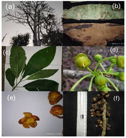
Figure 1: Collage of different morphological features of Dillenia pentagyna Roxb. (a) Habit (b) Bark (c) Leaves (d) Flower bud (e) Fruit (f) Dried fruit.
The plant specimen was authenticated from the prepared herbarium by experts at BSI, Shillong, and catalogued under Accession No. 099904 (Memo No. BSI/ERC/Tech/2023-24/1240; dated 09.08.2023). For this study, plant materials (Figure 1) were collected from Chasingre area of West Garo Hills, Meghalaya (25.56588°N, 90.23634°E). The complete taxonomic classification is provided in Table 1.
|
Table 1: Taxonomic classification of D. pentagyna. |
||
|
1 |
Kingdom |
Plantae |
|
2 |
Division |
Phanerogamic |
|
3 |
Sub-division |
Angiospermae |
|
4 |
Class |
Dicotyledonae |
|
5 |
Subclass |
Polypetalous |
|
6 |
Order |
Millennials |
|
7 |
Family |
Dilleniaceae |
|
8 |
Genus |
Dillenia |
|
9 |
Species |
pentagyna Roxb. |
|
10 |
BSI, Shillong Acc. No. |
099904 |
|
11 |
Memo No. |
BSI/ERC/Tech/2023-24/1240 |
|
12 |
Date |
09.08.2023 |
3.2. Qualitative Phytochemical Analysis of DPA
The preliminary analysis for phytochemicals in the aqueous extract of D. pentagyna stem bark (DPA) revealed an array of major phytochemical classes. The DPA displayed a notable combination of alkaloids, flavonoids, tannins, cardiac glycosides, coumarin, steroids and unspecified proteins and amino acids in the qualitative screening, indicative of a robust antioxidant profile and prospective pharmacological properties (Table 2).
|
Table 2: Screening for major phytochemicals from the aqueous extract of D. pentagyna bark (DPA). |
||
|
Phytochemical tested |
Result |
Biological activities |
|
Alkaloids |
+ |
Rheumatism, antispasmodic, analgesisc, gastrointestinal disorder[37],[38] |
|
Flavonoids |
+ |
ROS scavenging,[39],[40] stress responses in plants,[41] anticancer,[42] antihypertensive[43],[44] |
|
Phenols (Tannins) |
+ |
Antioxidant, [45] preservative, antimicrobial[46] |
|
Saponins |
+ |
Surfactant, hemolytic properties, stimulation of immune response[47] |
|
Cardiac Glycosides |
+ |
Inhibition of Na+/K+ ATPase pump,[48] anticancer, [49] neuroprotective[50] |
|
Steroids |
+ |
Lipid metabolism, [51] modulator of plant growth and development[52] |
|
Triterpenoids |
+ |
Growth and development of plants[53] |
|
Coumarins |
+ |
Anticancer, neuroprotective,[54] antimicrobial,[54],[55] anti-inflammatory,[55],[56] antiviral,[57] antidiabetic[58] |
|
Quinones |
+ |
Electron transport cofactor[59] |
|
Proteins and Amino Acids |
+ |
Influence nutritional and bioactive functions[60] |
3.3. Quantitative Estimation of Major Phytochemicals of DPA
3.3.1. TPC – The total phenolic content of the extract was assessed using the Folin–Ciocalteu assay, employing gallic acid as the calibration standard. The resulting standard curve exhibited strong linearity (y = 0.002x + 0.1045, R² = 0.9698), supporting the robustness of the analytical approach (Figure 2. a). Using the regression model and the measured absorbance of the extract, the TPC was calculated as 220.27 ± 0.72 mg GAE per gram of dry extract (Table 3).
This comparatively high phenolic level suggests that the extract has substantial reducing power, which is likely to play an important role in its overall antioxidant activity.
3.3.2. TFC – The total flavonoid content of the extract was quantified using the aluminium chloride colorimetric method, with quercetin serving as the reference standard. The quercetin calibration curve showed strong linearity (y = 0.0058x + 0.1366, R² = 0.9875), confirming the reliability of the measurement (Figure 2. b). Based on this curve, the flavonoid content of the extract was determined to be 123.76 ± 0.19 mg QE per gram of dry extract (Table 3).
3.3.3. TTC – Tannin levels in the extract were determined using the Folin–Ciocalteau method, with gallic acid employed as the reference standard. The calibration curve demonstrated strong linearity (y = 0.0207x + 0.0512, R² = 0.9794), confirming the suitability of the method (Figure 2. c). Using this regression model, the total tannin content (TTC) was calculated as 36.02 ± 0.06 mg GAE per gram of dry extract (Table 3).
3.3.4. Steroids Content – Total steroid content was quantified using β-sitosterol as the calibration standard. Based on the standard curve, the extract contained 0.93 ± 0.14 mg SE per gram of dry extract (Table 3).
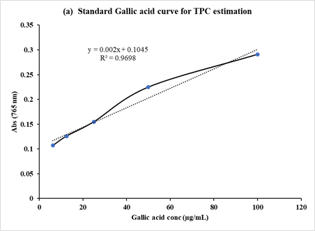
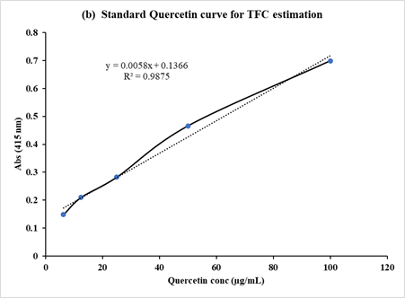
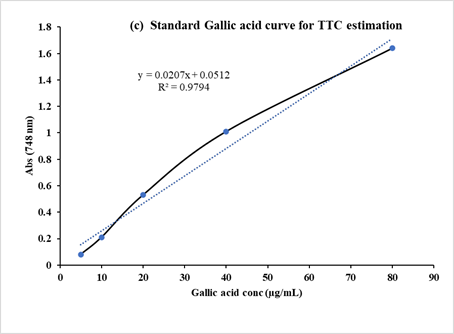
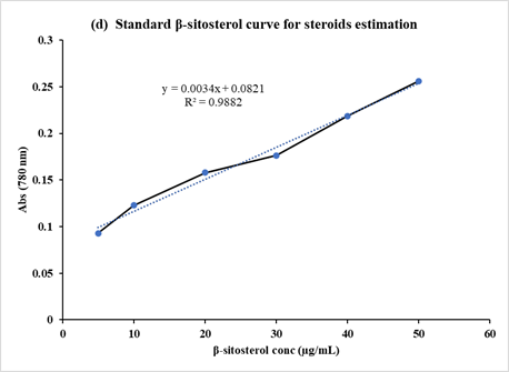
Figure 2: The standard calibration curves for the estimation of (a) TPC, (b) TFC, (c) TTC and (d) steroids.
|
Table 3. Quantitative estimation of major phytochemicals in the aqueous extract (DPA). |
||
|
Phytochemical |
Concentration (mg/g extract) |
Estimation technique |
|
TPC |
220.27±0.72 |
Spectrophotometry |
|
TFC |
123.76±0.19 |
Spectrophotometry |
|
TTC |
36.02±0.06 |
Spectrophotometry |
|
Steroids |
0.93±0.14 |
Spectrophotometry |
|
Coumarins |
2.192* |
Liquid chromatography |
|
Concentration represents Mean±SD, (n = 5). * n = 1. |
||
3.4. FTIR Analysis of DPA
The aqueous extract (DPA) was examined using Fourier-transform infrared (FTIR) spectroscopy to determine its functional groups and chemical bonding patterns. The absorption bands obtained from the FTIR spectrum (Figure 3) were evaluated to infer the functional groups likely associated with the major phytochemicals present in the extract. Distinct spectral peaks appeared at 3444 cm?¹, 2925 cm?¹, 1603 cm?¹, 1383 cm?¹, 1025 cm?¹, and 691 cm?¹, corresponding respectively to O–H stretching of hydroxyl groups, aliphatic C–H stretching, aromatic C=C stretching, aliphatic C–H bending, C–O stretching, and out-of-plane aromatic C–H bending (Table 4).
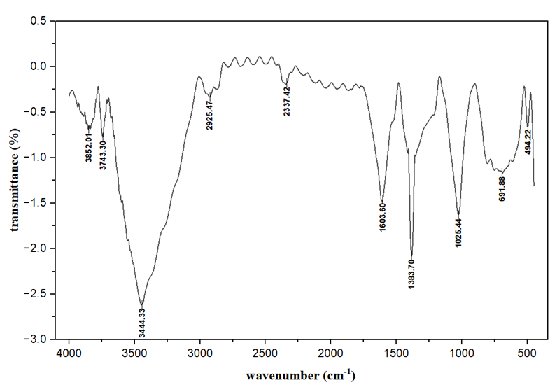
Figure 3: FTIR spectrum of the extract (DPA).
|
Table 4: FTIR peak values and functional groups in the aqueous extract (DPA). |
||||
|
Spectrum |
Wave number (cm-1) |
Probable Assignment |
Functional Group / Vibration |
Interpretation |
|
1 |
3852, 3743 |
O–H stretching (free) |
Hydroxyl group (alcohol or phenol) |
Sharp peaks suggest free (non-hydrogen-bonded) hydroxyl groups. |
|
2 |
3444 |
O–H stretching (H-bonded) |
Alcohols, phenols, or carboxylic acids |
Broad, strong band typical of hydrogen-bonded hydroxyl groups. |
|
3 |
2925 |
C–H stretching |
Aliphatic C–H (–CH?– or –CH?) |
Indicates presence of alkyl chains. |
|
4 |
2337 |
Possible CO? (asymmetric stretching) or overtone |
Atmospheric CO? or weak overtone |
May not correspond to a functional group in the sample itself. |
|
5 |
1603 |
C=C stretching / C=O stretching (aromatic or conjugated) |
Aromatic ring or conjugated carbonyl group |
Strong band, indicates aromatic or conjugated unsaturation. |
|
6 |
1383 |
C–H bending (deformation) |
–CH? or –CH?– groups |
Typical for aliphatic components. |
|
7 |
1025 |
C–O stretching |
Alcohols, phenols, ethers, or esters |
Suggests presence of C–O linkage. |
|
8 |
691 |
C–H bending (out-of-plane) |
Aromatic C–H (mono- or disubstituted benzene ring) |
Confirms aromatic ring presence. |
|
9 |
494 |
Metal–O or C–X stretching |
Possibly C–Br, C–Cl, or metal–oxygen |
Seen in halogenated or inorganic compounds. |
The broad, intense O–H band suggests a substantial presence of phenolic or flavonoid constituents. In addition, the C–O and C=C signals reinforce the presence of aromatic and oxygen-containing functional groups characteristic of secondary metabolites such as tannins, glycosides, and flavonoids. Collectively, these spectral features highlight the phytochemical richness of the aqueous extract and indicate that the stem bark of D. pentagyna harbor a diverse array of polar bioactive compounds that may contribute to its therapeutic properties.
3.5. UV-VIS Absorption Analysis of DPA and Standards
The UV–Visible absorption spectrum of DPA showed a pronounced absorption maximum around 270–280 nm (Figure 4a), a region typically associated with π→π* electronic transitions in aromatic rings and conjugated double-bond systems. This sharp and intense peak indicates a substantial presence of phenolic and flavonoid molecules within the extract. When compared with standard reference compounds (Figure 4b), including gallic acid, caffeic acid, coumaric acid, sinapic acid, rutin, and ascorbic acid, the extract exhibited spectral features most closely aligned with gallic acid and flavonoid-type structures. This similarity suggests that these classes of metabolites contribute significantly to the observed absorption pattern. Minor shifts in λ-max and the broadening of the absorption band relative to standards are likely due to the complex mixture of polyphenols, along with hydrogen bonding and solvent interactions characteristic of aqueous plant extracts. Overall, these spectral signatures point to a diverse composition of conjugated and aromatic constituents, consistent with the presence of phenolics, flavonoids, and other UV-active secondary metabolites known for their antioxidant activities.
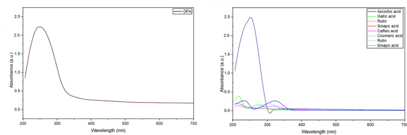
(a) (b)
Figure 4: UV-Vis Absorbance Spectra of DPA and common phytochemical standards. (a) The characteristic absorbance spectrum of DPA, showing a broad peak indicative of multiple compounds. (b) Absorbance spectra of individual phenolic and flavonoid standards (ascorbic acid, gallic acid, rutin, sinapic acid, caffeic acid, and coumaric acid), demonstrating their unique absorption maxima in the UV range and serving as reference data for identifying components within the crude extract.
Further support for these findings was provided by LC–HRMS and HPTLC analyses, which confirmed the presence of several phenolic acids and flavonoid derivatives corresponding to the UV–Vis signals. The LC–MS chromatogram of DPA revealed multiple peaks within the retention window typical of compounds such as gallic acid, caffeic acid, sinapic acid, and rutin, each known to exhibit strong absorption below 300 nm. Likewise, the HPTLC fingerprint showed distinct UV-active bands whose Rf values matched those of the standard references, providing additional validation of their occurrence in the extract. Together, the UV–Vis, LC–HRMS, and HPTLC results clearly demonstrate that the stem bark of D. pentagyna is rich in polyphenolic constituents, particularly hydroxylated aromatics and glycosylated flavonoids. These metabolites are well recognized for their potent antioxidant and therapeutic properties. The combined analytical evidence thus offers a comprehensive phytochemical profile of the aqueous extract and underscores its potential as a promising source of bioactive natural compounds.
3.6. HPTLC ‘Fingerprinting’ of DPA
The HPTLC profiling of major phytochemical constituents in the extract DPA was carried out on a silica gel 60 F254 plate using a mobile phase of toluene: ethyl acetate: methanol (8:1:1 v/v/v). Three different sample volumes (4 µL, 6 µL, 8 µL; tracks 1-3) were applied to check the consistency and concentration-dependent response of the analyte bands. The developed plates were visualized under UV light at 254 nm, 366 nm, and after derivatization with FeCl3 reagent for compound identification (Figure 5).
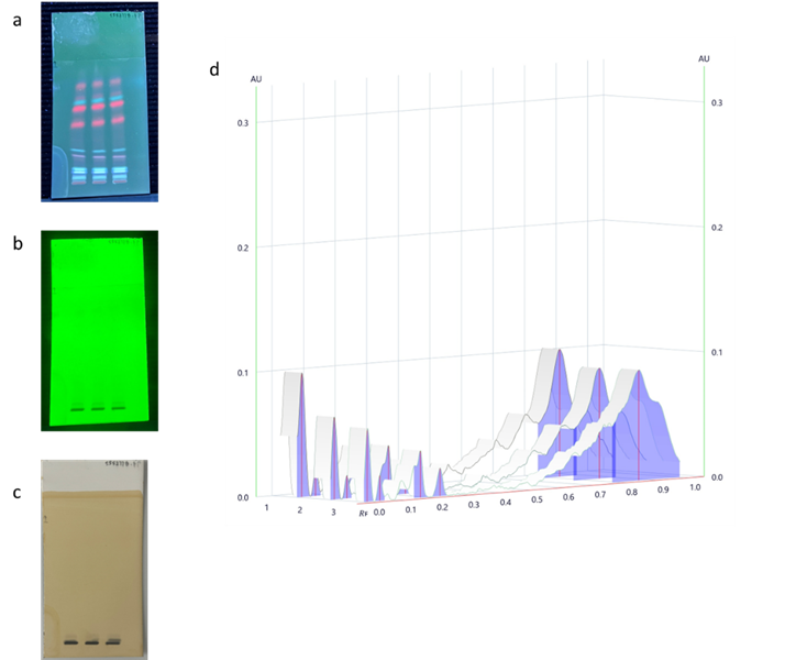
Figure 5: Thin Layer Chromatography (TLC) analysis of DPA. (a) TLC plate visualized under UV light at 366 nm (fluorescent compounds). (b) TLC plate visualized under UV light at 254 nm (absorbing compounds) (c) TLC plate derivatized with FeCl? reagent (detection of phenolic compounds). (d) Densitometric scan (3D view) of the TLC plate.
Under 254 nm UV, multiple compact and well-resolved dark bands appeared, indicating the presence of UV-absorbing compounds (Figure 5-b). A prominent spot with Rf value around 0.78-0.80 was observed across all three tracks, representing the major component of the extract. The band intensity increased proportionally with the application volume, confirming the linearity of response and reproducibility of the spotting and development process. Minor peaks observed at Rf ≈ 0.15–0.25 and 0.45–0.60 suggest the presence of secondary constituents in smaller quantities (Table 5).
|
Table 5: HPTLC derived spectral peak and Rf values of functional groups in DPA. |
|||
|
Wavelength 254 nm |
Wavelength 366 nm |
||
|
Peak no. |
Rf value |
Peak no. |
Rf value |
|
1 |
0.017 |
1 |
0.013 |
|
2 |
0.058 |
2 |
0.060 |
|
3 |
0.198 |
3 |
0.080 |
|
4 |
0.248 |
4 |
0.198 |
|
5 |
0.832 |
5 |
0.242 |
|
- |
- |
6 |
0.488 |
|
- |
- |
7 |
0.630 |
|
- |
- |
8 |
0.685 |
|
- |
- |
9 |
0.835 |
Under 366 nm UV, several fluorescent bands were visible, appearing mainly as blue and pink zones (Figure 5-a). These correspond to compounds with conjugated aromatic or phenolic structures, which exhibit fluorescence when excited at this wavelength. The densitometric data (Figure 5-d) confirm multiple peaks in the Rf range 0.03-0.07, with the most intense peak again at Rf ≈ 0.68-0.70, showing the primary active compound fluoresced strongly under UV.
After FeCl3 derivatization, three clear dark violet or bluish-black bands became visible, confirming the presence of phenolic compounds (Figure 5-c). FeCl3 reacts specifically with phenolic hydroxyl groups, producing colored complexes. This indicates that the sample contains polyphenolic or related antioxidant constituents. The identical Rf alignment of these spots across all volumes further supports the reproducibility of the method.
Overall, the HPTLC fingerprint of DPA is characterized by a major component at Rf ≈ 0.78 and several minor components at lower Rf values. The results show good separation, consistent peak intensity with increasing concentration, and stability under both UV and post-derivatization. These findings demonstrate the suitability of the chosen solvent system and confirm the presence of UV-active and phenolic compounds in the sample.
3.7. LC-HRMS Detection and Evaluation of Bioactive Compounds in DPA
The Liquid Chromatography–High Resolution Mass Spectrometry (LC–HRMS) analysis of the aqueous extract (DPA) revealed a highly complex and well-defined chromatographic pattern, as shown in the base peak chromatograms (BPCs) obtained in both positive and negative ionization modes. Numerous distinct peaks were distributed across the early, middle, and late retention windows, indicating a diverse mixture of phytochemicals spanning a broad range of polarities and molecular sizes. In the positive ionization mode (Figure 6a), a prominent sharp peak appeared at approximately 1.21 min, characteristic of small, highly polar compounds that elute rapidly due to strong interactions with the mobile phase. Additional peaks between 14.26 and 26.77 min reflected the presence of metabolites of moderate to low polarity. The negative ionization mode (Figure 6b) showed more pronounced peaks in the mid- and late-eluting regions, particularly at 14.27 and 26.47 min, suggesting the presence of higher-molecular-weight or more hydrophobic analytes.
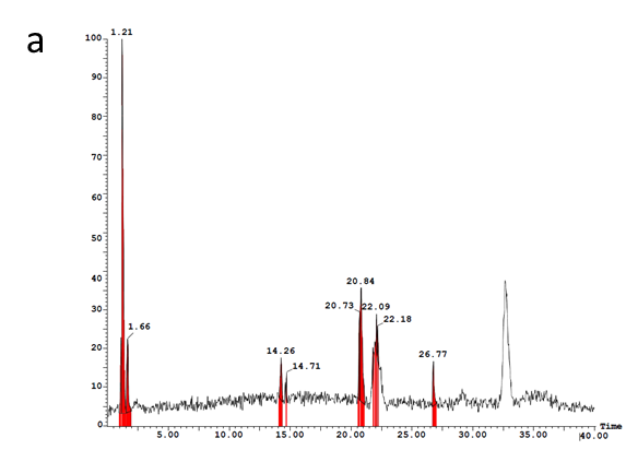
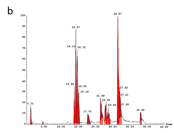
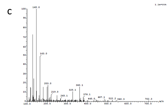
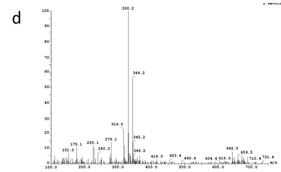

Figure 6. LC–HRMS profile of the extract (DPA): (a) BPC (base peak chromatogram) in positive ion mode; (b) BPC in negative ion mode; (c–e) corresponding MS spectra of major chromatographic peaks, illustrating the presence of diverse metabolites across different retention regions.
A total of thirteen compounds were tentatively identified using accurate mass measurements of precursor and product ions (m/z), supported by spectral database matches and literature reports. The earliest eluting peaks (RT 1.08–1.26 min) corresponded to small polar metabolites such as glycyl-glycine, L-glutamine, L-DOPA, and coumarin, indicating the presence of amino acid derivatives and other nitrogen-containing compounds (Table 6). The mid-retention region (RT 14.1–15.2 min) contained a cluster of bioactive molecules, including Scilli-N-desmethylpretazettine pyrenophorol, and 3-hydroxy-6-[[(E)-3-hydroxy-2,4-dimethylhept-4-enoyl]amino]-2,4-dimethyl-5-oxohexanoic acid, all showing precursor ions near m/z 330–344. These compounds are commonly associated with alkaloidal or polyketide-like structures and may contribute to the biological activity of the extract. Peaks detected between 20.6 and 23.2 min were consistent with dihydrorotenone, and karakoline, representing a mixture of flavonoid-related metabolites. A strong signal at approximately 26.47 min indicated the presence of co-eluting nonpolar constituents with high ionization efficiency, likely reflecting more hydrophobic secondary metabolites.
Late-eluting peaks (RT 32.6–33.1 min) were assigned to larger, structurally complex phytoconstituents, including HDMBOA + O-Hex, salvinorin A, a β-D-glucopyranoside derivative (C??H??O??), and a substituted cyclodecafuran analogue. These compounds typically belong to glycosylated phenolic or terpenoid classes and imply the presence of high-molecular-weight metabolites with potential pharmacological significance. Although the measured m/z values closely matched theoretical predictions, these identifications remain tentative and will require verification through co-elution with authentic standards and high-resolution MS/MS fragmentation analysis.
|
Table 6. LC–HRMS identified bioactive compounds from the extract (DPA). |
|||||
|
Sr. No |
Compound |
RT (min) |
Precursor ion (m/z) |
Product ions (m/z) |
Ref |
|
1 |
Glycyl-glycine |
1.174333 |
325.0281 |
325.02719 |
|
|
2 |
L-Glutamine |
1.226533 |
158.9346 |
158.92999 |
[61] |
|
3 |
LEVODOPA (L-DOPA) |
1.261333 |
115.0673 |
115.0693 |
[62] |
|
4 |
Coumarin |
1.261333 |
145.1243 |
145.12474 |
[63] |
|
5 |
Scilli-N-desmethylpretazettine |
14.12 |
330.1305 |
330.133 |
[64] |
|
6 |
Pyrenophorol |
14.2766 |
330.1955 |
330.1911 |
[65] |
|
7 |
3-hydroxy-6-[[(E)-3-hydroxy-2,4-dimethylhept-4-enoyl]amino]-2,4-dimethyl-5-oxohexanoic acid |
14.5898 |
344.2028 |
344.20676 |
|
|
8 |
Dihydrorotenone |
20.66242 |
148.9262 |
148.92986 |
[66] |
|
9 |
Karakoline |
21.98485 |
378.2628 |
378.26389 |
|
|
10 |
HDMBOA + O-Hex |
32.65112 |
432.1165 |
432.11371 |
|
|
11 |
Salvinorin A |
32.68592 |
431.174 |
431.17114 |
|
|
12 |
NCGC00380228-01 (C19H26O10 β-D-Glucopyranoside, 4-hydroxy-5-methyl-2-isopropylphenyl, 6-(2-carboxyacetate)) |
32.75552 |
432.1815 |
432.186 |
|
|
13 |
6-formyl-10-(hydroxymethyl)-5-methoxy-3-methylidene-2-oxo-2H,3H…cyclodeca[b]furan-4-yl 2-methylbutanoate |
33.10352 |
431.1414 |
431.14664 |
|
Overall, the LC–HRMS data reveal that the aqueous extract of D. pentagyna stem bark contains a broad spectrum of both polar and nonpolar bioactive molecules, reinforcing its phytochemical diversity and supporting its potential therapeutic value.
3.8. HPLC-Based Quantification of Coumarins
The concentration of coumarins in the DPA sample was determined using High-Performance Liquid Chromatography (HPLC) equipped with UV detection at 274 nm. The chromatographic procedure achieved clear resolution of analytes, yielding a well-defined coumarin peak at a retention time of approximately 4.32 minutes. This closely aligned with the retention time of the coumarin reference standard (4.15–4.25 minutes), thereby confirming the presence of coumarin in the sample. A calibration curve generated from standard solutions at 10, 20, and 40 µg/mL (Figure 7-a) demonstrated strong linearity between analyte concentration and peak area, described by the regression equation y = 898507x – 435035 with a correlation coefficient of R² = 0.9838, indicating reliable quantitative performance of the method.
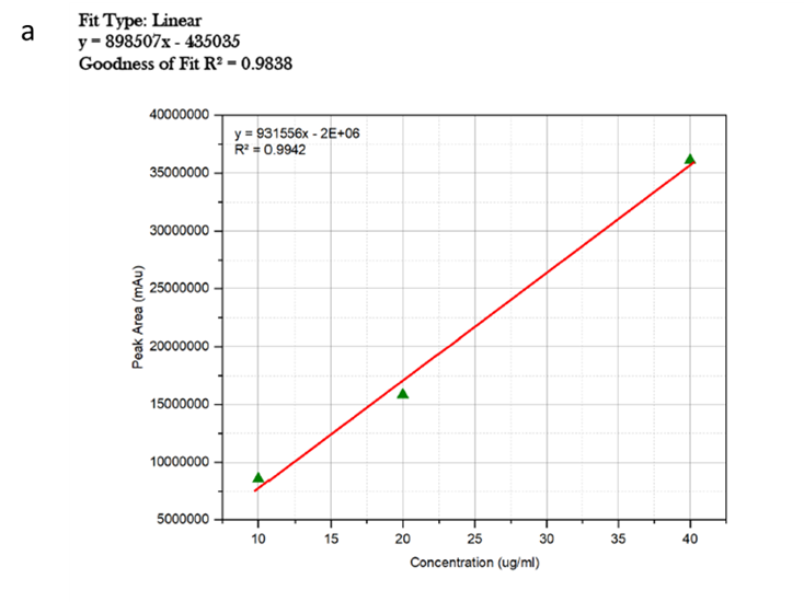
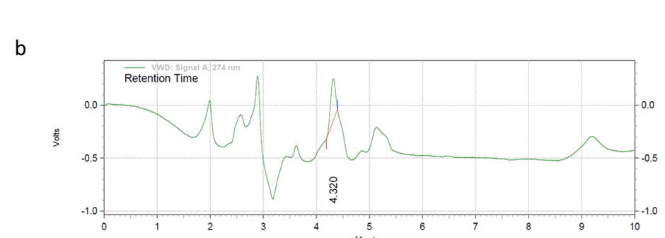
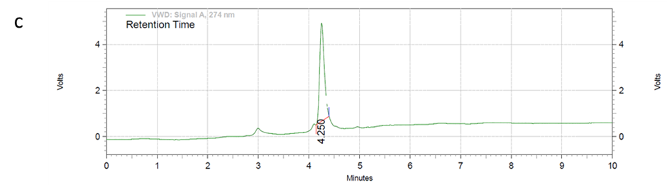
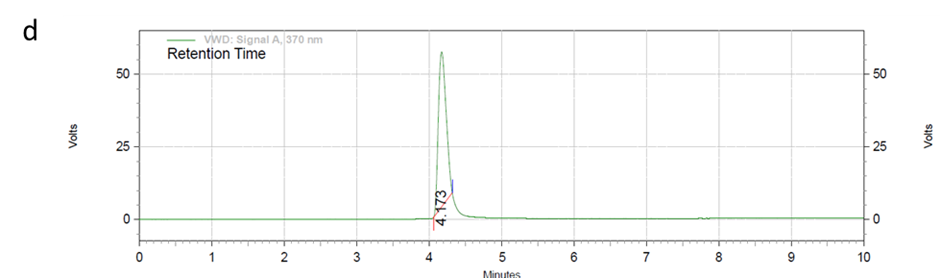
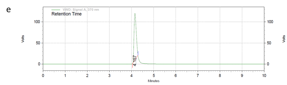
Figure 7: High-Performance Liquid Chromatography (HPLC) profiles for the identification of coumarins in the aqueous extract of D. pentagyna stem bark (DPA). a) Calibration curve showing the linear relationship between standard concentration (µg/mL). b) Chromatogram of DPA, showing various separated phytochemical peaks. (c-e) Chromatograms of the coumarin standard(s).
Analysis of the DPA sample (Figure 7-b) produced a coumarin peak area of 41,523, which corresponds to an estimated concentration of 2.192 µg/mL based on the calibration model. This indicates that coumarin is present at a low but measurable level in the sample. The close agreement in retention times and the symmetry of chromatographic peaks in both standards (Figure 7-c, -d, -e) and the sample suggest that the analytical parameters, including mobile phase composition (methanol:acetonitrile, 60:40), flow rate (1.0 mL/min), and the C18 stationary phase, were well optimized for selective separation and accurate quantification of coumarin.
3.9. Antioxidant Activity Evaluation
The antioxidant potential of DPA extract was evaluated using DPPH and ABTS radical scavenging assays. In the DPPH assay (Figures 8-a; 8-b), DPA exhibited a dose-dependent increase in percent radical scavenging activity (% RSA) across concentrations of 10 to 50 µg/mL. The linear regression analysis yielded the equation y = 1.6819x – 5.7416 with a strong coefficient of determination (R² = 0.9748), indicating a robust linear relationship between DPA concentration and % RSA. The IC50 value for DPA was calculated as 33.14 µg/mL, reflecting moderate antioxidant potency when compared to the reference antioxidant ascorbic acid, which showed a linear regression equation y = 1.6714x + 9.4831 (R² = 0.9768) and a lower IC50 of 24.24 µg/mL. The slightly lower IC50 and comparable R² of vitamin C indicate its superior free radical scavenging efficiency.
Similarly, in the ABTS assay (Figures 8-c; 8-d), DPA again demonstrated concentration-dependent antioxidant activity. The regression equation for DPA was y = 1.9641x – 25.585 with an R² of 0.8934, suggesting a good fit but slightly less linearity than observed in the DPPH assay. The IC50 value was 38.48 µg/mL, indicating a lower potency relative to the reference antioxidant Trolox, which showed a regression equation of y = 1.6983x + 8.8079 and a stronger correlation (R² = 0.9499) along with a significantly lower IC50 value of 24.25 µg/mL. These findings consistently demonstrate that while DPA possesses considerable antioxidant activity against both DPPH and ABTS radicals, its efficacy is moderately lower than established standards. The regression models and IC50 values together provide quantitative validation of DPA’s antioxidant capacity, supporting its potential as a natural free radical scavenger.
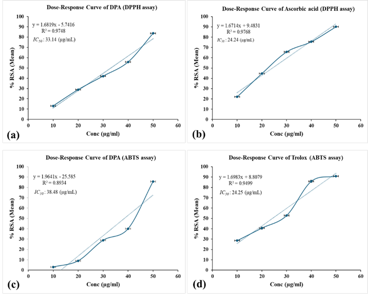
Figure 8 (a-b): DPPH radical-scavenging activity of the extract (DPA) and ascorbic acid. (c-d): ABTS radical-cation scavenging activity of DPA and Trolox.
3.10. In Vitro Antimicrobial Potentiality of DPA
Extrapolation of empirical results (Figure 9; Tables 7, 8) indicated that DPA influenced microbial growth inhibition in a dose-dependent manner. A ‘p’-value less than 0.05 of the DPA dosage (Mean±SD) as compared to the reference drug(s) is considered as having no significant difference in the effect i.e., the dosage has some effect against the particular microbial isolates. Consequently, DPA dosage of 10 mg/mL exhibited growth inhibition effect near to the standard reference drugs Ciprofloxacin 200 ppm (bacteria) and Itraconazole 350 ppm (fungi) on all tested microbial isolates except T. reesei (Table 7). The MIC determination of the extract (DPA) was found to be 0.09375 mg/mL against S. aureus, 0.1875 mg/mL against E. faecalis and S. typhi, and 0.375 mg/mL against P. aeruginosa among the bacterial strains. The MIC of DPA against the fungal strains A. niger and T. reesei were 0.1875 mg/mL and 0.75 mg/mL respectively (Table 8).
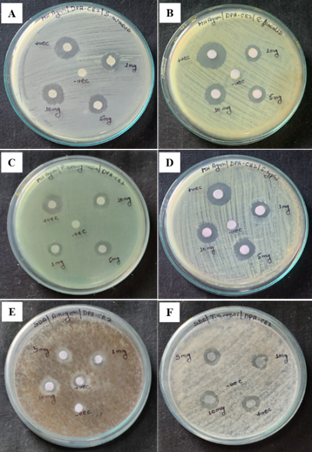
Figure 9: Photographs of antimicrobial activity across six microbial isolates showing zone of inhibition for DPA (1, 5, 10 mg/mL), negative control (distilled water) and positive control (Ciprofloxacin/Itraconazole). (A) S. aureus; (B) E. faecalis; (C) P. aeruginosa; (D) S. typhi; (E) A. niger; (F) T. reesei.
|
Table 7: Zone of inhibition exhibited by DPA and the reference drugs against select microbial isolates. |
||||||
|
Microbial isolates |
Inhibition Zone (mm) |
|||||
|
Ciprofloxacin |
Itraconazole |
DPA (1mg/mL) |
DPA (5mg/mL) |
DPA (10mg/mL) |
p value |
|
|
S. aureus |
10.25±0.25 |
… |
3.91±0.14 |
7.83±0.14 # |
10.83±0.14 ## |
0.0001#; 0.015 ## |
|
E. faecalis |
13.10±0.13 |
… |
1.91±0.14 |
6.33±0.14 |
12.41±0.14 * |
0.0012 * |
|
P. aeruginosa |
11.85±0.13 |
… |
1.75±0.25 |
5.33±0.14 |
12.88±0.23 ** |
0.0006 ** |
|
S. typhi |
10.30±0.18 |
… |
3.83±0.14 |
6.41±0.14 |
11.33±0.28 § |
0.0008 § |
|
A. niger |
… |
9.06±0.02 |
1.70±0.26 |
5.26±0.25 |
9.80±0.20 §§ |
0.01 §§ |
|
T. reesei |
… |
24.93±0.12 |
5.23±0.05 |
8.03±0.05 |
16.43±0.50 |
|
|
Data are presented as Mean ± Standard Deviation (SD); sample size (n) = 3 plates per microbial isolate. |
||||||
|
Table 8: Determination of MIC of DPA using broth dilution method. |
|||||||||
|
Microbial isolates |
MC |
Concentration (mg/mL) |
GC |
MIC (mg/mL) |
|||||
|
1 |
0.5 |
0.25 |
0.125 |
0.0625 |
0.0312 |
||||
|
S. aureus |
NT |
NT |
NT |
NT |
NT |
WT |
WT |
WT |
0.09375 |
|
E. faecalis |
NT |
NT |
NT |
NT |
WT |
WT |
WT |
WT |
0.1875 |
|
P. aeruginosa |
NT |
NT |
NT |
WT |
WT |
WT |
WT |
WT |
0.375 |
|
S. typhi |
NT |
NT |
NT |
NT |
WT |
WT |
WT |
WT |
0.1875 |
|
A. niger |
NT |
NT |
NT |
NT |
WT |
WT |
WT |
WT |
0.1875 |
|
T. reesei |
NT |
NT |
WT |
WT |
WT |
WT |
WT |
WT |
0.75 |
|
Abbreviations: MC - Media control; GC - Growth control; NT - No turbidity; WT - With turbidity |
|||||||||
4. DISCUSSION:
The field survey confirmed that Dillenia pentagyna is well established in the West Garo Hills of Meghalaya, and other parts of Northeast India. This ecological finding is important because the geographic origin and growing environment of medicinal plants can strongly influence their phytochemical composition and, in turn, their biological activity. Recording the plant’s vernacular names emphasizes its cultural and ethnobotanical importance in the region. At the same time, the taxonomic identification of the species by the Botanical Survey of India, ensures clarity and reliability in the subsequent chemical studies.
The notably high flavonoid level of the aqueous extract of Dillenia pentagyna (DPA) indicates that the extract contains abundant secondary metabolites, which are well recognized for their free-radical-scavenging, anti-inflammatory, and cytoprotective activities.[20] The presence of tannins suggests that they may still contribute meaningfully to the extract’s antioxidant activity, as well as its potential metal-chelating and antimicrobial effects. Although present at much lower levels than phenolics and flavonoids, the steroidal constituents of the aqueous extract (DPA) may still play a role in the extract’s bioactivity, particularly through their reported anti-inflammatory and membrane-stabilizing effects.
FTIR spectroscopy provided the first definitive evidence of the main functional groups in DPA. The broad, intense band at 3444 cm?¹ (O–H stretching), along with the corresponding C–O stretching signal at 1025 cm?¹, indicates a high abundance of hydroxyl-rich compounds such as phenols, flavonoids, and glycosides. Additional peaks at 1603 cm?¹ (C=C stretching) and 691 cm?¹ (out-of-plane C–H bending) point to aromatic rings and conjugated systems. Together, these features are typical of polyphenolic structures and confirm that the extract contains a substantial amount of polar, bioactive molecules.
These results were supported by the UV–Vis absorption spectrum, which showed a major absorption peak between 270–280 nm. This π→π* transition is characteristic of aromatic and conjugated compounds, especially phenolic acids and flavonoids.[67] The similarity between DPA’s absorption profile and those of gallic acid and rutin further suggests that these classes of metabolites make significant contributions to its chemical composition. Overall, the combined FTIR and UV–Vis data provide a strong phytochemical basis, indicating that the aqueous extract of D. pentagyna stem bark (DPA) is distinctly rich in polyphenols.
HPTLC profiling produced a clear, reproducible chemical fingerprint of the extract. Well-defined UV-absorbing bands at 254 nm and fluorescent bands at 366 nm reinforced the presence of aromatic and polyphenolic compounds identified by spectroscopy. The appearance of intense dark violet bands after FeCl? derivatization, a classic reaction for phenolics, offered direct chemical confirmation of multiple phenolic constituents in the DPA extract.[67],[68] Densitometric analysis revealed a major, high-Rf compound indicative of a relatively less-polar metabolite, along with several minor spots representing additional components, highlighting the extract’s chemical complexity.
LC–HRMS analysis provided the most comprehensive view of the extract’s chemical diversity, enabling the tentative identification of thirteen compounds. The elution patterns in both positive and negative ionization modes revealed a wide range of molecular polarities, from highly polar amino acid derivatives (e.g., L-glutamine, L-DOPA) to more hydrophobic and higher-molecular-weight compounds (e.g., Salvinorin A and β-D-glucopyranoside derivatives). The detection of alkaloids, such as Scilli-N-desmethylpretazettine and karakoline, further underscores the chemotaxonomic richness of D. pentagyna.
Coumarins content (2.192 mg/g extract) was relatively in moderate amount, and is comparable or higher to many reported plants.[54] The presence is noteworthy because these benzopyrone derivatives are associated with anti-inflammatory, anticoagulant, and antimicrobial activities.[56],[57],[55],[69] The consistent identification of coumarin across both LC–HRMS (RT = 1.261 min) and quantitative HPLC analyses reinforces the reliability and complementarity of the analytical approaches used.
In the antioxidant tests, DPA showed strong ability to neutralize free radicals with an IC?? value close to the standard antioxidants ascorbic acid and Trolox. This might be explained by the synergistic presence of several phenolic chemicals as elucidated by the phytochemical characterization earlier in the section. These results are consistent with those of Tene et al. (2021)[20] and Singh et al. (2023),[70] who found that D. pentagyna fractions rich in polyphenols were potent modulators of oxidative stress.
Additionally, DPA showed broad spectrum antibacterial activity encompassing gram-positive (S. aureus, E. faecalis) and gram-negative (P. aeruginosa, S. typhi) bacterial strains with relatively low MIC values ?1 mg/mL. A high potential for application in natural antibacterial formulations is suggested by this broad-spectrum inhibition. This aligns with previous findings with D. pentagyna and other therapeutic plants.[71],[72],[70] According to Chethan kumara et al. (2021),[73] polyphenolic and flavonoid chemicals most likely provide antibacterial activity by disrupting cell membranes or by inhibiting microbial enzymes. Antifungal efficacy of DPA against A. niger (MIC 0.1875 mg/mL) showed promising results whereas T. reesei was least responsive.
CONCLUSION:
This study brings together ecological observations and a range of analytical techniques to paint a clear picture of the phytochemical richness of Dillenia pentagyna. Our fieldwork confirmed that the plant grows abundantly in the West Garo Hills of Meghalaya, and careful taxonomic verification ensured that all subsequent analyses were based on correctly identified material. From the very first spectroscopic tests, FTIR and UV–Vis, it became evident that the aqueous extract of Dillenia pentagyna is packed with polyphenols and other hydroxyl-rich compounds.
Further chromatographic analyses strengthened this view. HPTLC produced a distinct chemical “fingerprint” of phenolic compounds, while the LC–HRMS profiling added even more depth by tentatively identifying thirteen diverse metabolites, ranging from polyphenols and alkaloids to amino acid derivatives and lipid-related molecules. HPLC allowed us to quantify coumarins. Notably, the level of coumarins in the stem bark points to D. pentagyna as a potentially overlooked natural source of this widely valued antioxidant.
Because of its high phenolics and flavonoid content, D. pentagyna has strong antioxidant properties, and promising broad spectrum antimicrobial potentiality.
Overall, our findings show that D. pentagyna is chemically diverse and rich in compounds known for their therapeutic potential. This multidimensional analysis not only confirms the plant’s value in traditional medicinal contexts but also lays the groundwork for future research into its biological effects and possible use in developing standardized herbal or nutraceutical products. To prove its therapeutic usefulness and safety for human applications, however, more research incorporating in vivo pharmacokinetics, molecular pathways, and clinical evaluations is required.
ABBREVIATIONS:
ABTS – 2',2'-Azinobis-(3-ethylbenzothiazoline-6-sulfonic acid); ANOVA – Analysis of Variance; BPC – Base Peak Chromatogram; BSI – Botanical Survey of India; Da – Dalton; DPPH – 2,2-Diphenyl-1-picrylhydrazyl; DW – Distilled water; DPA – Dillenia pentagyna Aqueous extract; ESI-MS detector – Electrospray ionization-mass spectrometry detector; ATR-FTIR – Attenuated Total Reflectance-Fourier-Transform Infrared; HPLC – High Performance/Pressure Liquid Chromatography; HPTLC – High-Performance Thin-Layer Chromatography; hr – Hour(s); IR – Infrared; LC-HRMS – Liquid Chromatography-High Resolution Mass Spectrometry; MHA – Mueller Hinton Agar; MoNA – MassBank of North America; MTCC – Microbial Type Culture Collection and Gene Bank; PDA detector – Photodiode Array Detector; Rf – Retention factor; Roxb. – Roxburgh; SRL – Sisco Research Laboratories Pvt Ltd, India; TEAC - Trolox Equivalent Antioxidant Capacity; UV-Vis – Ultra violet & visible.
ACKNOWLEDGEMENT
The authors express their gratitude to the Director, CytoGene Research & Development LLP, Lucknow, India, for extending essential research facilities (HPTLC, LC-HRMS and HPLC) for this work.
FINANCIAL SUPPORT AND SPONSORSHIP
There is no financial support or grant from funding agencies for this research work.
DECLARATION OF COMPETING INTEREST
The authors declare that there is no conflict of interest.
AVAILABILITY OF DATA AND MATERIALS
The datasets analysed during the current study are available from the corresponding author on reasonable request.
CREDIT AUTHORSHIP CONTRIBUTION STATEMENT
Lyngdoh T.S.: Conceptualization, Methodology, Investigation, Writing – Original draft, Editing, References.
Maibam D.D.: Conceptualization, Supervision, Writing – Critical Review, Editing.
REFERENCES


Toijam Sanjeev Lyngdoh, Maibam Damayanti Devi, Comprehensive Phytochemical Characterization and Antimicrobial Synergy of Dillenia pentagyna Aqueous Extract, Int. J. of Pharm. Sci., 2025, Vol 3, Issue 12, 1637-1665. https://doi.org/10.5281/zenodo.17868469
 10.5281/zenodo.17868469
