
We use cookies to ensure our website works properly and to personalise your experience. Cookies policy
Department of Life Sciences (Botany), Manipur University, Manipur, India 795003
Oxidative stress and inflammation are fundamental biological processes that play critical roles in the onset and progression of various acute and chronic diseases. Homalomena aromatica (Spreng.) Schott is an aromatic medicinal plant traditionally used for treating inflammatory conditions. The present study evaluated the effect of H. aromatica leaf aqueous extract against copper sulfate-induced oxidative stress and inflammation by integrating in vitro bioactivity screening with in vivo validation and safety assessment using zebrafish. The extract showed no acute or developmental toxicity at the limit dose of toxicity testing assigned by OECD Guideline 236. The extract showed a dose-dependent protection against CuSO4-induced lethality and morphological deformations. The extract also showed protection against CuSO4-induced intracellular ROS generation, neutrophil accumulation and lipid peroxidation in a dose-dependent manner. However, the extract did not show strong in vitro Cu2+-chelating activity. In addition, radical scavenging activity was also significantly lower than that of the standard ascorbic acid. In conclusion, despite limited in vitro metal-chelating and radical scavenging activities, the extract showed potent in vivo antioxidant and anti-inflammatory properties, effectively mitigating CuSO4-induced oxidative stress and inflammation in zebrafish. These findings highlight the importance of whole-organism validation, suggesting that antioxidant and anti-inflammatory effects may be mediated through biological modulation, rather than direct chemical scavenging mechanisms.
Oxidative stress and inflammation are fundamental biological processes that play critical roles in the onset and progression of various acute and chronic diseases. Recent studies have shown that dysregulation of redox homeostasis and persistent inflammatory responses contribute to cardiovascular and neurodegenerative diseases, metabolic disorders, cancer and aging-related complications.[1,2] Consequently, the identification of safe and effective agents that could reduce oxidative stress and inflammation has drawn much interest.
Medicinal plants have been used for centuries in traditional medicine systems for treating and managing inflammatory and oxidative stress-related disorders. Secondary metabolites produced by plants such as phenolics, flavonoids, alkaloids and terpenoids have been studied for their antioxidant and anti-inflammatory properties.[3] However, despite extensive research on medicinal plants, important knowledge gaps still remain. Numerous studies rely solely on assays conducted under cell-free in vitro conditions which do not accurately predict biological efficacy within cells or whole organisms.[4] The scientific evaluation of medicinal plants, must therefore, integrate in vitro bioactivity screening with in vivo validation and safety assessment.
Homalomena aromatica (Spreng.) Schott is a perennial aromatic medicinal herb belonging to the family Araceae (order Alismatales). According to the Plants of the World Online, the species is accepted and native across parts of South and Southeast Asia, including Assam and the East Himalayan region, Bangladesh, Myanmar, Thailand, Vietnam, Laos, Cambodia and southern China.[5] The plant is widely valued for its aromatic rhizomes which are commercially used to extract a volatile aromatic essential oil.[6] Previous studies have reported the use of H. aromatica leaves and rhizomes for treating infections and inflammatory conditions such as joint pain, common cold, skin infections and gastrointestinal ailments.[7]
Therefore, the aim of this study was to evaluate the antioxidant and anti-inflammatory potential of H. aromatica leaf aqueous extract under in vitro as well as in vivo conditions. The present study used chemically-induced oxidative stress and inflammation model in zebrafish larvae using copper sulfate pentahydrate (CuSO4.5H2O).[8] The safety profile of the plant extract was also assessed using zebrafish embryos and larvae.
Zebrafish (Danio rerio) larvae were selected as the primary in vivo model in this study owing to their optical transparency, thereby enabling non-invasive visualization of reactive oxygen species (ROS) production and immune cell migration.[9,10] In addition, zebrafish show a strong genetic and physiological conservation with humans, with at least one zebrafish ortholog for approximately 70% of human protein-coding genes, including genes involved in redox regulation and inflammatory signaling.[11] Furthermore, zebrafish embryos and larvae are well-established models for toxicity testing. The Fish Embryo Acute Toxicity (FET) test is standardized under the Organisation for Economic Co-operation and Development (OECD) Test Guideline 236, providing internationally accepted criteria for evaluating lethality, developmental toxicity and sublethal morphological endpoints.[12] The above attributes validate the selection of zebrafish embryos and larvae as a scientifically robust model for integrated antioxidant, anti-inflammatory and safety evaluation.
2. MATERIALS AND METHODS
2.1. Collection of plants and extract preparation
Plants were collected from Jiribam District of Manipur, India. A voucher specimen was prepared and identified at the Department of Life Sciences (Botany), Manipur University with accession number 001039 and deposited at the Manipur University Museum of Plants. The leaves were thoroughly washed in running tap water, then finally rinsed with distilled water. They were shade dried for a few hours, then cut into small sections and allowed to shade dry until completely dried. The dried samples were then ground using an electric grinder.
800 mL of milli-Q water was added to 40 g leaves and constantly agitated at 150 rpm for 24 hours. The extract was then filtered using Whatman 1 filter paper and concentrated using a lyophiliser. The concentrated leaf extract (HALAE) was stored at -20 ? for further use.
2.2. Chemicals and reagents
2,2-diphenyl-1-picryl-hydrazyl (DPPH), 2,2´-azinobis-(3-ethylbenzothiazoline-6-sulphonate) (ABTS), ascorbic acid, copper sulphate pentahydrate (CuSO4.5H2O) and Ethylenediaminetetraacetic acid (EDTA) were obtained from Sisco Research Laboratories (SRL), India. 3,4-Dichloroaniline (DCA) was obtained from Sigma-Aldrich, USA. Pyrocatechol violet (PV) was obtained from TCI, Japan. Methanol was obtained from HiMedia, India. All other reagents were of analytical grade. In the following sections, copper sulphate pentahydrate (CuSO4.5H2O) is referred to as CuSO4.
2.3. Zebrafish maintenance and embryo collection
Adult wild-type zebrafish were obtained from a local vendor in India and acclimatized in glass aquariums containing aerated dechlorinated water at a temperature of 28 ± 2 ? and a 14 hours light/ 10 hours dark photoperiod. They were fed thrice daily with commercial fish food. The fully mature adult males and females were separated one day prior to breeding.
The fish were transferred to a breeding tank in the ratio of three males to three females. A net was used to protect the newly spawned eggs. The tank was maintained at a temperature of 28 ± 2 ? and 14 hours light/ 10 hours dark photoperiod. Spawning and fertilization were instigated at the onset of the first light. The embryos were collected using a Pasteur pipette and transferred to petri dishes containing egg water (milli-Q water containing 60 µg/mL Instant Ocean salt).
The embryos were washed twice with egg water. Healthy 6 hours post-fertilization (hpf) embryos were selected under a stereozoom microscope. For further experiments in larvae, the 6 hpf embryos were reared in 1x E3 medium until hatched. Only spontaneously hatched larvae at 72 hpf were used for treatment. The embryos were staged according to Kimmel et al.[13]
2.4. Safety assessment using zebrafish
The toxicological evaluation was performed following the OECD guideline number 236.[12] 6 hpf embryos were transferred to 6-well plates, 10 in each well. The embryos were divided into three treatment groups: Egg water (negative control), 4 µg/mL of DCA (positive control) and different concentrations of HALAE prepared in egg water i.e. 100, 250, 500, 750, 1000, 1250, 1500 µg/mL. The final volume of the treatment solution was 2 mL per well. Semi-static method was followed and the embryos were transferred to fresh solution of the same test concentration every 24 hours. The embryos were exposed for 96 hours and observations were recorded every 24 hours. Coagulated embryos and dead larvae were removed. Mortality rate (%) at 96 hpf, hatching rate (%) at 72 hpf and heartbeat rate [beats per minute (bpm)] at 48 hpf were taken as endpoint observations. The hatched dead larvae were also taken into account for determining the hatching rate. The heartbear rate was recorded only for those embryos with a clearly visible heartbeat.
Mortality rate (%) was calculated using the following equation:
Mortality rate (%) = Total number of dead embryos/larvae at 96 hpfTotal number of embryos at the start of treatment ×100

Hatching rate (%) was calculated using the following equation:
Hatching rate (%) = Total number of hatched larvae at 72 hpfTotal number of embryos at the start of treatment × 100

All treatments were done in triplicate. Each replicate consisted of ten embryos. The results are expressed as mean ± standard error of mean (S.E.M.).
LC50 was calculated using probit analysis.
2.5. Effect of HALAE against CuSO4-induced acute toxicity
The effect of HALAE against CuSO4-induced acute toxicity was assessed following Nguyen et al.[8] with certain modifications. Spontaneously hatched 72 hpf larvae were transferred to 24-well plates, 20 in each well. The larvae were incubated in 1980 µL of different concentrations of HALAE (2, 4, 6, 8, 10, 15, 20 µg/mL) for 1 hour. Then, 20 µL of 20 mM CuSO4 (prepared in milli-Q water) was added to each well so that each well contained a final concentration of 20 µM of CuSO4 in a final volume of 2 mL per well. At 24 hours post-treatment (hpt), mortality was taken as the endpoint. Larvae treated with egg water and 20 µM CuSO4 were taken as negative and positive controls respectively. All treatments were done in triplicate. The experiments were repeated thrice. The results are expressed as mean ± standard error of mean (S.E.M.).
In a follow-up experiment, the effect of the highest tested dose of HALAE i.e. 20 µg/mL was assessed against different concentrations of CuSO4 (10, 20, 30, 40, 50 µM) following the above procedure. EC50 was determined by non-linear regression analysis.
All treatments were done in triplicate. The experiments were repeated thrice. The results are expressed as mean ± standard error of mean (S.E.M.).
Zebrafish maintenance and all the experiments were done in accordance to the guidelines of the Institutional Animal Ethics Committee.
2.6. Cu2+-chelating activity
The in vitro Cu2+-chelating activity was assessed following the protocol described by Nguyen et al.[8] with slight modifications. EDTA was used as standard. In brief, a reaction mixture containing 30 µL of different concentrations of EDTA (1.46, 2.92, 4.38, 5.84, 7.31, 8.77 ng/mL) or HALAE (0.25, 0.5, 0.75, 1.0, 1.5, 2.0 mg/mL) or water (control), 200 µL of 50 mM sodium acetate buffer (pH 6.0) and 30 µL of 1 mM CuSO4 was added to each well of a 96-well plate. It was allowed to react for two minutes, after which 8.5 µL of 2 mM PV was added. The reaction mixtures were then incubated for 10 minutes on a horizontal shaker. Absorbance was measured at 632 nm using a UV-Visible spectrophotometer.
The Cu2+-chelating activity was calculated using the following equation:
Cu2+-chelating activity %=A0-A1A0×100
Where, A0 is the absorbance of the control and A1 is the absorbance of the test sample.
EC50 was calculated using linear regression analysis.
All reactions were done in triplicate. The experiments were repeated thrice. The results are expressed as mean ± standard error of mean (S.E.M.).
2.7. In vivo antioxidant activity
Intracellular ROS levels were assessed using the 2’,7’-dichlorodihydrofluorescein diacetate (DCFH-DA) dye.[8] Following cellular uptake, DCFH-DA is deacetylated by intracellular esterases to yield the non-fluorescent compound 2’,7’-dichlorodihydrofluorescein (DCFH) which is subsequently oxidized by ROS to the fluorescent 2’,7’-dichlorofluorescein (DCF). The fluorescence intensity was used to assess level of oxidative stress. Spontaneously hatched 72 hpf zebrafish larvae were transferred to 24-well plates, six in each well. The larvae were incubated in 1980 µL of different concentrations of HALAE (4, 8, 20 µg/mL) for 1 hour. Then, 10 µL of 20 mM CuSO4 (prepared in milli-Q water) was added to each well so that each well contained a final concentration of 10 µM of CuSO4 in a final volume of 2 mL per well. After 24 hours of incubation, the larvae were washed thrice with egg water and then incubated with 20 µg/mL DCFH-DA solution for thirty minutes in the dark. After incubation, the larvae were washed thrice with egg water to remove the excess dye and anaesthetized with 0.003% tricaine methane-sulfonate prepared in egg water. DCF fluorescence was visualized using a fluorescence microscope. Larvae treated with egg water and 10 µM CuSO4 were taken as negative and oxidative stress controls respectively. Larvae that were pre-incubated with 20 µg/mL gallic acid and further treated with 10 µM CuSO4 for 24 hours were taken as positive control.
2.7. In vitro antioxidant activity
The in vitro antioxidant activity was evaluated using the DPPH[14] and ABTS[15] radical scavenging assays. Ascorbic acid was used as standard.
2.7.1. ABTS radical scavenging assay
The ABTS radical cation was generated by mixing 7 mM ABTS solution with 2.6 mM potassium persulfate in equal volumes (1:1, v/v), then incubated in the dark at room temperature for 12 hours. The resulting ABTS cation stock solution was diluted with ethanol to obtain an absorbance of 0.70 ± 0.02 at 734 nm. 1 mL of different concentrations of ascorbic acid (2, 4, 6, 8, 10 µg/mL) and HALAE (20, 50, 100, 150, 200 µg/mL) were mixed with 2 mL of the working ABTS solution and incubated in the dark for 30 minutes. Absorbance was measured at 734 nm using a UV-Visible spectrophotometer.
2.7.2. DPPH radical scavenging assay
A 0.1 mM DPPH solution prepared in methanol was used as the working solution. 1 mL of different concentrations of ascorbic acid (2, 4, 6, 8, 10, 12 µg/mL) and HALAE (100, 200, 300, 400, 500 µg/mL) were mixed with 2 mL of the working DPPH solution and incubated in the dark for 30 minutes. Absorbance was measured at 517 nm using a UV-Visible spectrophotometer.
2.7.3. Calculation
The radical scavenging activity (%) was calculated using the following equation:
Scavenging activity %=A0-A1A0×100
Where, A0 is the absorbance of the control (radical solution without sample) and A1 is the absorbance of the test sample.
IC50 was calculated using linear regression analysis.
All reactions were done in triplicate. The experiments were repeated thrice. The results are expressed as mean ± standard error of mean (S.E.M.).
2.8. In vivo anti-inflammatory activity evaluation
2.8.1. Assessment of neutrophil accumulation
The neutrophil accumulation in zebrafish larvae was observed using Sudan Black B staining.[16,17] Briefly, larvae were subjected to the same experimental design as in the ROS generation assessment. Following this, the larvae were anaesthetized in 0.003% tricaine methane-sulfonate and fixed in 4% paraformaldehyde (PFA) for 24 hours. After this, the larvae were washed in 1X Phosphate-buffered saline (PBS) three times for five minutes. Then, the larvae were incubated in Sudan Black B working solution in the dark for 1 hour, then washed with 70% ethanol three times, then washed with depigmentation solution for 5 minutes. The accumulation of neutrophils was then observed under a stereozoom microscope.
2.8.2. Evaluation of lipid peroxidation
Lipid peroxidation was evaluated using the TBARS method.
[18] Briefly, larvae were subjected to the same experimental design as in the ROS generation assessment. Following this, the larvae were anaesthetized in 0.003% tricaine methane-sulfonate and homogenized in 1X PBS. 150 µL of homogenate was added to 150 µL of 10% trichloroacetic acid (TCA) and centrifuged at 10 g at 4? for ten minutes. 150 µL of the supernatant was added to 150 µL of 0.67% Thiobarbituric acid (TBA) and boiled at 100? for thirty minutes, then cooled to room temperature. Absorbance was measured at 532 nm using a UV/Visible spectrophotometer.
2.8. Statistical analysis
Results were analyzed and visualized using GraphPad Prism 8.0.2, IBM SPSS Statistics V21.0 and MS Excel 2021. The means were compared using one-way analysis of variance (ANOVA) followed by Tukey’s post hoc test at α equals 0.05. The No Observed Adverse Effect Concentration (NOAEC) and Lowest Observed Adverse Effect Concentration (LOAEC) were computed as the highest tested concentration that shows no statistically significant adverse effect compared with the negative control and the lowest tested concentration that shows a statistically significant adverse effect relative to the negative control.
3. RESULTS
3.1. Safety assessment using zebrafish
The safety assessment of HALAE was done using zebrafish. It was clearly observed that at 96 hpf, HALAE was not lethal to zebrafish embryos even at the highest tested dose of 1500 µg/mL (Figure 1 [A]), which far surpasses the limit dose of toxicity testing 100 µg/mL assigned by OECD.[12] However, at 72 hpf, a distinct hatching delay was observed in the embryos treated with 1000, 1250 and 1500 µg/mL of HALAE as compared to the negative control embryos (Figure 1 [B]). In addition, at 48 hpf, a statistically significant decrease in heartbeat rate was observed in the embryos treated with 1000, 1250 and 1500 µg/mL of HALAE as compared to that of the negative control embryos (Figure 1 [C]).
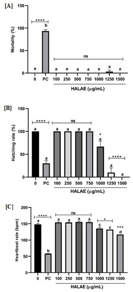
Figure 1: Embryo and developmental toxicity of HALAE in zebrafish embryos: [A]: mortality (%) at 96 hpf; [B]: hatching rate (%) at 72 hpf; [C]: heartbeat rate (bpm) at 48 hpf respectively of zebrafish embryos treated with different concentrations of HALAE.
Each bar represents the mean ± S.E.M. Statistical differences among groups were analyzed using one-way ANOVA followed by Tukey’s post-hoc test, α equals 0.05. Significance levels in the figure represent the difference of each column from the negative control column and are presented as: * indicates p<0.05, *** indicates p<0.001, **** indicates p<0.0001, ns indicates not significant. Columns with significantly different means are represented by different alphabets. bpm: beats per minute; hpf: hours post fertilization; PC: Positive control (4 µg/mL DCA).
The estimated values of LC10, LC50 and LC90 determined from the mortality rate of zebrafish embryos treated with varying concentrations of HALAE are given in Table 1. The NOAEC and LOAEC with respect to hatching rate and heartbeat rate are given in Table 2. However, these values cannot be determined for mortality because no lethality of HALAE was observed in the embryos.
Table 1. LC10, LC50 and LC90 values of HALAE in zebrafish embryos.
|
HALAE |
Estimated LC value |
Lower bound |
Upper bound |
|
LC10 |
2302.00 |
Cannot be determined |
Cannot be determined |
|
LC50 |
3536.53 |
Cannot be determined |
Cannot be determined |
|
LC90 |
4771.06 |
Cannot be determined |
Cannot be determined |
Table 2. NOAEC and LOAEC of HALAE in zebrafish embryos with respect to mortality, hatching rate and heartbeat rate.
|
HALAE |
NOAEC (µg/mL) |
LOAEC (µg/mL) |
|
Mortality |
Cannot be determined |
Cannot be determined |
|
Hatching rate |
750 |
1000 |
|
Heartbeat rate |
750 |
1000 |
NOAEC: No Observed Adverse Effect Concentration; LOAEC: Lowest Observed Adverse Effect Concentration.
3.2. Effect of HALAE against CuSO4-induced acute toxicity
In this study, 72 hpf zebrafish larvae exposed to a lethal dose of 20 µM CuSO4 showed a mortality rate of 81.11% while the larvae that were co-treated with varying concentrations of HALAE showed a dose-dependent decrease in mortality rate (Figure 2 [A]). The mortality rates of the larvae pre-treated with different concentrations of HALAE were as follows: 76.11% (0.5µg/mL); 69.44% (2 µg/mL); 58.33% (4 µg/mL); 43.33% (6 µg/mL); 30.56% (8 µg/mL); 26.67% (10 µg/mL); 3.89% (15 µg/mL) and 0.56% (20 µg/mL). The mortality rates of larvae treated with the lowest tested doses i.e. 0.5 and 2 µg/mL showed no significant difference from that of the larvae treated with only CuSO4 (Figure 2 [A]). A significantly lower mortality rate was observed in the larvae treated with the higher doses starting from 4 µg/mL to 20 µg/mL. At the highest tested doses i.e. 15 and 20 µg/mL, the mortality rates were not significantly different from that of the negative control, showing efficient protection (Figure 2 [A]). Protective effect of the highest tested dose i.e. 20 µg/mL was evaluated against varying concentrations of CuSO4. A dose-dependent increase in mortality rate was observed with increase in CuSO4 concentration. At 40 and 50 µM, 97.22% and 99.44% mortality rates were observed (Figure 2 [B]). Larvae treated with 20 µg/mL of HALAE showed significant protection even at the higher doses of CuSO4 (Figure 2 [B]). A mortality rate of 69.44% was observed in the larvae co-treated with 20 µg/mL and 50 µM CuSO4, which is significantly lower than the mortality observed in the larvae treated with only 50 µM CuSO4 (Figure 2 [B]).
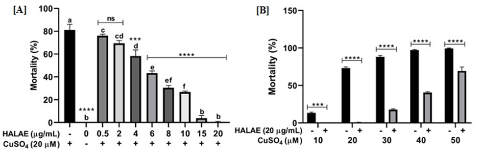
Figure 2: Protective effect of HALAE against CuSO4-induced mortality in 72 hpf zebrafish larvae at 24 hpt. [A]: Percentage mortality of zebrafish larvae pre-incubated with different concentrations of HALAE and treated with 20 µM CuSO4; [B]: Percentage mortality of zebrafish larvae pre-incubated with 20 µg/mL HALAE and treated with different concentrations of CuSO4.
Each bar represents the mean ± S.E.M. of three different experiments performed in triplicate. *** indicates p<0.001, **** indicates p<0.0001, ns indicates not significant at α equals 0.05. Columns with significantly different means are represented by different alphabets. hpf: hours post-fertilization; hpt: hours post-treatment.
Larvae treated with HALAE also showed protection against CuSO4-induced morphological deformation. Larvae exposed to 20 µM CuSO4 showed a distinct spine curvature, which showed visible reduction when co-treated with HALAE in a dose-dependent manner (Figure 3).
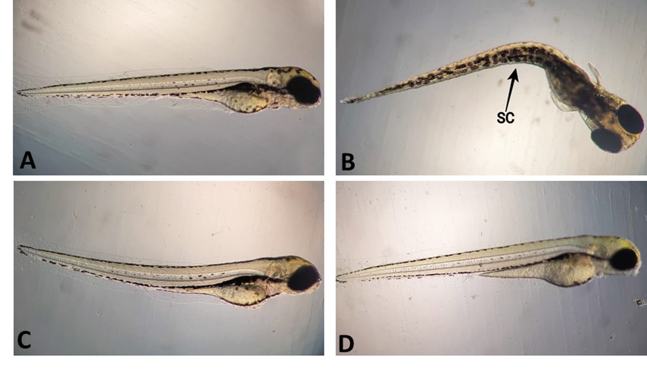
Figure 3: Representative images of protective effect of HALAE against CuSO4-induced morphological defects in 72 hpf zebrafish larvae at 24 hpt. [A]: Control larva with normal morphology; [B]: Positive control larva treated with 20 µM CuSO4 showing spine curvature; [C]: Larva treated with 6 µg/mL HALAE showing slightly curved spine; [D]: Larva treated with 20 µg/mL HALAE showing normal morphology similar to control. hpf: hours post fertilisation; hpt: hours post treatment; SC: Spine curvature.
3.3. Cu2+-chelating activity
The in vitro Cu2+-chelating activity of HALAE was found to be low. The highest concentration tested, i.e. 2 mg/mL showed a chelating ability of (56.35 ± 1.59) %. On the other hand, EDTA showed high in vitro Cu2+-chelating activity with the highest tested concentration, i.e. 8.77 ng/mL showing (98.05 ± 0.08) % chelating activity (Table 4). Therefore, EC50 value of EDTA was considerably lower, i.e. (3.99 ± 0.5) ng/mL as compared to that of HALAE which was found to be (1.55 ± 9.75) mg/mL.
Table 4. In vitro Cu2+-chelating activity (%) of varying concentrations of EDTA and HALAE.
|
EDTA (ng/mL) |
Cu2+-chelating activity (%) |
HALAE (mg/mL) |
Cu2+-chelating activity (%) |
|
1.46 |
30.87 ± 0.35 |
0.25 |
11.45 ± 0.56 |
|
2.92 |
42.80 ± 1.44 |
0.50 |
20.08 ± 0.51 |
|
4.38 |
60.23 ± 2.05 |
0.75 |
28.29 ± 0.32 |
|
5.84 |
79.55 ± 1.20 |
1.00 |
37.09 ± 0.17 |
|
7.31 |
92.60 ± 0.86 |
1.50 |
48.37 ± 0.33 |
|
8.77 |
98.05 ± 0.14 |
2.00 |
59.25 ± 0.30 |
Data are presented as mean ± S.E.M.
3.4. In vivo antioxidant activity
HALAE showed protective effect against CuSO4-induced oxidative stress. A clear reduction in fluorescence intensity, which is indicative of intracellular ROS generation, is observed in larvae treated with HALAE in a dose-dependent manner (Figure 4).
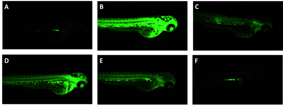
Figure 4: HALAE protects 72 hpf zebrafish larvae against CuSO4-induced ROS generation. [A]: Egg water; [B]: 10 µM CuSO4; [C]: Gallic acid 20 µg/mL; [D]: HALAE 4 µg/mL; [E]: HALAE 8 µg/mL; [F]: HALAE 20 µg/mL. hpf: hours post fertilization
3.5. In vitro antioxidant activity
The ABTS cation radical scavenging activity of HALAE was higher as compared to the DPPH scavenging activity. The ABTS scavenging activity of HALAE ranged from (7.32 ± 0.96) % at 20 µg/mL to (84.94 ± 0.22) % at 200 µg/mL (Table 1) while the DPPH scavenging activity ranged from (19.14 ± 0.39) % at 100 µg/mL to (79.68 ± 0.30) % at 500 µg/mL (Table 2). However, the radical scavenging activity of ascorbic acid was higher for both ABTS and DPPH with values ranging from (18.51 ± 1.48) % at 2 µg/mL to (99.29 ± 0.45) % at 10 µg/mL for ABTS (Table 1) and (7.76 ± 1.27) % at 2 µg/mL to (53.96 ± 0.62) % at 12 µg/mL for DPPH (Table 2). Therefore, ascorbic acid recorded the lowest IC50 values for both the ABTS and DPPH scavenging activities as compared to HALAE (Table 3).
Table 1. ABTS radical scavenging activity (%) of varying concentrations of ascorbic acid and HALAE.
|
Ascorbic acid (µg/mL) |
Radical scavenging activity (%) |
HALAE (µg/mL) |
Radical scavenging activity (%) |
|
2 |
18.51 ± 1.48 |
20 |
7.32 ± 0.96 |
|
4 |
41.36 ± 1.53 |
50 |
22.38 ± 0.33 |
|
6 |
61.03 ± 1.59 |
100 |
47.42 ± 0.84 |
|
8 |
81.57 ± 1.42 |
150 |
67.78 ± 0.44 |
|
10 |
99.29 ± 0.45 |
200 |
84.94 ± 0.22 |
Data are presented as mean ± S.E.M.
Table 2. DPPH radical scavenging activity (%) of varying concentrations of ascorbic acid and HALAE.
|
Ascorbic acid (µg/mL) |
Radical scavenging activity (%) |
HALAE (µg/mL) |
Radical scavenging activity (%) |
|
2 |
7.76 ± 1.27 |
100 |
19.14 ± 0.39 |
|
4 |
17.49 ± 0.57 |
200 |
35.60 ± 2.23 |
|
6 |
25.81 ± 0.74 |
300 |
51.01 ± 0.32 |
|
8 |
34.48 ± 0.65 |
400 |
65.84 ± 0.30 |
|
10 |
44.39 ± 0.51 |
500 |
79.68 ± 0.30 |
|
12 |
53.96 ± 0.62 |
- |
- |
Data are presented as mean ± S.E.M.
Table 3. IC50 values of ascorbic acid and HALAE for the ABTS and DPPH scavenging activities.
|
IC50 (µg/mL) |
Ascorbic acid |
HALAE |
|
ABTS |
4.97 ± 0.05 |
109.40 ± 0.30 |
|
DPPH |
11.32 ± 0.15 |
303.90 ± 2.70 |
Data are presented as mean ± S.E.M.
3.6. In vivo anti-inflammatory activity evaluation
3.6.1. Assessment of neutrophil accumulation
HALAE showed effective protection against neutrophil accumulation in CuSO4-treated zebrafish larvae. A dose-dependent reduction in neutrophil accumulation, particularly in the brain area, was observed (Figure 5). However, in the cardiac region, neutrophil accumulation is still observed in the larvae co-incubated with HALAE.
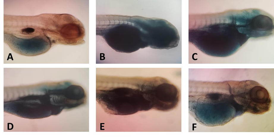
Figure 5: Photographs showing varying rates of neutrophil accumulation in 72 hpf zebrafish stimulated with 10 µM CuSO4 and treated with HALAE. [A]: Egg water; [B]: 10 µM CuSO4; [C]: HALAE 4 µg/mL; [D]: HALAE 8 µg/mL; [E]: HALAE 15 µg/mL; [F]: HALAE 20 µg/mL. hpf: hours post fertilization
3.6.2. Evaluation of lipid peroxidation
HALAE showed significant protection against CuSO4-induced lipid peroxidation. The lowest tested dose of 2 µg/mL did not show protection against lipid peroxidation. However, from 4 µg/mL to the highest tested dose of 20 µg/mL, a significant reduction in lipid peroxidation as compared to the larvae treated with only 10 µM CuSO4 is observed (Figure 6).
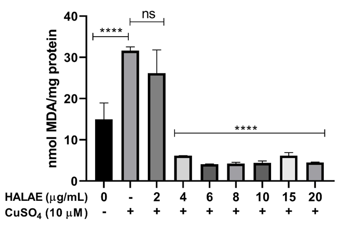
Figure 6: Protective effect of HALAE against CuSO4-induced lipid peroxidation in 72 hpf zebrafish larvae at 24 hpt. Each bar represents the mean ± S.E.M. of three different experiments performed in triplicate. **** indicates p<0.0001, ns indicates not significant at α equals 0.05. hpf: hours post-fertilization; hpt: hours post-treatment.
4. DISCUSSION
Safety evaluation is crucial before studying the potential bioactivities of plant extracts. The safety profile of HALAE was assessed using zebrafish embryos. Toxicological evaluation using zebrafish has been extensively used in natural product and pharmacological research for determining extracts with acceptable safety margins.[19] The NOAEC and LOAEC of HALAE with respect to hatching and heartbeat rates were found to be 700 and 1000 µg/mL. No statistically significant mortality rate was observed even at the highest tested dose of 1500 µg/mL. This clearly indicates that HALAE does not show acute nor developmental toxicity at the limit dose of toxicity testing as given by OECD Guideline 236.[12] Although sublethal toxicity is observed at the higher doses of 1000, 1250 and 1500 µg/mL, these concentrations are substantially higher than the limit dose. Sublethal toxic effects at high doses may be due to non-specific physiological stress rather than true toxic liability.[20] Therefore, the absence of toxicity within the OECD recommended range supports the overall safety of the extract while the sublethal toxic effects observed at the higher doses emphasize the need for careful dosage selection before evaluating the bioactivities of plant extracts.
After establishing the safety profile of HALAE, further studies were conducted both in vitro and in vivo using CuSO4-induced oxidative stress and inflammation model in zebrafish. Previous studies using zebrafish have reported that low concentrations of copper induce oxidative stress and inflammatory responses while high concentrations of copper lead to developmental defects and lethality.[21],[22] These observations align with the findings of the present study where exposure to high dose of CuSO4 resulted in high mortality and morphological defects while exposure to lower dose of CuSO4 resulted in increased intracellular ROS production, neutrophil recruitment and lipid peroxidation. This validates the suitability of this model for evaluating oxidative stress and inflammation.
In the present study, mortality following exposure to lethal dose of CuSO4 was taken as the initial endpoint observation. It was observed that pre-incubation with HALAE significantly reduced CuSO4-induced mortality and morphological deformation in a dose-dependent manner. Following this, this study found that HALAE showed dose-dependent protection against intracellular ROS generation, neutrophil recruitment and lipid peroxidation. Previous studies have also reported protective effects of plant extracts and plant-derived compounds against CuSO4 toxicity. β-sitosterol, a common phytosterol, has been reported to exhibit potent antioxidant and anti-inflammatory properties against CuSO4-induced oxidative stress and inflammatory model in zebrafish by upregulating antioxidant genes sod and gpx4b and downregulating pro-inflammatory genes il-8 and myd88.[23] Acacetin, a plant flavonoid, has shown protective effect in CuSO4-treated zebrafish larvae by increasing antioxidant gene expression (GPx, GST and GR) while downregulating pro-inflammatory gene expression (COX-2, TNF-α and IL-1).[24] Leaf ethanol extract of Clerodendrum cyrthophyllum Turcz showed protective effect against CuSO4-induced oxidative stress by downregulating hsp70 and gadd45bb while upregulating sod. It was also effective against CuSO4-induced inflammation by downregulating cox-2, pla2, c3a, mpo, il-8 and il-1β.[8] Methanol extract of Jussiaea repens L. stem with leaves increased catalase and SOD activities in CuSO4-treated zebrafish larvae.[16]
However, HALAE showed weak in vitro Cu2+-chelating activity and comparatively low ABTS and DPPH scavenging activities. This difference between limited in vitro activity and potent in vivo efficacy reinforces the importance of whole-organism models for evaluating potential bioactivities of plant extracts.
5. CONCLUSION
The present study showed that the leaf aqueous extract of H. aromatica showed significant in vivo antioxidant and anti-inflammatory potential despite weak in vitro metal chelating and radical scavenging activities. This highlights the importance of integrating whole-organism evaluations while studying complex plant extracts. In addition, the leaf extract was found to be non-toxic according to OECD guidelines. Therefore, future prospects include determining the mechanisms by which the H. aromatica leaf aqueous extract exerts protective effects against oxidative stress and inflammation and bioactivity-guided isolation of the active fractions and subsequent identification and characterization of the active compounds.
ACKNOWLEDGEMENTS
The authors thank the University Grants Commission (UGC), India for providing financial support through Junior Research Fellowship.
AUTHOR CONTRIBUTIONS
DECLARATION OF CONFLICT OF INTERESTS
The authors declare that there is no conflict of interest.
REFERENCES


Sangeeta Yanglem, Birjit Singh Waikhom, Thangal Yumnamcha, Maibam Damayanti Devi, Leaf Aqueous Extract of Homalomena aromatica (Spreng.) Schott Alleviates Copper sulfate-induced Oxidative Stress and Inflammation in Zebrafish (Danio rerio) Larvae, Int. J. of Pharm. Sci., 2025, Vol 3, Issue 12, 4117-4130. https://doi.org/10.5281/zenodo.18097056
 10.5281/zenodo.18097056