School of Pharmacy, YBN University, Ranchi Jharkhand 834010.
Introduction: From ancient times people relied on herbal medicines for the treatment of various chronic and acute diseases. Advancement in Chemistry and Biology has enabled research to know how natural substances interact with human body and find new compounds for treatment of various diseases. Free radicals cause several diseases including cancer, neurodenerative disorders and inflammatory conditions [4][5]. Objective: The aims of my research work to find a herbal source of antioxidants which can replace artificial antioxidants. Materials and Methods: Butea Monosoerma leaves extraction is done by Soxhlet Extraction using hydroethanolic solvent (30:70). Phytochemical investigation is done for alkaloids, triterpenoids, tannins, phenols, flavonoids and glycosides. After quantitative estimation of phenol was done by Folin–Ciocalteu method using gallic acid as standard and for flavonoids it was done by colorimetric method using rutin as standard. Their in-vitro antioxidant potential was evaluated using DPPH, H2O2, NITRIC OXIDE standard antioxidants. Results and discussion: Phytochemical investigation revealed that Butea Monosoerma leaves contain alkaloids, tannins, phenols and flavonoids. The total phenolic content of hydroethanolic extract of Butea Monosperma (HEEBM) was 80.67mg gallic acid equivalent (GAE)/g of extract. The total flavonoid content was 83.547mg rutin equivalent/g of extract. The extract significantly inhibited NO production, shows DPPH radical scavenging and also hydrogen peroxide scavenging activity. Conclusion: The HEEBM leaves exhibited significant antioxidant activity as indicated by its high phenolic and flavonoid content and strong free radical scavenging potential. It can be used as a herbal antioxidant after further studies for isolation and characterization of active compounds.
Herbal medicine, also called herbalism, involves using plants or plant-based substances for healing. These natural remedies can be consumed or applied directly to the skin. For thousands of years, people from a wide range of cultures have relied on herbs to treat illnesses and support the body’s natural functions. Civilizations across history have turned to plants as essential sources of medicine. As a result, unique herbal treatments emerged in various regions around the world. Since the earliest stages of human development, natural remedies have played a vital role in health and healing. Our ancient ancestors would chew on specific plants to ease discomfort or cover wounds with leaves to help them heal faster. Before modern medicine, nature was the primary source of treatment for sickness and injuries [1].?Today’s advancements in chemistry and biology have enabled researchers to understand how natural substances interact with the human body at a detailed level. These scientific tools have also helped identify how certain compounds can work together, offering great potential for creating new treatments for serious illnesses like cancer and dementia. The progress in modern chemistry has opened up exciting possibilities in the exploration and application of natural products in medicine [2] [3].?Living organisms are constantly exposed to an oxidative environment that produces various free radicals, such as superoxide, hydroxyl radicals, nitric oxide, and peroxynitrite. A growing body of research highlights the contribution of these reactive molecules to the onset and progression of several diseases, including cancer, neurodegenerative disorders, and certain inflammatory conditions [4][5].?Due to their ability to combat free radicals, antioxidants have attracted significant attention. In this regard, researchers around the globe are exploring the antibacterial and antioxidant activities of different medicinal plants. This growing interest is largely driven by the health risks linked to synthetic preservatives and artificial antioxidants [6].? Butea monosperma thrives in warm, humid climates and is widely distributed across South and Southeast Asian nations and belongs to the family Fabaceae [7].?Common names include flame-of-the-forest, palash and bastard teak. It is a small-sized dry-season deciduous tree,?growing to 15 m (49 ft) tall. It is a fast-growing tree, young trees have a growth rate of a few feet per year. The leaves are pinnate, with an 8–16 cm (3.1–6.3 in) petiole and three leaflets, each leaflet 10–20 cm (3.9–7.9 in) long. The leaves are typically thick and tough in texture, making it unpalatable to grazing animals like cattle. In earlier times, people commonly used these durable leaves as natural plates, serving meals on them long before the advent of plastic dishware [8].?The dried flowers and bark of the stem are rich in valuable bioactive compounds, including flavonoids such as medicarpin and plasonin, as well as alkaloids like butrin and isobutrin. These constituents are known for their diverse therapeutic and medicinal applications. Ethanol-based extracts from the petals and seeds of Butea frondosa have demonstrated properties such as antiestrogenic, antiimplantation, and antifertility effects [9, 10].?The petroleum ether extract of Butea monosperma flowers has demonstrated anticonvulsant activity[11,12].Hence, the primary aim of this study was to assess the total phenolic and flavonoid content, along with evaluating the antioxidant and antibacterial properties of hydroethanolic (70:30 ethanol\water)extracts derived from the leaves of Butea monosperma.
2. MATERIALS AND METHOD
2.1 Chemicals
Gallic acid, Folin–Ciocalteu’s reagent, 1, 1-Diphenyl-2-picrylhydrazyl (DPPH), Sodium nitroprusside were purchased from SR Scientifics (Tirupathi, India). All other chemical reagents used were of analytical grade.
2.2 Collection of plant material
Fresh flowers and stems of Butea monosperma were gathered from Ranchi, Jharkhand, India, during the summer season. The plant materials were promptly packed after collection and stored in a cool, dark place until further processing.?
2.3 Extraction of crude drug
Hot Soxhlet Extraction Method
In this procedure, leaves of Butea monosperma were collected, thoroughly washed, and rinsed. After cleaning, they were shade-dried and then ground into a coarse powder using a mechanical grinder. The powdered plant material was subjected to successive extraction using a hydroethanolic solvent in a 70:30 ratio (ethanol\ water).? The powdered sample was placed inside a thimble, which was inserted into the extraction chamber of a Soxhlet apparatus. The solvent was heated in a flask, and its vapors traveled up to the condenser, where they condensed and dripped back into the thimble containing the plant material. This allowed continuous contact between the solvent and the sample, facilitating efficient extraction.? Once the solvent level in the chamber reached the top of the siphon tube, it automatically drained back into the flask, carrying the extracted compounds with it. This cycle was repeated until a drop of the solvent no longer left any residue upon evaporation, indicating completion of the extraction. The resulting extract was then filtered, concentrated to dryness, weighed, and stored for later use. The percentage yield of the extract was determined using the following formula.?
Yield (%) = Mass of the residue obtained× 100
Mass of plant material taken
2.4 Phytochemical Investigation?
A comprehensive qualitative phytochemical screening was carried out to detect the presence or absence of various phytoconstituents. The appearance of color changes or formation of precipitates served as indicators for the presence of specific compounds. These tests were conducted using well-established standard protocols.?
Test for Alkaloids
Dragendorff’s Test: One milliliter of the extract, dissolved in alcohol, was mixed thoroughly with a few drops of acetic acid followed by the addition of Dragendorff’s reagent. The formation of an orange-red precipitate confirmed the presence of alkaloids.?
Wagner’s Test: One milliliter of the extract, previously dissolved in acetic acid, was treated with a few drops of Wagner’s reagent. The appearance of a reddish-brown precipitate indicated the presence of alkaloids.?
Mayer’s Test: To 1 ml of the extract in acetic acid, a few drops of Mayer’s reagent were added. The formation of a pale white or dull white precipitate suggested the presence of alkaloids.?
Hager’s Test: Approximately 1–2 ml of the extract dissolved in acetic acid was mixed with 3 ml of Hager’s reagent. The development of a yellow precipitate was indicative of alkaloid content.?
Tests for Triterpenoids and Steroids
Libermann-Burchard Test: The extract was first dissolved in chloroform, followed by the addition of 1 ml of acetic acid and 1 ml of acetic anhydride. The mixture was gently heated using a water bath and then allowed to cool. A few drops of concentrated sulfuric acid were carefully added along the inner wall of the test tube. The appearance of a bluish-green coloration indicated the presence of steroids.
Salkowski Test: The extract was mixed with chloroform, and an equal volume of concentrated sulfuric acid was added to the solution. The development of a bluish-red to cherry-red color in the chloroform layer, along with green fluorescence in the acid layer, confirmed the presence of steroids.?
Tests for Tannins and Phenolic Compounds
Ferric Chloride Test: A portion of the extract was dissolved in distilled water, and a few drops of a dilute ferric chloride solution were added. The appearance of a dark blue coloration indicated the presence of tannins.
Gelatin Test: A measured amount of the extract was dissolved in distilled water, followed by the addition of 2 ml of a 1% gelatin solution containing 10% sodium chloride. The formation of a white precipitate signified the presence of phenolic compounds.?
Lead Acetate Test: The extract was dissolved in distilled water, and a few drops of lead acetate solution were added. The resulting white precipitate confirmed the presence of phenolic compounds.?
Test for Flavonoids
Shinoda’s Test: To 1 ml of the alcoholic extract, a few small pieces of magnesium metal and several drops of concentrated hydrochloric acid were added. The mixture was then gently heated in a water bath. The development of a red to pink coloration confirmed the presence of flavonoids.
Tests for Glycosides
Borntrager’s Test: About 3 ml of the test solution was mixed with dilute sulfuric acid, then boiled for 5 minutes and filtered. After cooling, an equal amount of benzene or chloroform was added, and the mixture was shaken thoroughly. The organic layer was separated, and ammonia was added. A pink to red coloration in the ammoniacal layer indicated the presence of anthraquinone glycosides.
Keller-Killiani Test: To 2 ml of the test sample, 3 ml of glacial acetic acid and one drop of 5% ferric chloride solution were added in a test tube. Then, 0.5 ml of concentrated sulfuric acid was carefully introduced along the side of the test tube. The appearance of a blue color in the acetic acid layer confirmed the presence of cardiac glycosides.
2.5 Quantitative Phytochemical assay
?Estimation of Total Phenolic Content (TPC)
The total phenolic content of the extracts was assessed using the Folin–Ciocalteu method. Gallic acid served as the standard, and results were expressed in terms of milligrams of gallic acid equivalent (GAE) per gram of extract. Standard solutions of gallic acid were prepared in ethanol at concentrations of 0.01, 0.02, 0.03, 0.04, and 0.05 mg/ml. Plant extract solutions were also prepared in ethanol at concentrations of 0.1 mg/ml and 1 mg/ml.? A volume of 0.5 ml from each sample was added to test tubes, followed by 2.5 ml of 10-fold diluted Folin–Ciocalteu reagent. After that, 2 ml of 7.5% sodium carbonate solution was added. The test tubes were sealed with parafilm and left to react at room temperature for 1 hour. The absorbance of the resulting blue-colored solution was measured at 765 nm using a spectrophotometer. ?Each sample analysis was carried out in triplicate. The Folin–Ciocalteu reagent reacts with reducing agents, such as polyphenols, leading to the formation of a blue complex which can be quantified spectrophotometrically.? A standard calibration curve was constructed using the gallic acid standards, and the regression equation obtained was: y = 0.005x + 2.570 with a correlation coefficient R² = 0.992, indicating a strong linear relationship. The concentration of phenolics in the plant samples was calculated by substituting the sample absorbance (y) into the regression equation [13].
?Determination of Total Flavonoid Content
Total flavonoid content was determined using a colorimetric method described by Dewanto et al. A measured volume of the diluted plant extract or rutin standard solution was combined with 75 µL of sodium nitrite (NaNO?) solution. The mixture was allowed to react for 6 minutes before the addition of 0.15 mL of aluminum chloride (AlCl?) solution at a concentration of 100 g/L. After 5 minutes, 0.5 mL of sodium hydroxide (NaOH) was added to the mixture. The final volume was brought up to 2.5 mL using distilled water, and the contents were mixed thoroughly.? The absorbance of the resulting solution was measured at 510 nm using a spectrophotometer. A blank was prepared using the same reagents, but without the sample. The total flavonoid content was calculated using a rutin calibration curve and expressed as milligrams of rutin equivalent per gram of dry weight (mg rutin/g DW).? Each sample was analyzed in triplicate [14]. The concentration of flavonoids in the samples was estimated using the linear regression equation derived from the rutin standard curve:? y = 0.001x - 0.019, with a correlation coefficient of R² = 0.993, indicating strong linearity. The absorbance value (y) from the test sample was substituted into the regression equation to calculate the flavonoid concentration.?
2.6 Assessment of antioxidant potential in vitro
DPPH Radical Scavenging Activity:
The antioxidant activity was evaluated using the 2, 2-diphenyl-1-picrylhydrazyl (DPPH) free radical assay. DPPH is a stable free radical that exhibits a deep violet color. When antioxidants are present, they donate hydrogen atoms to the DPPH radicals, reducing them to the non-radical form (DPPH-H), which leads to a decrease in absorbance. The extent of this color reduction reflects the ability of the antioxidant to scavenge free radicals.? To determine the scavenging activity, varying concentrations (5–20 mg/mL) of each sample were prepared in water and ethanol. Each sample solution was mixed with 1 mL of a 0.2 mM DPPH solution in ethanol. The mixture was shaken thoroughly and incubated in the dark for 30 minutes. After the reaction period, the absorbance was measured at 517 nm using a blank as the reference.?
The percentage of DPPH radical inhibition was calculated using the formula:?
Inhibition (%) = 100 × (Absorbance of blank – Absorbance of sample)
Absorbance of blank
Where blank represents the absorbance of the control (without test extract), and sample is the absorbance in the presence of the sample.?
Nitric Oxide Scavenging Assay
A solution of sodium nitroprusside (5 mM) prepared in phosphate-buffered saline was combined with varying concentrations of the plant extract dissolved in ethanol. The mixture was incubated at 25°C for 30 minutes. A control sample containing the same volume of ethanol but without the extract was also prepared. After incubation, 1.5 ml of the reaction mixture was taken and mixed with an equal volume (1.5 ml) of Griess reagent, which contained 1% sulfanilamide, 2% phosphoric acid, and 0.1% N-(1-naphthyl) ethylenediamine dihydrochloride. The formation of a pink-colored chromophore due to the diazotization reaction between nitrite and sulfanilamide, followed by a coupling reaction with N-(1-naphthyl) ethylenediamine, was measured spectrophotometrically at 546 nm. The nitric oxide scavenging activity was calculated in comparison to a standard [15].?
Hydrogen Peroxide Scavenging Assay?
A 40 mM hydrogen peroxide (H?O?) solution was prepared using phosphate buffer at pH 7.4. To this, 2 ml of the plant extract (ranging from 20 to 100 µg/ml) dissolved in phosphate buffer was added to 1 ml of the H?O? solution. After a reaction time of 10 minutes, the absorbance was measured at 245 nm using a blank containing only phosphate buffer without hydrogen peroxide. The hydrogen peroxide scavenging activity of the extract and the reference compound, ascorbic acid, was evaluated using the following formula:?
% Inhibition = (Absorbance of control – Absorbance of sample) × 100
Absorbance of control
3. RESULT & DISCUSSION
3.1 Phytochemical Investigation
Table 1: Qualitative Phytochemical Analyses Test for Alkaloids
|
S. No. |
Experiment |
Result |
|
1. |
Dragendorff’s test |
-ve |
|
2. |
Wagner’s Test |
-ve |
|
3. |
Mayer’s Test |
-ve |
|
4. |
Hager’s Test |
-ve |
Test for Triterpenoids and Steroids
|
S. No. |
Experiment |
Result |
|
1. |
Libermann-Burchard Test |
+ve |
|
2. |
Salkowski Test |
+ve |
Tests for Tannins and Phenolic Compounds
|
S. No. |
Experiment |
Result |
|
1. |
Ferric Chloride Test |
+ve |
|
2. |
Gelatin Test |
+ve |
|
3. |
Lead Acetate Test |
+ve |
Test for Flavonoids
|
S. No. |
Experiment |
Result |
|
1. |
Shinoda’s Test |
+ve |
Tests for Glycosides
|
S. No. |
Experiment |
Result |
|
1. |
Borntrager’s Test |
-ve |
|
2. |
Keller-Killiani Test |
-ve |
3.2 Total Phenolic Constituent of Butea Monosperma Extract
Total Phenolic Constituent of Butea Monosperma hydroethanolic Extract is 80.67 mg GAE/g.
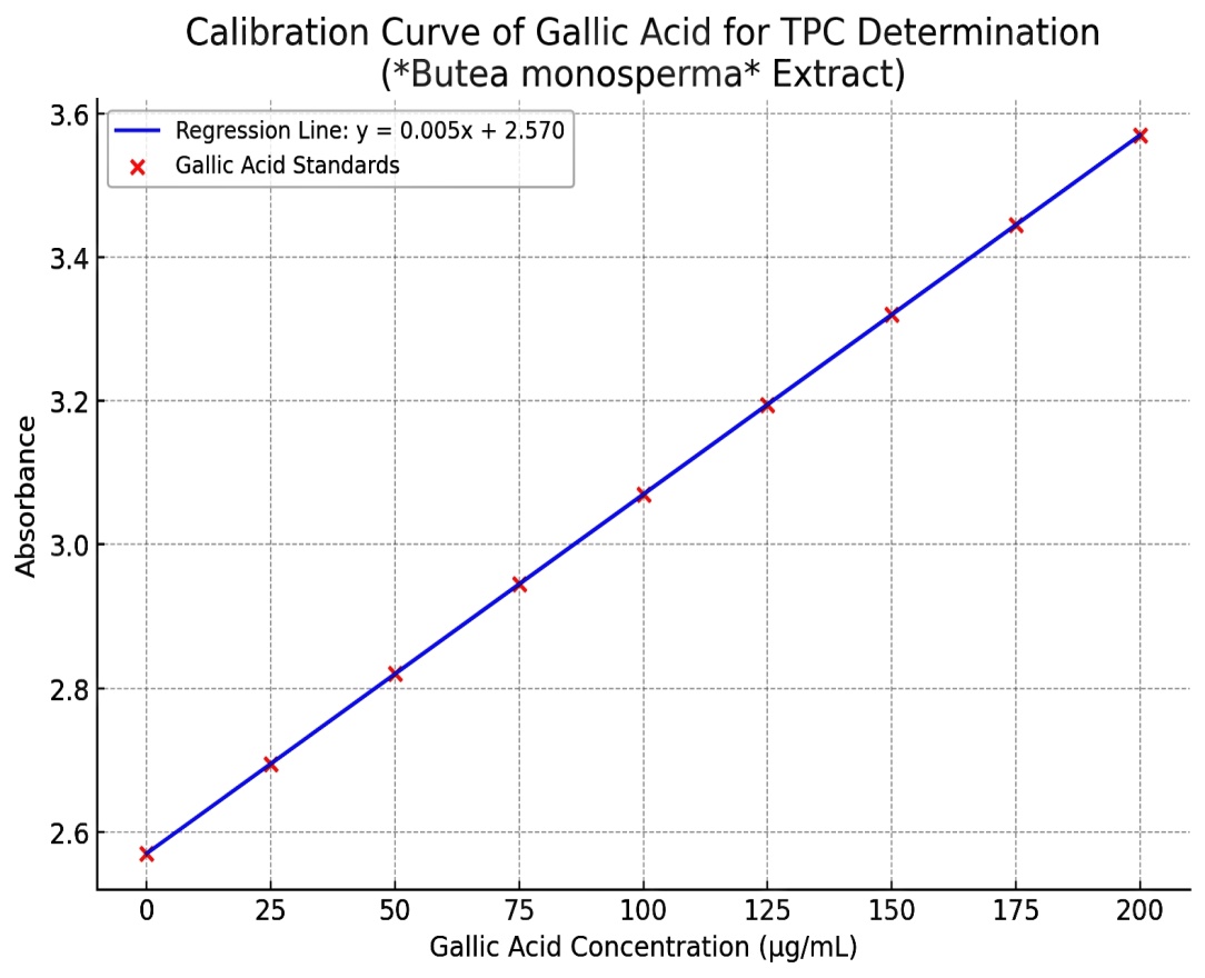
Fig, 1: Standard curve of Gallic acid
3.3 Total Flavonoid Constituent of Butea Monosperma Extract
Total Flavonoid Constituent of Butea Monosperma hydroethanolic Extract is 83.547 mg Rutin/g
Standard Curve Equation: y = 0.001x - 0.019
Coefficient of Determination (R²): 0.9930
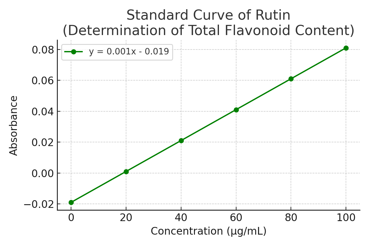
Fig, 2: Standard curve of rutin
3.4 DPPH Antioxidant Assay
Table 2: DPPH radical Scavenging activity of Std. Ascorbic acid
y = 0.3769x + 44.513
R² = 0.9689
|
Ascorbic acid (std) |
||
|
S. No. |
Concentration (µg/mL) |
% Scavenging Activity |
|
1. |
10 |
48.28 |
|
2. |
20 |
52.05 |
|
3. |
30 |
55.82 |
|
4. |
40 |
59.59 |
|
5. |
50 |
63.36 |
|
6. |
60 |
67.13 |
|
7. |
70 |
70.90 |
|
8. |
80 |
74.66 |
|
9. |
90 |
78.43 |
|
10. |
100 |
82.20 |
|
IC50 |
62.24 µg/ml |
|
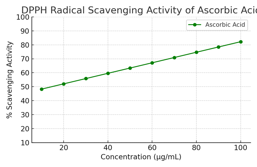
Fig, 3: Standard Curve of Ascorbic Acid
DPPH radical scavenging activity of hydroethanolic extract of Butea Monosperma
Table 3: DPPH radical scavenging activity of hydroethanolic extract of Butea Monosperma
|
Hydroethanolic Extract of Butea Monosperma(HEEBM) |
||||
|
S. No. |
Concentration (µg/mL) |
% Scavenging |
Absorbance |
|
|
1. |
10 |
45.93 |
0.541 |
|
|
2. |
20 |
48.61 |
0.514 |
|
|
3. |
30 |
51.3 |
0.487 |
|
|
4. |
40 |
53.98 |
0.46 |
|
|
5. |
50 |
56.66 |
0.433 |
|
|
6. |
60 |
59.34 |
0.407 |
|
|
IC50 |
56.71µg/ml |
|||
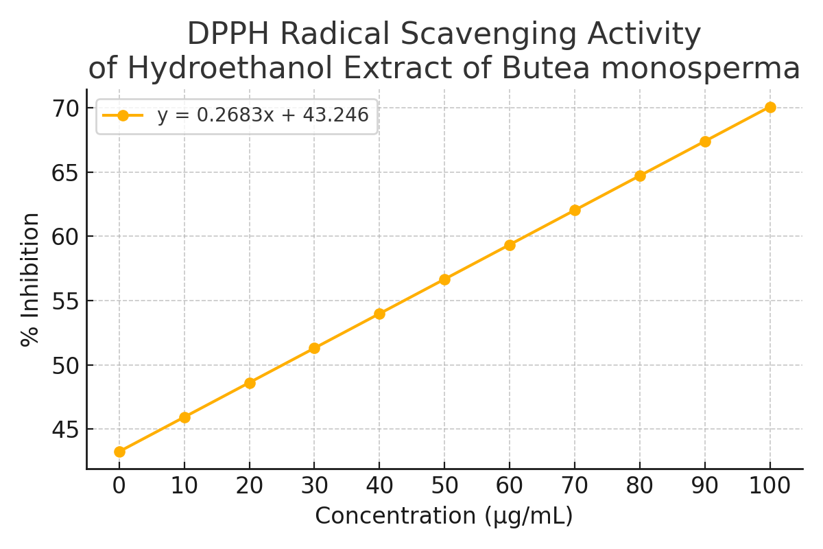
Fig, 4: DPPH radical scavenging activity of HEEBM
3.5 H2O2 Antioxidant Assay
Table 4: H2O2 radical scavenging activity of Std. Ascorbic Acid
|
Ascorbic acid (std.) |
|||
|
S. No. |
Concentration (µg/mL) |
% Scavenging Activity |
|
|
1. |
10 |
43.60 |
|
|
2. |
20 |
51.99 |
|
|
3. |
30 |
60.38 |
|
|
4. |
40 |
68.77 |
|
|
5. |
50 |
77.16 |
|
|
6. |
60 |
85.55 |
|
|
7. |
70 |
93.94 |
|
|
IC50 |
77.14µg.ml |
||
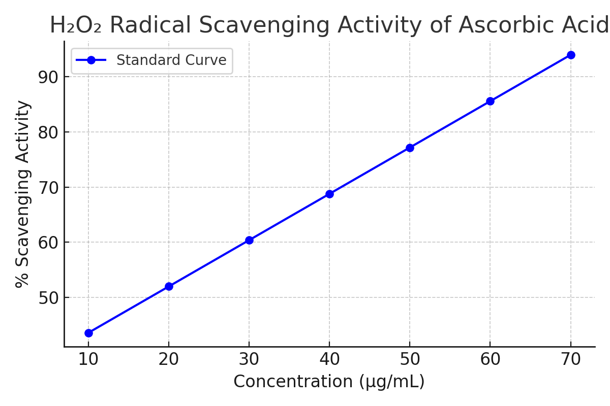
Fig, 5: H2O2 radical scavenging activity of Std. Ascorbic acid
3.6 H2O2 radical scavenging activity of Hydroethanolic extract of Butea monosperma
Table 5: H2O2 radical scavenging activity of HEEBM
|
Hydroethanolic extract of Butea monosperma(HEEBM) |
|||
|
S. No. |
Concentration (µg/mL) |
% Inhibition |
|
|
1. |
10 |
28.63 |
|
|
2. |
20 |
36.06 |
|
|
3. |
30 |
43.48 |
|
|
4. |
40 |
50.91 |
|
|
5. |
50 |
58.34 |
|
|
6. |
60 |
65.77 |
|
|
7. |
70 |
73.2 |
|
|
8. |
80 |
80.63 |
|
|
9. |
90 |
88.06 |
|
|
10. |
100 |
95.49 |
|
|
IC50 |
58 .452µg/ml |
||
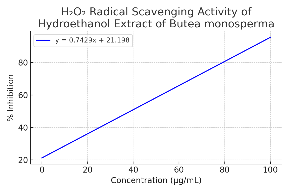
Fig, 6: H2O2 radical scavenging activity of HEEBM
3.7 Nitric Oxide Assay
Table 6: NO radical scavenging activity of Ascorbic acid
|
Ascorbic acid (std.) |
|||
|
S. No. |
Concentration(µg/mL) |
% Inhibition |
|
|
1. |
10 |
48.08 |
|
|
2. |
20 |
52.77 |
|
|
3. |
30 |
57.46 |
|
|
4. |
40 |
62.15 |
|
|
5. |
50 |
66.84 |
|
|
6. |
60 |
71.53 |
|
|
7. |
70 |
76.22 |
|
|
8. |
80 |
80.91 |
|
|
9. |
90 |
85.6 |
|
|
10. |
100 |
90.29 |
|
|
IC50 |
68.38µg/ml |
||
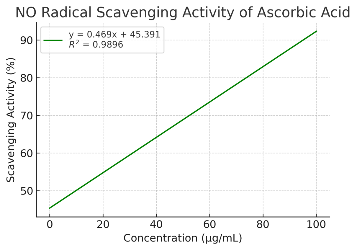
Fig. 7: NO radical scavenging activity of Ascorbic acid
NO radical scavenging activity of Hydroethanolic extract of Butea monosperma
Table 7: NO radical scavenging activity of Hydroethanolic extract of Butea Monosperma
|
HEEBM |
|||
|
S. No. |
Concentration (µg/mL) |
Inhibition (%) |
|
|
|
10 |
23.25 |
|
|
|
20 |
28.78 |
|
|
|
30 |
34.3 |
|
|
|
40 |
39.83 |
|
|
|
50 |
45.36 |
|
|
|
60 |
50.89 |
|
|
|
70 |
56.42 |
|
|
|
80 |
61.94 |
|
|
|
90 |
67.47 |
|
|
|
100 |
73.0 |
|
|
IC50 |
45.23µg/ml |
||
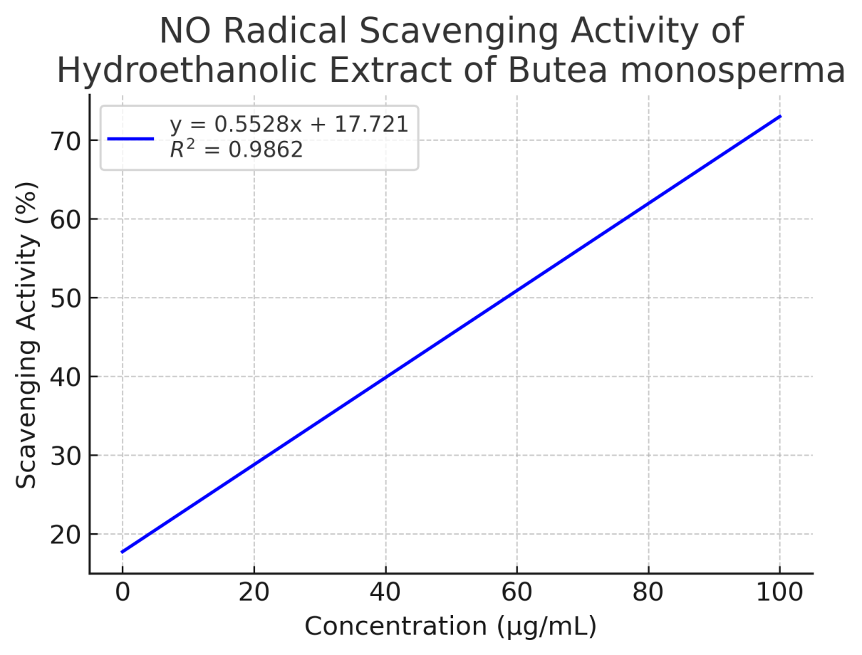
Fig, 8: NO radical scavenging activity of HEEBM
Phytochemical screening of Hydroethanolic extract of Butea Monosperma revealed the presence of alkaloids, tannins, triterpenoids, phenols and flavonoids.The total phenolic content of HEEBM was determined using the Folin–Ciocalteu assay and calculated from a gallic acid calibration curve. The phenolic content was found to be 80.67 mg gallic acid equivalent (GAE)/g of extract. The total flavonoid content, expressed in rutin equivalents, was 83.547 mg RE/g of extract.
Incubation of Sodium nitroprusside in phosphate buffer saline at 25°C for 30 mins resulted in the generation of nitric oxide(NO).The Hydroethanolic extract of Butea Monosperma (HEEBM) significantly inhibited NO production in this system, indicating its potential as a nitric oxide scavenger. In the DPPH radical scavenging assay, the IC50 values for HEEBM and the standard antioxidant, ascorbic acid were found to be 56.71µg/ml and 62.24 µg/ml respectively (Table2&3), demonstrating strong free radical scavenging potential of the extract.
Additionally, HEEBM exhibited hydrogen peroxide (H?O?) scavenging activity in a concentration-dependent manner (Table 5).
5. CONCLUSION
The hydroethanolic extract of Butea monosperma leaves exhibited significant antioxidant activity, as indicated by its high total phenolic and flavonoid content, and strong free radical scavenging potential in DPPH and hydrogen peroxide assays. The findings support the traditional use of Butea monosperma in herbal medicine and warrant further studies for isolation and characterization of active compounds.
REFERENCES


Gahna Kumari*, Sangita Kumari, Nazish Farhan, Phytochemical Investigation and Evaluation of In-Vitro Antioxidant Activity of Butea Monosperma Leaves, Int. J. of Pharm. Sci., 2025, Vol 3, Issue 7, 1530-1542. https://doi.org/10.5281/zenodo.15860773
 10.5281/zenodo.15860773
