

We use cookies to ensure our website works properly and to personalise your experience. Cookies policy
1.Pharm D Intern, Ezhuthachan College of Pharmaceutical Sciences, Marayamuttom, Neyyattinkara, Thiruvananthapuram, Kerala, India
2.Assistant Professor, Department of Pharmacy Practice, Ezhuthachan College of Pharmaceutical Sciences, Marayamuttom, Neyyattinkara, Thiruvananthapuram, Kerala, India
3.Consultant Surgical Oncologist, NIMS Medicity, Aralumoodu, Neyyattinkara, Thiruvananthapuram, Kerala, India
4.Principal / Head of Department, Pharmacy Practice, Ezhuthachan College of Pharmaceutical Sciences, Marayamuttom, Neyyattinkara, Thiruvananthapuram, Kerala, India
Wilkie syndrome, also known as Superior Mesenteric Artery (SMA) syndrome, is a rare gastrointestinal disorder caused by compression of the third part of the duodenum between the superior mesenteric artery and the abdominal aorta due to narrowing of the aortomesenteric angle. A case of a 24-year-old male who presented with severe abdominal pain, nausea, recurrent vomiting, reduced oral intake, and significant weight loss. Clinical evaluation suggested upper gastrointestinal obstruction, while laboratory investigations revealed elevated inflammatory markers, including C-reactive protein (88.6 mg/L). Contrast-enhanced computed tomography confirmed compression of the third part of the duodenum with characteristic features of Wilkie syndrome. Initial conservative management with nutritional support and symptomatic therapy was unsuccessful, necessitating laparoscopic duodenojejunostomy. The patient had an uneventful postoperative recovery with complete resolution of symptoms and successful resumption of oral intake. This case highlights the importance of considering Wilkie syndrome in young patients. presenting with persistent upper gastrointestinal obstructive symptoms and significant weight loss, as early diagnosis and timely surgical intervention can lead to favorable clinical outcomes.
Wilkie syndrome, also known as Superior Mesenteric Artery (SMA) syndrome, is an uncommon gastrointestinal disorder characterized by extrinsic compression of the third part of the duodenum between the superior mesenteric artery and the abdominal aorta. Under normal physiological conditions, the fat pad surrounding the mesenteric vessels maintains an adequate aortomesenteric angle and distance, allowing free passage of the duodenum. However, reduction of this angle due to loss of retroperitoneal fat results in duodenal compression, leading to partial or complete obstruction. Although rare, Wilkie syndrome should be considered as an important differential diagnosis in patients presenting with symptoms of chronic or acute upper gastrointestinal obstruction. (1)
The clinical manifestations of Wilkie syndrome are often variable and nonspecific, which contributes to diagnostic delays. Patients may present with postprandial epigastric pain, early satiety, nausea, recurrent vomiting, abdominal distension, and progressive weight loss. Symptoms may worsen after meals due to increased duodenal compression, resulting in reduced oral intake and further nutritional deterioration, creating a vicious cycle of weight loss and worsening obstruction. Since these symptoms closely resemble other gastrointestinal disorders such as gastroparesis, peptic ulcer disease, functional dyspepsia, and other causes of intestinal obstruction, a high index of clinical suspicion is essential for early diagnosis. (20
The development of Wilkie syndrome is primarily associated with conditions that result in significant loss of the mesenteric fat cushion or alteration of normal anatomical relationships. Common predisposing factors include rapid weight loss, severe malnutrition, chronic illnesses causing cachexia, prolonged immobilization, trauma, burns, and spinal corrective procedures. Loss of the protective fat pad decreases the aortomesenteric angle and distance, causing the superior mesenteric artery to compress the underlying duodenum against the vertebral column. This anatomical alteration leads to impaired gastric and duodenal emptying, resulting in symptoms of proximal intestinal obstruction. (3)
Accurate diagnosis of Wilkie syndrome requires correlation between clinical presentation and radiological findings. Contrast-enhanced computed tomography (CECT) with sagittal reconstruction is considered the preferred diagnostic modality as it provides detailed visualization of the duodenal compression site and allows precise measurement of the aortomesenteric angle and distance. Radiological confirmation is generally based on demonstration of narrowing of the aortomesenteric angle along with compression and dilatation of the proximal duodenum. Early diagnosis through appropriate imaging is essential to prevent complications associated with prolonged obstruction and nutritional depletion. (4)
Management strategies for Wilkie syndrome depend on the severity of symptoms, duration of illness, nutritional status, and response to initial therapy. Conservative treatment is usually considered as the first-line approach and includes nutritional rehabilitation aimed at restoring the retroperitoneal fat pad, gradual weight gain, dietary modification, nasogastric decompression, and correction of fluid and electrolyte disturbances. However, patients who fail to respond to conservative measures or develop persistent obstructive symptoms may require surgical intervention. Among available surgical procedures, laparoscopic duodenojejunostomy has become the preferred approach due to its high clinical success rate, reduced invasiveness, shorter recovery period, and favorable long-term outcomes. (1)
Despite advances in diagnostic imaging and treatment approaches, Wilkie syndrome remains a challenging condition due to its rarity and nonspecific presentation. Delayed diagnosis may result in prolonged symptoms, malnutrition, electrolyte abnormalities, and reduced quality of life. Reporting individual cases contributes to increasing clinical awareness and improving recognition of this uncommon disorder among healthcare professionals. (6)
Here, we report a rare case of Wilkie syndrome in a 24-year-old male who presented with severe abdominal pain, recurrent vomiting, reduced oral intake, and significant weight loss. The diagnosis was confirmed through contrast-enhanced computed tomography demonstrating characteristic duodenal compression. The patient initially received conservative management; however, due to persistent symptoms and failure of medical therapy, laparoscopic duodenojejunostomy was performed, resulting in significant clinical improvement. This case highlights the importance of early diagnosis, multidisciplinary management, and timely surgical intervention in patients with persistent Upper Gastrointestinal obstructive symptoms suggestive of Wilkie syndrome.
CASE REPORT
The case involved a 24-year-old male patient who was admitted to the Surgical Oncology Department with complaints of severe abdominal pain, frequent bouts of vomiting, nausea, and poor oral tolerance. It was noted that there had been progressive worsening of the patient’s symptoms, thus impacting his nutritional state and quality of daily living. It was also noted that he had a history of significant weight loss before his admission, and this is an important risk factor for SMA’s.
Clinical examination showed that the patient was conscious and oriented. The physical examination of the abdomen showed upper abdominal tenderness and features of proximal gastrointestinal obstruction. The patient had various laboratory investigations done, such as complete blood count, renal function test, liver function test, serum electrolytes, and inflammatory markers. It was found that the CRP level was increased (88.6 mg/L).
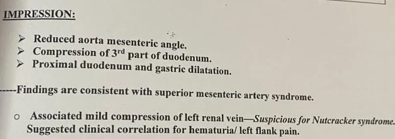
Figure 1. CECT Abdomen report
CECT scan of the abdomen was done to ascertain the underlying cause of obstruction. Figure 1 CECT imaging showed that there was compression of the third part of the duodenum between the superior mesenteric artery and abdominal aorta, with proximal duodenal and gastric dilatation. Reduced aortomesenteric angle and distance were features of Superior Mesenteric Artery Syndrome(Wilkie’s syndrome).
Initially, conservative treatment was done using bowel rest, correction of fluids and electrolytes, nutritional support, and symptomatic management. But the patient had symptoms of obstruction without improvement of his ability to take orally.
The patient was given the following treatment including INJ. CEFOPERAZONE SULBACTAM 1.5g to treat any underlying infection, INJ. PARACETAMOL 1g was given to treat the pain, INJ. PANTOPRAZOLE 40mg to treat any gastric irritations caused by administration of drugs, INJ. TRAMADOL 50mg to help reduce the pain, INJ. IBUPROFEN 100mg was given as stat medicine to help with pain relief, INJ. ONDANSETRON 4mg was administered to reduce vomiting, NEB. LEVOSALBUTAMOL 0.63mg was given to help with difficulty in breathing, TAB. DEFLAZACORT 6mg was given to help reduce the inflammation on the legs, CAP. RACECADOTRIL was given to reduce diarrhea and CAP. VSL#3 was given to improve the gut microbiome.
Due to the failure of conservative treatment and obstruction of the duodenum, a decision to do surgical management was taken.
The patient was operated on and done duodenojejunostomy via laparoscopy under general anesthesia. In this case, the obstructed part of the duodenum was bypassed by joining the duodenum and jejunum side to side.
Induction of anesthesia perioperatively was done using Propofol, while Fentanyl was used as analgesia, Atracurium as neuromuscular blocking agent, Midazolam for sedation, Dexmedetomidine for support of sedation, and Ondansetron as prophylactic to vomiting and nausea after surgery.
The Patient Got Clinically Better And Was Discharged With TAB.FAROPENAM+ Clavulanic Acid (300+125mg), Tab. pantoprazole 40mg, Tab. Paracetamol 650mg, TAB. Etoricoxib 90mg And cap. Methylcobalamin 1500mcg +Thiamine 10mg+Folic Acid 1.5mg +Pyridoxine 3mg +Alpha Lipoic Acid 100mg
Postoperative care was uncomplicated. Pain control, recovery of bowel movement, fluid management, and any complication related to surgery were monitored. Introduction of feeding gradually was achieved with significant improvement of vomiting, abdominal pain, and general well-being of the patient. Stable condition with appropriate advice for improvement of nutrition and follow up surgical consultations was achieved before discharge of the patient.
CLINICAL OUTCOME
The patient responded well after undergoing laparoscopic duodenojejunostomy with symptoms such as vomiting and abdominal pain subsiding completely. Oral feeding was slowly started without any postoperative complications being noted. The patient was discharged in a stable state with instructions regarding nutrition.
DISCUSSION
Wilkie’s syndrome, also known as Superior Mesenteric Artery (SMA) syndrome, is an uncommon gastrointestinal disorder characterized by compression of the third part of the duodenum between the superior mesenteric artery and the abdominal aorta. This compression occurs due to a reduction in the aortomesenteric angle and distance, usually caused by loss of the intervening mesenteric fat pad. Although rare, Wilkie’s syndrome is an important cause of proximal intestinal obstruction, and diagnosis is often delayed due to its nonspecific symptoms and similarity with other gastrointestinal disorders. (2)
In the present case, a 24-year-old male presented with severe abdominal pain, recurrent vomiting, reduced oral intake, and significant weight loss, suggestive of upper gastrointestinal obstruction. Contrast-enhanced computed tomography (CECT) of the abdomen demonstrated characteristic compression of the third part of the duodenum, confirming the diagnosis of Wilkie’s syndrome. The patient initially received conservative management; however, persistence of symptoms and inadequate clinical improvement necessitated surgical intervention with laparoscopic duodenojejunostomy, following which the patient showed symptomatic improvement and resumed oral feeding.
The pathogenesis of Wilkie’s syndrome is primarily associated with reduction of the retroperitoneal fat cushion that maintains the normal anatomical relationship between the superior mesenteric artery and the abdominal aorta. Factors such as rapid weight loss, malnutrition, chronic illness, prolonged immobilization, trauma, and spinal surgery may predispose patients to narrowing of the aortomesenteric angle, resulting in duodenal compression. In the present case, reduced oral intake and progressive weight loss may have contributed to depletion of the mesenteric fat pad and development of obstructive symptoms. (3)
Welsch et al. reported that Wilkie’s syndrome predominantly affects young and underweight individuals and commonly presents with symptoms including nausea, vomiting, abdominal pain, and weight loss. They emphasized that computed tomography angiography is an important diagnostic tool for demonstrating reduced aortomesenteric angle and decreased aortomesenteric distance. Similar clinical and radiological findings were observed in the present case, where a young male patient with weight loss and recurrent vomiting was diagnosed through CECT abdomen showing duodenal compression. However, unlike some cases managed successfully with conservative therapy, the present patient required surgical intervention due to persistent symptoms and failure of medical management. (2)
Management of Wilkie’s syndrome depends on symptom severity, nutritional status, and response to initial conservative measures. Nutritional rehabilitation, weight restoration, dietary modification, nasogastric decompression, and correction of fluid and electrolyte imbalance are considered first-line approaches. However, patients who do not respond adequately to conservative therapy may require surgical correction. Lee et al. reported that laparoscopic duodenojejunostomy provides favorable outcomes with high success rates and minimal invasiveness. In accordance with these findings, the present patient underwent laparoscopic duodenojejunostomy after failure of conservative management and achieved significant clinical improvement. (5)
Radiological assessment is essential for confirming Wilkie’s syndrome and differentiating it from other causes of duodenal obstruction. Unal et al. demonstrated that contrast-enhanced computed tomography with sagittal reconstruction accurately identifies duodenal compression and allows measurement of the aortomesenteric angle and distance, which are important diagnostic parameters. In the present case, CECT abdomen played a crucial role in establishing the diagnosis and guiding further therapeutic decisions. (4)
Recent case reports have highlighted that Wilkie’s syndrome may present with variable clinical severity and that delayed diagnosis can result in prolonged symptoms, nutritional compromise, and deterioration in quality of life. Surgical intervention, particularly laparoscopic duodenojejunostomy, has shown successful outcomes in patients who fail conservative treatment. The favorable postoperative recovery observed in the present case supports the effectiveness of timely surgical management and emphasizes the importance of early recognition of this rare condition. (6)
This case highlights the diagnostic and therapeutic challenges associated with Wilkie’s syndrome due to its rarity and overlapping clinical features with other gastrointestinal disorders. A high index of suspicion, appropriate imaging evaluation, and multidisciplinary management are essential for achieving optimal outcomes. Early diagnosis and timely intervention can prevent complications related to chronic obstruction and improve recovery in affected patients.
CONCLUSION
Wilkie’s syndrome is a rare but clinically significant gastrointestinal disorder that can result in persistent duodenal obstruction, nutritional decline, and reduced quality of life if not recognized early. This case demonstrates the diagnostic challenges associated with Wilkie’s syndrome, as its symptoms often overlap with more common gastrointestinal conditions. The present case highlights that persistent abdominal pain, recurrent vomiting, poor oral intake, and unexplained weight loss in young patients should prompt consideration of this rare condition.
From a clinical perspective, early recognition and appropriate radiological evaluation are essential for establishing the diagnosis and preventing complications associated with prolonged obstruction and malnutrition. Contrast-enhanced computed tomography plays a crucial role in confirming duodenal compression and guiding further management decisions. In patients who fail conservative measures, surgical intervention, particularly laparoscopic duodenojejunostomy, can provide effective symptom relief and improve clinical outcomes.
This case emphasizes the importance of maintaining clinical awareness of uncommon gastrointestinal disorders that may be overlooked due to their rarity and nonspecific presentation. A multidisciplinary approach involving physicians, surgeons, radiologists, and pharmacists is essential for optimizing nutritional support, symptom management, and overall patient care. Greater awareness and timely intervention may contribute to improved recognition and outcomes in future patients with Wilkie’s syndrome.
Further studies and accumulation of clinical experience are needed to better understand the factors influencing treatment response and to establish standardized management strategies for this rare condition.
REFERENCES


Darsana Radhika, Grace Raju, G.S. Jeevan , Shaiju Dharan A Rare Case Of Wilkie’s Syndrome Presenting As Acute Duodenal Obstruction In A Young Male Successfully Managed With Laparoscopic Duodenojejunostomy: A Case Report, Int. J. of Pharm. Sci., 2026, Vol 4, Issue 8, 2096-2102. https://doi.org/ 10.5281/zenodo.21905752
 10.5281/zenodo.21905752