

We use cookies to ensure our website works properly and to personalise your experience. Cookies policy
1,2,3,4 Viva Institute Of Pharmacy, Department Of Pharmacology, Virar, Maharashtra, India.
Ipriflavone is a semi-synthetic and modified drug which is administered for its benefits in osteoporosis and bone fracture worldwide. It has been widely studied and known for its dual actions on bone metabolism regulation by suppressing resorption and simultaneously inducing bone formation mechanisms, as per the available literature. While, most of the literature follows and highlights its anti-osteoporotic properties, little is known about its therapeutic scope in other diseases revolving around systems like hepatic, neurological, oncological, etc. Oxidative stress and inflammatory conditions are associated with most of these diseases, which are also prone to resistance to standard treatments to a certain extent in some cases. Hence, a constant need and demand for alternatives showcasing similar therapeutic potential remains in the medicinal field. Thus, this review discusses and highlights the multifaceted potential findings of Ipriflavone assessed in certain animal model and laboratory studies, performed to explore its benefits in different pathological conditions, other than its well-known antiosteoporosis effects.
Ipriflavone is a type of isoflavone derivative belonging to the flavonoid class of polyphenolic compounds, synthesised from modifications made to daidzein.[1] It was originally developed as an anabolic agent but, was then investigated for an application in cardiovascular diseases, however, was later studied and established as an anti-osteoporotic drug [2,3]. Since 1989, Ipriflavone has been approved and marketed in more than 22 countries for the use in prevention and management of osteoporosis, while also remaining as an available over-the-counter in several jurisdictions [4]. Ipriflavone is also known as 7-isopropoxy-3-phenyl-4H-1-benzopyran-4-one and 7-isopropoxyisoflavone.[5] It has a lipophilic nature with very low aqueous solubility showcasing 0.0009 mg/ml at 37-degree Celsius, with increasing solubility as per the increasing temperature. [6] It is mainly metabolised by liver due to first pass metabolism, where, its isopropyl group is oxidised or hydroxylation of the-β ring takes place, while, also undergoing phase II glucuronidation or sulfation processes, resulting into 7 metabolites, namely M1 to M7, reported in humans and animals. [7,8] Some studies have shown that oral bioavailability of ipriflavone in rats averages approximately 24%, while, also subject to considerable variability, along with hepatic and intestinal first-pass effects further reducing systemic exposure.[5] Renal excretion is the primary elimination route, through which, Ipriflavone and all its metabolites are completely eliminated from the body within 120 hours of time period, following no tissue or bone accumulation has been demonstrated. [1]
The primary indication of ipriflavone is for prevention and treatment of postmenopausal osteoporosis, including attenuation of spinal bone mineral density (BMD) loss, Paget's disease of bone, where it preferentially inhibits excessive bone resorption, as well as, secondary osteoporosis associated with conditions such as primary hyperparathyroidism and as an adjunctive for management of senile osteoporosis.[9] with its standard clinical dosing regimen consisting of 200 mg orally three times daily (total 600 mg/day), typically co-administered with calcium supplementation (500–1000 mg/day) [10,4] It inhibits the renal excretion process of calcium levels, while increasing the calcium-stimulated calcitonin secretion, and also reported to improve osteopenia induced due to low calcium and low vitamin D diet [11]. Ipriflavone is henceforth, expected to decrease the bone resorption process in experimental animal models osteoporosis, as well as, in osteoporotic patients [12,13]
The overall safety profile of ipriflavone has been characterized as favourably safe to use in most clinical investigations. Ipriflavone is regarded as having low acute toxicity and is generally well tolerated in long-term treatments indicated by a pooled study analysis of smaller clinical trials involving 2,769 patients, where the frequency of adverse events in ipriflavone-treated subjects (14.5%) was comparable to or lower than that in placebo recipients (16.1%) [14,4]. Reported adverse effects are predominantly mild and mostly include for effects like gastrointestinal disturbances, while smaller incidence exists for skin abnormalities, central and peripheral nervous system disturbances, whereas, serious toxicities have been uncommon. [15,4] However, the most clinically notable serious adverse finding is the induction of lymphocytopenia due to ipriflavone treatment. The landmark Ipriflavone Multicenter European Fracture Study (IMEFS), was a double-blind, placebo-controlled trial for 474 postmenopausal women of four years, reported that approximately 13.2% (31 of 234) of ipriflavone-treated subjects developed subclinical lymphocytopenia, when compared to a negligible incidence within the placebo group. Of these, 52% recovered spontaneously within one year and 81% within two years of discontinuing the drug.[16] Acute toxicity studies in animal models demonstrated that ipriflavone possesses low levels of acute toxicity, indicated by its high LD₅₀ values, following a single oral administration of up to 10,000 mg/kg in ICR mice and Sprague Dawley rats, with 5000 mg/kg in in ICR mice and Sprague Dawley rats for subcutaneous administration and 2500 mg/kg in ICR mice and Sprague Dawley rats for intraperitoneal administration while , only sporadic mortality was observed in mice groups, whereas, no deaths occurred in rats during the 14-day observation period. Transient reductions of spontaneous motor activity and diarrhea were the only principal toxic manifestations, but was resolved within a few days. The oral LD₅₀ values were estimated to be greater than 10,000 mg/kg in rats and greater than 2,500 mg/kg in mice which showcases a wide margin of safety for ipriflavone dosing regimen.[17]
While there have been many studies of ipriflavone in-vivo and in-vitro for different diseases, the primary indication of use for Ipriflavone remains as an anti-osteoporic drug for its beneficial effects on bone metabolism.[1] Nonetheless, this review discusses about the multifaceted potential of ipriflavone in diseases other than its well-known effects for osteoporosis, explored by the different animal models findings and in-vitro studies conducted.
Oxidative stress, as a result of excessive reactive oxygen species (ROS) production, is a central contributor to the pathogenesis of many diseases ranging from metabolic disorders like diabetes to neurodegenerative disorders like Alzheimer’s disease.[18,19] Elevated ROS levels induce lipid peroxidation, while overwhelming the antioxidant defence system consisting of enzymes like SOD, CAT, GSH, GST, GPx, etc. which ultimately contributes to diseases. [20,21] The generation of ROS is mainly linked with mitochondrial dysfunction.[22] which can be mitigated by active radical scavenging and/or restoration of endogenous antioxidant levels of body by treatment of some exogenous compounds.[23]
Scopolamine administration causes Alzheimer’s disease–like state with dementia in rodent In-vivo models by increasing oxidative stress which is characterized by elevated ROS production followed by lipid peroxidation in the brain tissue.[24] A study conducted on evaluation of memory impairment in wistar rats group administered with scopolamine caused depletion of antioxidant enzymes levels, where, Ipriflavone (50 mg/kg) treatment significantly increased the depleted antioxidant levels, specifically, GST, GSH, GPx, followed by significant decrease of elevated MDA and NO levels, as compared to disease control animals receiving scopolamine.[25] These results were consistent with another similar study of scopolamine treated rats in Alzheimer’s model, where Ipriflavone 50 mg/kg alone and in combination 25 mg/kg with berberine significantly reduced the TBARS and NO levels, while also significantly increasing the GSH, GST, GPx enzyme levels.[26] In REM-sleep deprived rats treated with scopolamine, Ipriflavone along with vitamin D treatment indicated synergistically better reduction in elevated MDA and NO levels, along with significant increase in low GSH levels, than administered alone.[27] A study regarding environmental effects of heavy metal and trace elements influence on rats showcasing neurotoxicity and dementia as a symptom of Alzheimer’s disease, reported an altered neuropathological biomarkers like increased TBARS and NO levels with decreased SOD, GPX, GST, and GSH levels which were significantly restored by Ipriflavone.[28] The study using targeted ipriflavone nanoparticles from poly-lactic-co-glycolic acid (PLGA) and with Tet-1 peptide in streptozoctocin administered intracerebroventricularly to induce cognitive deficits in diabetic rats , showed to have better antioxidant potential than free ipriflavone.[29] This effect is also seen against dexamethasone (DEXA)-induced osteoporosis in rats, where glucocorticoid exposure induced oxidative stress by significantly increasing the levels of MDA, NO levels, while decreasing the SOD, GSH, GPx, GST levels, which were countered by Ipriflavone 50 mg/kg administration.[30] Ipriflavone also proved to be a potential antioxidant in rats subjected to high fat diet (HFD) model to induce insulin resistance and NASH (non-alcoholic steatohepatitis), where it reduced the expressions of MDA while increasing SOD and GSH significantly.[31] These results correspond to another study on NASH induced by HFD in obese dams and their neonates, where ipriflavone administered as natural food supplement ameliorated the negative effects by restoring the antioxidants SOD, GSH, CAT levels.[32]
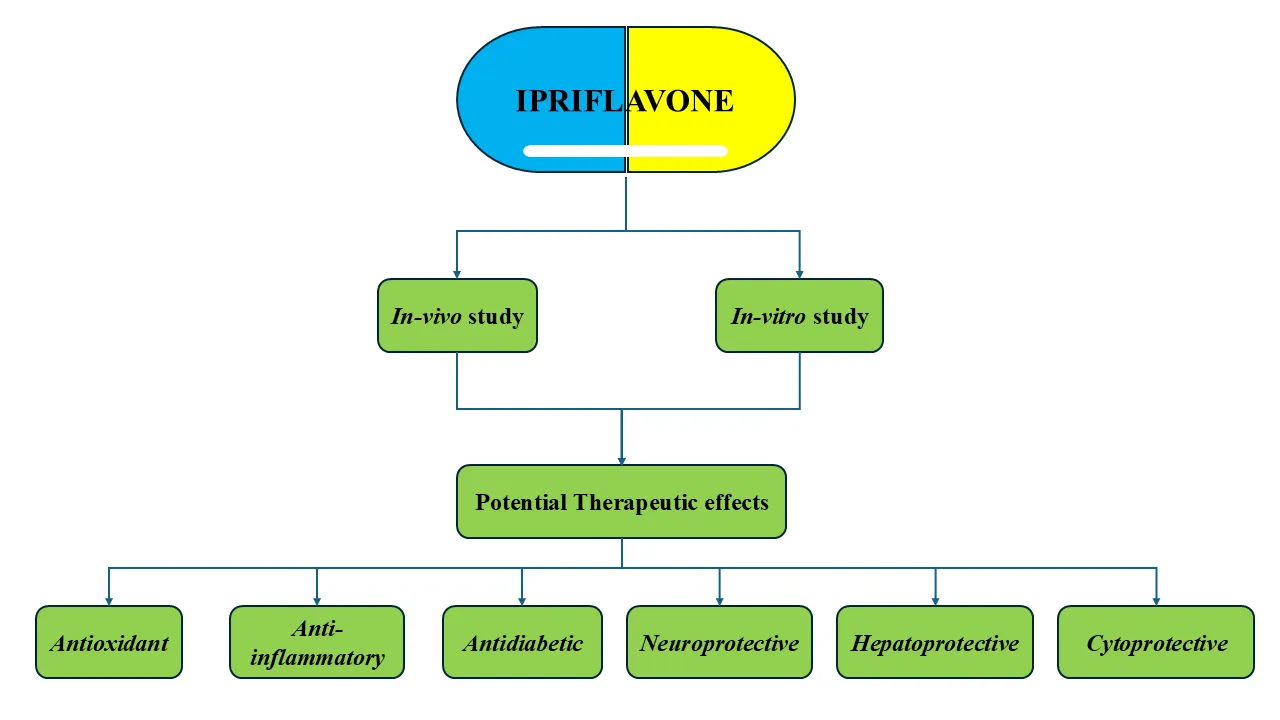
Figure 1: Therapeutic potential of Ipriflavone
Thus, these studies emphasise on how Ipriflavone can act as a potential antioxidant against many in-vivo models. Conversely, it has also been reported that Ipriflavone 50 mg/kg increased lipopolysaccharides (LPS)-induced NO production specifically in the brain cortex, cerebellum regions, liver, heart, kidneys, blood, lungs along with skeletal tissues in rats, despite its known to reduce NO release from RAW-264.7 cells as previously reported.[33]
The antioxidant potential of compounds can also be evaluated in some in-vitro studies by DPPH assay or H2DCF-DA (DCFH-DA) assay. [34,35] A study conducted to explore the potential of ipriflavone in opposition of cytotoxic and mutagenic effects induced by cyclophosphamide observed in bone marrow cells of mice by the micronucleus assay, also conducted an in-vitro assay for understanding the mechanism of Ipriflavone’s ROS scavenger capacity by DPPH method using rutin as a standard, where Ipriflavone was proposed to have a small amount of ROS scavenging action as compared to rutin which was found to be unsatisfactory.[36] However, Another study conducted on antiaging effects of Pseudoalteromonas flavipulchra STILL-33 extract’s fraction 1 (Ipriflavone 75 µM as one of the major components) had remarkably high antioxidant activities observed in in-vitro H2DCF-DA assay, which increased the longevity of the fission yeast Schizosaccharomyces pombe, by combating ROS.[37] Although, the Ipriflavone was one of the major component of fraction 1 along with L-arginine, the study states that it was responsible for antioxidant action by reducing ROS at an intracellular level regardless of H2O2-induced oxidative stress.[37] Also, this fraction was responsible for regulation of Pap1 expression following increased ctt1+ gene expression which is single gene encoding for antioxidant catalase (CAT) enzyme, which was associated with reduced levels of intracellular ROS of yeast used.[37] However, more study is required to assess and quantify the part of Ipriflavone’s antioxidant effect, as L-arginine is also a well-known antioxidant compound.[38] Another study on mice model of Hypoxia-Induced Gastrointestinal Mucosal Ferroptosis, where the subjects were treated by Aquilaria malaccensis Lam. exosome-like nanoparticles (AELNs) containing Ipriflavone as a component, observed that Ipriflavone 100 nM, 1, 5, and 10 µM tested significantly reduced polyunsaturated fatty acid-phospholipids, namely PUFA-PCs (phosphatidylcholines) and PUFA-Pes (phosphatidylethanolamines) along with ROS, 4-HNE and LPO levels by downregulating NADPH oxidase 4 (NOX4), and arachidonate 5-lipoxygenase (ALOX5) by inhibiting HIF- α.[39] Here also, Aquilaria malaccensis Lam. was reported to have antioxidant action, which also demands further study on Ipriflavone’s individual effect in this model. Another study incorporated the anti-osteoporotic drug Ipriflavone with Bioactive mesoporous glass nanospheres (nanoMBG-IPs) as well as only Bioactive mesoporous glass nanospheres (nanoMBG) in CD4+ Th2 lymphocytes, where increase in ROS was observed after treatment with only nanoMBGs, whereas, it was reduced by treatment of Ipriflavone loaded nanospheres (nanoMBG-IPs).[40] A study on human neuroblastoma SH-SY5Y cells model explored how Ipriflavone demonstrated neuroprotective effects by reduced H2O2 induced elevated reactive oxygen species (ROS) levels.[41] This study states the potential therapeutic effect of Ipriflavone as an potential antioxidant in in-vitro studies related to humans more efficiently.
Nevertheless, all these studies still emphasise on in-vitro as well as in-vivo antioxidant potential of Ipriflavone, where if both of them are taken into careful consideration along with further study needed to assess the potential effects of ipriflavone when administered alone, they indicate how Ipriflavone can be used as treatment for other disease models, widening the scope of its pharmacotherapy against oxidative stress induced diseases.
Inflammation plays a pivotal role in the pathogenesis of numerous acute and chronic diseases by mediating a complex interplay between immune system activation, increased oxidative stress, caused by many harmful stimuli like toxins, pathogens, stress or metabolic imbalance, releasing some commonly found pro-inflammatory cytokines like tumor necrosis factor-α (TNF-α), interleukin-1β (IL-1β), and interleukin-6 (IL-6), amplifying the inflammatory response and contributing to cell injury in diseases like obesity, type 2 diabetes, atherosclerosis, asthma and other neurodegenerative diseases. [42,43]
Signaling pathways mediated by Nuclear factor kappa B (NFkb) and the NLRP3 inflammasome play critical roles in disease progression caused by oxidative stress, cytokines, pathogens, and cellular injury, which leads to high pro-inflammatory mediator levels, chemokines, and adhesion molecules.[44,45] Persistent NF-κB activation further amplifies inflammatory responses and tissue injury and acts as a priming step for NLRP3 inflammasome, with mitochondrial dysfunction causing ROS generation, ATP release, or danger-associated molecular patterns (DAMPs), the NLRP3 inflammasome recruits ASC and procaspase-1, resulting in caspase-1 activation and maturation of IL-1β and IL-18, thereby exacerbating neuroinflammation and inflammatory tissue damage.[44,45,46] Furthermore, studies in APP/PS1 Alzheimer’s disease mouse models demonstrated that NLRP3 activation contributes significantly to microglial activation, neuroinflammation, and cognitive impairment, whereas inhibition or deletion of NLRP3 ameliorates disease pathology.[47]
Signal Transducer and Activator of Transcription 1 is a key inflammatory transcription factor which can be activated mainly by Interferon gamma signaling. It promotes expression of inflammatory mediators including iNOS, COX, VCAM, and ICAM, thereby contributing to inflammation and tissue injury associated with various inflammatory diseases, making it an important therapeutic target.[48] Reactive Oxygen Species (ROS) activating STAT1 within the IFN signaling pathway is called the "STAT-ROS cycle". This pathway upregulates the mitochondrial complexes by STAT1, which includes increasing ROS consequentially and sustaining STAT1 activity, which was observed in cancer cells.[49]
In a study done on rats induced with Alzheimer like disease by scopolamine administration reported a significant reduction due to Ipriflavone 50 mg/kg alone; 25 mg/kg in combination combined with berberine in elevated iNOS and NO levels. [26] Similarly, in mice with diabetic cognitive impairment, induced by streptozotocin, the elevated iNOS levels were reduced by Ipriflavone 20 and 40 mg/kg administration.[50] This anti-inflammatory effect was achieved through antagonistic action on GR/NF- κB/NLRP3/ASC/Caspase- 1 pathway acting in primary microglia of the type 1 and type 2 diabetic mice by Ipriflavone. [50] A significant reduction of inflammatory cell infiltration and proinflammatory cytokine interleukin-1β levels in a osteointegration model of C57BL/6 male mice tibias with implantations, was observed by Ipriflavone administration, which included similar mechanism of action of suppression of NLRP3 pathway in a dose dependent manner without affecting the priming stage and by protecting mitochondrial integrity, however, the reductions in TNF-α and IL-6 were not significant.[51] Conversely, Rats with dementia, induced by a mixture of aluminium, cadmium, and fluoride for 90 days followed by Ipriflavone for a further 30 days, where these metal-treated animals exhibiting abnormal behaviours also showcased significant reductions in all of the three inflammation parameter levels (TNF- α, IL-6, and IL-1β).[28] The anti-inflammatory effect of Ipriflavone is also observed in the Insulin resistance induced brain tissue dysfunction in obese albino dams and their neonates caused by high fat diet.[51]
Similarly, these effects are also carried out and confirmed in-vitro studies. The study with tibial implantation mouse model with intra-peritoneal injection of Ipriflavone, confirmed its anti-inflammatory potential by in-vitro mouse peritoneal macrophages exposed to biomaterial-associated inflammatory stimulation, which demonstrated significant activation of the NLRP3 inflammasome pathway, where Ipriflavone treatment suppressed inflammasome activation and the accumulated intracellular ROS, while also preserving mitochondrial function, inhibiting downstream IL-1β release.[51] Hence, these findings suggest that Ipriflavone exerted anti-inflammatory effects by ROS-dependent suppression of NLRP3 inflammasome signalling in macrophages, providing a mechanistic basis for its observed enhancement of early peri-implant bone healing in-vivo model.[51] An in-vitro study of Ipriflavone 200 mg/kg/day in OA mouse model, showcased suppressed KIAA1199-mediated hyaluronan (HA) degradation, where the low molecular fragmented HA is supposed to be proinflammatory than preserved high molecular weight HA in bovine cartilage explant culture. [52]
Likewise, a study on primary osteoarthritic chondrocytes derived from patient’s knees, Ipriflavone, amongst other candidate molecules, was evaluated in LPS-stimulated osteoarthritic chondrocytes in-vitro. However, its anti-inflammatory effects were not directly assessed through cytokine levels analysis, instead, the study concluded that Ipriflavone was proved to be non-detrimental or non-cytotoxic.[53] Also, an in-silico study done to identify genes and pathways associated with HIV-associated encephalitis (HIVE) and propose possible therapeutic targets, performed bioinformatic drug–gene interaction analysis and identified Ipriflavone as a compound that interacted with STAT1 gene, However, this association is derived from database-based prediction and has not been experimentally validated in disease-specific models such as HIV-associated encephalitis in vivo or in vitro. [54] Nonetheless, it shows unexplored potential as a anti-inflammatory modulator of STAT1, along with these studies taken into consideration, it highlights the potential anti-inflammatory effects of Ipriflavone, which appears as a vantage point for exploration in other studies, requiring further research regarding its actual mechanism of action and dose dependent efficacy./
Diabetes mellitus is a chronic metabolic disorder which is characterized by persistent hyperglycaemic conditions with impaired insulin secretion, insulin resistance, and/or both.[55] Type 1 diabetes is related to autoimmune destruction of pancreatic β-cells, whereas, type 2 diabetes is associated with insulin resistance and β-cell dysfunction.[55] Oxidative stress, inflammation, obesity, mitochondrial dysfunction, and advanced glycation end products play major roles in diabetes progression and its complications.[55] Hyperglycaemia increases reactive oxygen species production which results in cellular damage and impaired insulin signalling.[56] The in-vivo commonly used models of streptozotocin or alloxan-induced diabetic rodents help evaluate certain in-vivo parameters such as fasting blood glucose, oral glucose tolerance test, HbA1c, serum insulin, lipid profile, antioxidant enzymes, and organ histopathology consisting of pancreas, liver, and kidney tissues.[57,58] Whereas, The in-vitro studies mainly assess α-amylase, α-glucosidase, and DPP-IV inhibitory activities along with antioxidant assays to determine the antidiabetic potential of compounds. These experimental approaches are widely used for screening and evaluating novel antidiabetic agents.[59]
In a diabetic mouse model induced by streptozotocin, Ipriflavone 200 mg/kg administration was reported to exhibit notable metabolic effects along with its bone-protective actions on intervertebral disc degeneration of mice lumbar spine, where, the treatment with Ipriflavone significantly reduced fasting blood glucose levels and lowered HbA1c levels, indicating improved short-term as well as long-term glycaemic control, while, also improving serum insulin levels, preserving body weight levels compared to diabetic controls, suggesting a certain partial corrective antidiabetic effect.[60] This effect was also seen in dementia-induced rat model by mixture of heavy metals, where, Ipriflavone treatment significantly ameliorated metabolic abnormalities characterized by elevated serum glucose level, serum insulin level, and insulin resistance index (HOMA-IR), while also, improving β-cell function (HOMA-β).[28] Furthermore, Ipriflavone restored impaired cerebral glucose homeostasis by increasing brain glucose levels and enhancing insulin receptor gene expression and also elevated insulin-degrading enzyme (IDE) levels, which are critically involved in both insulin metabolism and amyloid-β clearance, which, indicates the therapeutic potential of Ipriflavone in mitigating insulin resistance-associated metabolic dysfunctions, particularly those linked to neurodegenerative disorders such as Alzheimer’s disease.[28] However, in another study with streptozotocin induced rats treated with TC-80 (Ipriflavone) orally through admixture in feed, the bodyweight and plasma glucose levels were found to have a non-significant change, whereas, the plasma Triglyceride and NEFA (Non-Esterified Fatty Acids) levels significantly decreased over 3 weeks, while NEFA increased a little after 6 weeks along with triglycerides.[61] Similar results were also seen in another study with rats developing diabetes due to streptozotocin, where, the plasma glucose levels decreased but was found to be not significant. The other antidiabetic parameters assessed were also found to be non-significant by Ipriflavone 25, 90, and ~270 mg/kg/day administration.[62]
A study conducted on the streptozotocin (STZ)-induced diabetic rat model for pharmacokinetic parameters assessment of Ipriflavone 20 mg/kg i.v. and 200 mg/kg oral included diabetes-related parameters, which were reported as mainly baseline, regardless, the study assessed the altered pharmacokinetic parameters due to streptozotocin which were significantly modulated by Ipriflavone treatment.[63] Some of these studies had certain limitations as Ipriflavone was evaluated primarily for bone-protective effects or altered pharmacokinetics in a streptozotocin-induced diabetic model without comprehensive assessment regarding metabolic or glycaemic endpoints and parameters required to fully assess antidiabetic action, which results in underexplored potential of antidiabetic action of Ipriflavone. Apart from that, the variable in-vivo findings also need to be performed and validated in in-vitro models, which is lacking and can be addressed in future research regarding the multipurpose use of Ipriflavone as an antidiabetic drug.
Neurodegenerative and other neurological disorders, including Alzheimer’s disease, are well characterized by complex interplay of oxidative stress, neuro-inflammation, mitochondrial dysfunction, impaired neurotransmission, insulin resistance, and apoptotic pathways.[64,65] Oxidative stress plays a pivotal role in neuronal injury through excessive generation of reactive oxygen and nitrogen species, lipid peroxidation, protein oxidation, reduced antioxidant defence systems, like glutathione (GSH), glutathione peroxidase (GPx), glutathione-S-transferase (GST), and glucose-6-phosphate dehydrogenase (G6PD), whereas, elevated malondialdehyde (MDA) and thiobarbituric acid reactive substances (TBARS) indicate enhanced lipid peroxidation and membrane damage and excessive nitric oxide (NO), produced due to nitric oxide synthase (iNOS), further aggravates reactive species stress and mitochondrial dysfunction.[64] Mitochondrial dysfunction in neurodegeneration results into reduced ATP production, disrupted calcium homeostasis, causing oxidative stress and loss of neuronal signalling and survival, which, further activates apoptotic pathways, resulting in progressive neuronal injury. Hence, mitochondria is a key regulator of neuronal survival and neurodegenerative disease progression. [64,65]
Neuroinflammation is another hallmark of these disorders and is mediated by elevated pro-inflammatory cytokines such as interleukin-6 (IL-6), interleukin-1 beta (IL-1β), and tumor necrosis factor-alpha (TNF-α), which activate downstream inflammatory signalling pathways including nuclear factor-kappa B (NF-κB) and NLRP3 inflammasome.[66,67] Chronic activation contributes to neuronal degeneration, synaptic dysfunction, and cognitive impairment.[66,67] In Alzheimer’s disease, accumulation of amyloid-beta peptides (Aβ42 and Aβ40), along with hyperphosphorylated tau protein, can be major pathological hallmarks of neuronal plaques and neurofibrillary tangles.[68] Insulin resistance and decreased insulin-degrading enzyme (IDE) cam further exacerbate amyloid accumulation and neuronal dysfunction.[65] Altered lipid metabolism, including cholesterol dysregulation and phospholipid homeostasis, also influences amyloid genesis and membrane fluidity, thereby affecting neuronal signalling and synaptic plasticity.[69]
Disturbances in neurotransmitter metabolism are mediated through increased monoamine oxidase (MAO) and acetylcholinesterase (AChE) activities, resulting in depleted monoaminergic and cholinergic neurotransmission, causing memory deficits and cognitive decline in neurodegenerative diseases.[70,71] Multiple intracellular signalling pathways, including phosphoinositide-3 kinase (PI3K)/Akt and mitogen-activated protein kinase (MAPK), regulate neuronal survival, apoptosis, and inflammatory responses. Activation of the PI3K/Akt pathway promotes neuroprotection and cell survival, whereas dysregulated MAPK signalling enhances oxidative stress, tau phosphorylation, and neuronal apoptosis. [72,73] Furthermore, neurotrophic signalling mediated through cAMP response element-binding protein (CREB) and brain-derived neurotrophic factor (BDNF) is a pathway that has critically been involved in synaptic plasticity, learning, memory, and neuronal regeneration.[74,75] Hence, reduced CREB-BDNF signalling has been implicated in many neurodegenerative disorders.[74] ADAM17, an important α-secretase, exerts neuroprotective effects by promoting certain non-amyloidogenic effects of amyloid precursor protein which leads to reduced amyloid-beta levels generated.[76] Collectively, these interconnected molecular pathways contribute to the progression of neurodegenerative and psychiatric disorders, and modulation of these pathways represents a promising therapeutic strategy for neuroprotection by potential compounds.
These findings can be supported by observations of neurobehavioral studies, playing a crucial role in the in vivo evaluation of neuroprotective agents by evaluation of functional, cognitive, and behavioural changes associated with neuronal alterations.[77] These studies provide valuable insight and highlight the efficacy of therapeutic compounds in experimental animal models used for neurodegenerative and neurological disorders. The Morris Water Maze (MWM) test is widely used to evaluate spatial learning and memory, acting as an important indicator of hippocampal integrity and cognitive function.[77] Similarly, the Open Field Test (OFT) is also one of extensively utilized test to assess locomotor activity, exploratory behaviour, and anxiety-like responses, which are frequently altered in neuropsychiatric and neurodegenerative conditions.[77] Another commonly used neurobehavioral test called Novel Object Recognition (NOR) test paradigm is used for assessing recognition from memory based on the natural tendency of rodents to have a preference for exploring novel objects over familiar ones, while, serving as a sensitive indicator highlighting learning and memory deficits associated with hippocampal and cortical dysfunction in various neurological disorders.[78] Improvement in NOR performance following drug treatment reflects preservation of cognitive function and supports the neuroprotective potential of the test compound. [78] Together, MWM, OFT and NOR offer reliable and sensitive measures that can help determine behavioural impairments and the potential neuroprotective effects of investigational drugs.
In the study investigating Ipriflavone as a non-steroidal glucocorticoid receptor antagonist against diabetic cognitive impairment in mice, behavioural and biochemical assessments together demonstrated the neuroprotective effect of Ipriflavone 20 mg/kg and 40 mg/kg, where behavioural tests like the Morris water maze test showed shortened escape latency and increased target-quadrant exploration/platform crossings, indicating improved hippocampus-dependent memory than diseased mice, while, Y-maze test showed better short-term memory for spatial working, and mice spend more time investigating the novel object, indicating preserved memory retention, with improved exploratory behaviour by increasing time and distance in the centre area; however, alteration of total locomotor activity was found to be not significant.[50] Biochemically, reduced tau hyperphosphorylation was observed with restored PI3K/AKT/GSK3β signalling by Ipriflavone which was associated with neuronal survival.[50] An increased synaptic proteins such as PSD95, synaptophysin, and VAMP2, alongside CREB, BDNF, and TrkB signalling, indicated preservation of synaptic function, while, the hippocampal long-term potentiation (LTP) and Golgi-Cox staining and electrophysiological recordings further confirmed synaptic function and dendritic spine density improvements, Additionally, suppression of inflammatory mediators including NF-κB, iNOS, TNF-α, IL-1β, NLRP3, ASC, and Caspase-1 in hippocampal tissues and microglia.[50] Collectively, these parameters established the multifaceted neuroprotective activity of Ipriflavone, against diabetic cognitive impairment through anti-inflammatory, anti-tauopathic, and synapto-protective mechanisms mediated largely via glucocorticoid receptor antagonism.
Similarly, another study investigating ipriflavone against neurodegeneration and dementia induced in rats due to environmental metal mixture, Ipriflavone (IP) 50 mg/kg was found to have a significant neuroprotective effect, which was evaluated using the Morris Water Maze (MWM), where IP improved spatial learning and memory and reduced escape latency with improved retention performance against metal-exposed rats, while, biochemical level, the study assessed oxidative stress markers including TBARS, NO, SOD, GPX, GST, and GSH, showing that IP attenuated oxidative damage by reducing lipid peroxidation and restoring antioxidant defence systems, with improvement in Neuroinflammatory status by significant reduction of TNF-α, IL-6, and IL-1β levels.[28] Additionally, the study also measured neurotransmission-related enzymes such as acetylcholinesterase (AChE) and monoamine oxidase (MAO), as well as insulin resistance markers including insulin, insulin receptor, and insulin-degrading enzyme, which was associated with improvement in cholinergic signaling and brain insulin sensitivity. [28] Furthermore, a decrease in hippocampal amyloid-β42 and tau protein accumulation was found with downregulated amyloid precursor protein and tau mRNA expression, supported by protected neuronal architecture, collectively confirming that Ipriflavone exerts multifaceted neuroprotective effects against metal-induced dementia via antioxidant, anti-inflammatory, anti-amyloidogenic, and neuromodulatory mechanisms. [28]
Such neuroprotective effects were also seen in the study done on rats with scopolamine-induced cognitive deficits, which assessed spatial learning and memory using the Morris Water Maze test, where the Ipriflavone alone 50 mg/kg and in mixture 25mg/kg with berberine improved scopolamine-induced cognitive deficits by significant decrease in escape latency and with non-significant increase in platform crossings. [26] Biochemical analysis included significance achieved in Ipriflavone and Ipriflavone-berberine treated group, in decreased oxidative stress markers (TBARS, NO), increased antioxidant defence levels (GSH, GPx, GST), decreased AChE levels, decreased metabolic marker (G6PD), and membrane lipid profile (significant decrease in cholesterol, with non-significant increased phospholipids, and a significant decrease in cholesterol/phospholipid ratio), as compared to normal and scopolamine-diseased rats. [26] Also, Alzheimer-related markers such as Aβ-42, Aβ-40, IDE, and iNOS were also measured and showed improvement with Berberine and Ipriflavone administration, while, gene expression analysis demonstrated downregulation of G6PD, iNOS, and Tau, with upregulation of ADAM-17, which reduced neurodegeneration and amyloidogenic activity and was consistent with results obtained from histopathology, which further confirmed in-vivo neuroprotection through preservation of neuronal architecture with some apoptotic neurons, less vacuolation, and reduced microglial activation in rats treated with Ipriflavone alone and in combination with berberine. [26]
Ipriflavone’s neuroprotective action was observed to be consistent with another study using Scopolamine-induced amnesia in REM sleep deprived rats, which assessed behavioural parameters by Morris Water Maze test, showcasing decreased escape latency, increased time spent in the target quadrant of probe trial, and improved direct swimming track/path pattern, indicating improved spatial learning and memory. [27] Biochemical parameters included decreased pro-oxidative stress markers (MDA, NO), with increased antioxidant defence levels (GSH, SOD, CAT), decreased AChE levels, increased brain-derived neurotrophic factor (BDNF), and significant decrease in brain cholesterol, observed in Ipriflavone alone and Vitamin-D combination treated groups, as compared to normal and scopolamine-treated REM sleep deprived rats. [27] Additionally, histopathological examination of the hippocampal CA3 region also revealed healthy neurons vs degenerating pyknotic neurons, neuronal loss, and tissue architecture. [27]
In a study involving use of Ipriflavone, demonstrated marked neuroprotective effects, against insulin resistance–induced brain dysfunction in obese dams and their neonates, where administration of Ipriflavone significantly reduced oxidative stress, evidenced by decreased TBARS, while enhancing antioxidant defences through increased GSH, CAT, SOD levels.[32] It also improved cholinergic neurotransmission by increasing acetylcholine and decreasing acetylcholinesterase activity. [32] Furthermore, Ipriflavone use also lowered Alzheimer’s-related neurodegenerative markers including β-amyloid 42, β-secretase, hyperphosphorylated tau, and APOE4, alongside reducing inflammatory mediators such as COX-II, with reduced pro-inflammatory cytokine levels. [32]
Another study investigated the effects of phytoestrogens (like Ipriflavone 2 mg/50 µL) on mitochondrial structure and function in the hippocampal CA1 region of ovariectomized rats by using ovariectomy-induced estrogen deficiency as a model of neurodegeneration, where, they reported mitochondrial ultrastructural damage, which was observed with increased mitochondrial volume density and surface area, along with reduced specific surface density and particle dispersity, and a significant decline in ATP content in the hippocampus, due to the ovariectomy procedure, while, treatment with estrogen, genistein, and Ipriflavone showcased significant reversal of these alterations, restoring mitochondrial morphology and improving ATP production, near to control levels. [79] The authors concluded that phytoestrogens, including Ipriflavone 2 mg/50 µL can exert neuroprotective effects by preserving mitochondrial integrity and function in the hippocampus, while also suggesting a role in reducing neuronal vulnerability, which was associated with estrogen deficiency. [79]
Overall, these findings suggest that Ipriflavone ameliorates neuronal deficits in neurodegenerative diseases like alzeimers by tackling oxidative stress, inflammation, mitochondrial impairment, metabolic dysfunction, and neuronal damage in-vivo animal models.
The in-vitro testing of Ipriflavone has also been done in some studies assessing its neuroprotective potential. A study conducted on SH-SY5Y cells exposed to H₂O₂ and Aβ reported that Ipriflavone 10⁻¹⁰ M and 10⁻⁸ M showed significant neuroprotective effects by improving cell viability which was reduced due to toxic insults, also, it significantly decreased intracellular ROS generation, indicating attenuated oxidative stress, and preserved mitochondrial membrane potential (MMP), and suggesting protection of mitochondrial function, and at the molecular level, it modulated MAPK signaling pathways by suppressing stress-related JNK and p38 phosphorylation while enhancing pro-survival ERK activation, collectively supporting its antioxidant, anti-apoptotic, and neuroprotective actions against Alzheimer’s disease-related neuronal toxicity and damage, evidenced by its reported in-vivo effects. [41]
In the comparative acetylcholinesterase inhibition study conducted in-vitro, Ipriflavone demonstrated significant anti-acetylcholinesterase activity which supports its potential as a cognitive-enhancing and neuroprotective agent relevant against Alzheimer’s disease.[80] When compared with natural extracts of Ginkgo biloba and berberry, Ipriflavone showcased measurable inhibition of acetylcholinesterase (AChE), the enzyme responsible for acetylcholine breakdown and hence, suggesting a suitable role in increasing cholinergic neurotransmission.[80] This activity was evaluated alongside Aricept® (donepezil) used as the reference standard, where, Aricept was the most potent inhibitor, nonetheless, Ipriflavone exhibited a notable inhibitory effect among the tested compounds.[80] These findings suggest that Ipriflavone may contribute to neuroprotection to a certain degree, through cholinergic pathway modulation, and hence, potentially improving memory and cognitive function by preserving acetylcholine availability in the synaptic cleft, as evidenced by the results obtained and compared to in-vivo models.
Hence, the above-mentioned studies indicate and highlight the potential of Ipriflavone as a neuromodulator in neurodegenerative disorders, particularly dementia and Alzheimer like diseases, and thus, points at unexplored potential to be assessed and evaluated in future research for other neurological and neurodegenerative diseases, to truly understand the scope of multifaceted neuroprotective effects of Ipriflavone.
Serum total cholesterol (TC), triglycerides (TG), low-density lipoprotein cholesterol (LDL-C), alanine aminotransferase (ALT), aspartate aminotransferase (AST), reduced glutathione (GSH), and superoxide dismutase (SOD) are most widely used biochemical markers for assessment of liver function, hepatic injury, and antioxidant mechanism.[81] Elevated serum TC, TG, and LDL-C levels are commonly associated with dysfunctional lipid metabolism leading to hepatic steatosis observed in liver disorders.[82] Elevation of sensitive hepatic enzymes like ALT and AST levels indicate liver injury or altered membrane permeability, while depletion of GSH and reduced SOD activity causes oxidative damage promoting liver diseases.[81] Therefore, these parameters are routinely evaluated in hepatoprotective studies and reflects upon the improvement in liver function and hepatoprotective efficacy of therapeutic agents against hepatic injury.
Ipriflavone demonstrated significant hepatoprotective activity in a study conducted on high-fat diet–induced non-alcoholic steatohepatitis in rats, where Ipriflavone treatment markedly reduced alanine aminotransferase (ALT) and aspartate aminotransferase (AST), indicating attenuation of hepatocellular injury, while, also lowered serum triglycerides, total cholesterol levels.[31] Additionally, Ipriflavone reduced pro-oxidant malondialdehyde (MDA) and elevated reduced glutathione (GSH) and superoxide dismutase (SOD) defence. [31] Histopathological findings further confirmed hepatoprotection, showcasing restoration of normal hepatic lobular architecture, reduced fat associated degeneration, necrosis, inflammation, and hemorrhage, However, the study also noticed certain mild residual fatty changes in sporadic hepatocytes and slight central vein congestion, despite overall hepatoprotective effects, hence, indicating partial recovery rather than complete reversal of hepatic damage. [31]
A study evaluating dietary supplementation of Ipriflavone 250, 500, 750 mg/kg demonstrated a beneficial reduction in homeostasis of hepatic iron stores, in wild-type mice, while, also an increased serum hepcidin levels, as well as, hepatic hepcidin-to-liver iron ratio, was observed suggesting improved systemic iron metabolism and regulation.[83] Consequently, Ipriflavone also decreased intestinal ferroportin and DMT1 expression, which may have contributed to limiting intestinal iron absorption and lower hepatic iron burden.[83] These findings support the potential use of Ipriflavone in hepatic iron overload and iron-induced oxidative toxicity state. However, Ipriflavone did not significantly reduce liver or splenic iron stores in Th3+/− mice which indicates limited efficacy in β-thalassemia induced iron overload.[83] Additionally, a modest reduction in mean corpuscular volume (MCV) was reported, suggesting mild iron-restricted erythropoiesis, but without any anaemia observed.[83]
Another study conducted in ovariectomized (OVX) rats, Ipriflavone 100 mg/kg/day demonstrated beneficial effects on hepatic lipid metabolism, particularly by reversing the estrogen deficient increase in liver cholesterol, induced by ovariectomy procedure, particularly by significantly reducing liver cholesterol levels, suggesting a protective effect against hepatic lipid disturbances associated in menopause.[84] These findings indicate that Ipriflavone may improve estrogen-deficient liver dysfunction, with cholesterol-lowering effects comparable to soy protein and estradiol. However, Ipriflavone failed to prevent OVX-induced abdominal fat accumulation, as well as, to significantly reduce triglyceride levels, thus, Ipriflavone may appear beneficial for reducing hepatic cholesterol deposition in OVX rats, with its metabolic effects being more specific to cholesterol regulation rather than adiposity management. On the other hand, Ipriflavone 300 mg/kg, 1500 mg/kg and 3000 mg/kg were found to increase triglyceride levels, at large doses in rats reported by a study exploring its teratogenicity.[85]
Cytochrome P450 enzymes have a crucial central role in hepatic metabolism of drugs, xenobiotics, and endogenous compounds, thereby influencing both pharmacological response and toxicity, while, having significant therapeutic relevance, particularly, in reducing the formation of reactive metabolites responsible for hepatocellular injury through inhibition of CYP-mediated bioactivation and suppressing oxidative stress, lipid peroxidation, inflammation, and necrosis, like processes, thereby preserving liver function and architecture. Thereby, the role of Ipriflavone can be of potential value in hepatic modulation. [86,87,88]
Ipriflavone 17.8 µM has shown to have such liver metabolism modulating effects, particularly, through significant inhibition of CYP1A2 and CYP2C9, along with similar effects seen by its metabolite 7-Hydroxy-isoflavone, but with additional weak inhibition of CYP3A effect in the in-vitro study.[86] Similarly, another study on human liver microsomal enzymes reported weak inhibition of CYP3A4 by Ipriflavone, however, inhibition of CYP2C19 was found to be non-significant, contrary to first study. [87] Additionally, another liver enzyme CYP19 by a study conducted to assess the aromatase inhibition potential of Ipriflavone 5, 10, and 50 µM as aromatase inhibitor.[88]
While in-vivo hepatoprotective effects have been assessed in animals, its liver enzyme modulatory effect has also been explored in-vitro. Still, future research is required for truly assessing the hepato-protection effect of Ipriflavone in other in-vivo and in-vitro liver-disease models, while, also assessing scope of enzyme modulation effect in other drugs toxicology.
Cell survival and death pathways are central to the evaluation of both cytoprotective and cancer preventive effects of drugs in in vitro and in vivo studies.[89] Excessive reactive oxygen species (ROS) generation leads to lipid peroxidation, mitochondrial dysfunction, DNA damage, mutagenicity, and apoptotic signalling pathway activation, making it a prime target for cytoprotective study. Chronic ROS-induced DNA damage may result in genomic instability, DNA mutations, and progressive cellular dysfunction leading to apoptosis.[90] Therefore, assessment of intracellular ROS levels and DNA damage can be an important marker of cytoprotective effect, where, reduction of ROS levels, prevention of DNA fragmentation as well as mutagenicity, suppression of caspase, and inhibition of apoptosis can indicate a drug’s cytoprotective effect against cellular damage. Similarly, improvement in cell viability assays reflects cyto-protection in vitro, while, decreased apoptotic cell count and reduced pro-oxidative biomarkers can support a protective pharmacological response in in-vivo models.[91,92]
Molecular signalling pathways can further provide mechanistic insights, like the Epidermal Growth Factor Receptor pathway regulates cell proliferation, survival, differentiation, and repair, where, activation of EGFR can promote tissue regeneration and survival in injured cells, while, abnormal overactivation contributes to uncontrolled proliferation in cancer. [93] Likewise, mTOR signalling is regarded as a key regulator of protein synthesis, metabolism, autophagy, and cell growth processes.[94] Controlled activation of mTOR pathway may support survival and regeneration processes in damaged tissues, whereas its dysregulation is strongly connected with tumor progression and proliferation, making it an important anticancer therapy target.[94] Pyroptosis has gained recent importance as a target for caspase-mediated inflammatory form of programmed cell death process, which when suppressed, can protect normal healthy cells from inflammation state, while, its induction in tumor cells may enhance anticancer activity.[95] Similarly, the Hedgehog signalling pathway regulates embryonic development, stem-cell renewal, and tissue repair, where, its activation may aid regeneration and cellular recovery, but, aberrant activation is implicated in tumorigenesis and metastasis.[96] Thus, studies performed on pharmacological agents showcasing modulation of cell processes like apoptosis, ROS generation, DNA integrity, cell viability, EGFR/mTOR functioning, pyroptosis, and Hedgehog signalling, collectively provides a strong mechanistic evidence showcasing the cytoprotective potential.
Ipriflavone 1.71, 8.54, and 42.85 mg/kg demonstrated a significant cytoprotective effect against cyclophosphamide-induced genetic injury in mice, primarily through its antimutagenic and anticytotoxic actions in an in vivo study using the mouse bone marrow micronucleus assay, where the decreased frequency of micronucleated polychromatic erythrocytes (MNPCEs) markers indicated protection against DNA/chromosomal damage, while the increase in PCE/(PCE+NCE) ratio indicated protection against bone marrow cytotoxicity.[36] Hence, Ipriflavone significantly reduced micronucleus formation in the pre-treatment and post-treatment phases, meanwhile, improving the erythrocyte ratio, suggesting protection against cyclophosphamide-induced chromosomal damage and cytotoxicity.[36] An in vitro mechanistic assessment for exploring antioxidant potential was also investigated using the DPPH free-radical scavenging assay, where, ipriflavone showed minimal DPPH scavenging activity.[36] Contrary to these findings, another study conducted on Swiss albino mice bone marrow micronucleus assay found that repeated clinically low and high dose of Ipriflavone 1.71 mg/kg, 8.57 mg/kg and 42.85 mg/kg induced cytotoxicity, but not mutagenicity. [97] While, a study conducted for evaluating cytotoxic and mutagenic effects of Ipriflavone 1.71, 8.57, and 42.85 mg/kg in vivo bone marrow and peripheral blood erythrocytes, in the micronucleus test, along with the human peripheral blood lymphocytes used in-vitro for 5 and 10 µg/mL dose in cytokinesis-blocked micronucleus assay (CBMN assay), reported that Ipriflavone was found to have no mutagenic or cytotoxic effects, however, micronucleus was observed during in-vitro testing, indicating significant mutagenicity.[98] These inconsistent results findings between in vitro and in vivo studies been reported, with mutagenic/cytotoxic effects observed might reflect differences occurred due to doses, exposure duration, metabolism, and model systems variations. Therefore, further well-designed studies are warranted in future to clarify the dose-dependent safety profile of Ipriflavone.
The potential anticancer action of Ipriflavone is also evaluated in some in-vivo and in-vitro models. A study conducted on in-vivo model of cancer-associated hypocholesterolemia in female Sprague-Dawley rats with breast cancer induced by methyl-N-nitrosourea, reported a lowest palpable tumor incidence and numbers, increased serum cholesterol, especially non-HDL cholesterol, with no significant effect on serum HDL cholesterol, triglycerides, liver total cholesterol, TBARS and FRA levels, on administration of Ipriflavone in combination with Genistin.[99] Another study demonstrated that Ipriflavone administered at 6 and 12 mg/mouse/day to nude mouse model of osteolytic bone metastasis with human breast cancer cells observed a significant reduction in osteolytic lesion formation and bone destruction caused due to metastatic breast cancer, where, the effect was attributed to suppression of tumor invasion altogether with inhibition of osteoclast-mediated bone resorption and osteoclast activity associated with tumour-induced bone destruction.[100] In vitro, Ipriflavone 1, 10, and 50 µM exerted direct antiproliferative effects on MDA-MB-231 human breast cancer cells where it significantly reduced viable cell numbers in a dose-dependent manner and also suppressed DNA synthesis as demonstrated by decreased [³H]-thymidine incorporation, while, mechanistically, Ipriflavone was found to inhibit ligand-induced phosphorylation of EGFR at Tyr845 residue, without altering total EGFR expression levels, however, it did not significantly induce apoptosis or DNA fragmentation, indicating that its anticancer activity is primarily observed due to growth-inhibitory (cytostatic) effect, rather than pro-apoptotic/cytotoxic. Hence, these findings suggest that Ipriflavone may have a dual therapeutic cytoprotective role in limiting skeletal degradation, associated with attenuating of metastatic progression in breast cancer of bone.[100]
Similarly, An study conducted on patient-derived xenograft (PDX) mouse 100 mg/kg/day in-vivo model as well as 60 µM in-vitro Human ESCC cell lines, particularly KYSE450 and KYSE510, reported a decrease suppressed tumour growth, size, weight, as well as proliferation, colony formation, and inhibition of mTOR signalling with reduced phosphorylation of downstream p70S6K and S6 ribosomal protein molecules, along with observed cell cycle arrest, which showcased anti-cancer and hence, cell-protective effect.[101]
In a recent in vivo study, Ipriflavone 200 mg/kg demonstrated a significant cytoprotective effect against diabetes-associated intervertebral disc degeneration (IVDD) mouse model, which combined lumbar spine instability with streptozotocin-induced diabetes, where, treatment with Ipriflavone improved vertebral and endplate microarchitecture, reduced vertebral osteoporosis, preserved intervertebral disc structure, and attenuated extracellular matrix degradation, which was evidenced by increased expression of the matrix components Aggrecan and Collagen II, altogether with reduced expression of the catabolic enzymes MMP-13 and ADAMTS-4. Mechanistically, ipriflavone suppressed NLRP3 inflammasome-mediated pyroptosis process, as indicated by decreased NLRP3, ASC, caspase-1, and cleaved gasdermin-D (N-GSDMD) expression, these findings suggest that the cytoprotective activity of ipriflavone is mediated through preservation of extracellular matrix homeostasis, with inhibition of inflammatory pyroptotic cell death, following improvement of vertebral bone health. However, the study was limited to an animal model, and the precise upstream molecular targets regulating NLRP3 pathway activation were not elucidated. Furthermore, no dedicated in vitro mechanistic experiments or long-term safety evaluations were performed, limiting direct translation of the findings to clinical settings.[60]
A study investigated the cytocompatibility of ipriflavone and ipriflavone-loaded poly(D,L-lactide-co-glycolide) (PLGA) nanoparticles for use as potential therapeutic approach, in an in-vitro osteoarthritis study using primary human osteoarthritic chondrocytes, where, Ipriflavone exhibited a certain level of cytoprotective profile by maintaining cellular viability and preventing treatment-induced apoptosis, where, the cell viability was observed above 70% following exposure to 5 and 10 μM Ipriflavone, while in Annexin V-FITC flow cytometric analysis, it demonstrated no significant increase in apoptotic cell death. Furthermore, both unloaded and ipriflavone-loaded PLGA nanoparticles showed excellent cytocompatibility across the tested concentrations, indicating no compromise of cellular safety, in addition to its observed protective effect of cell viability preservation and suppression of apoptosis, which suggests maintenance of chondrocyte integrity. However, the study did not directly evaluate the mechanism and pathways like anti-inflammatory pathways or oxidative stress markers responsible for cytoprotection effect noticed. Moreover, studies and the findings were limited to short-term in-vitro cytocompatibility assessments, requiring further exploration with in-vivo research.[102]
Another study demonstrated the chondroprotective potential of Ipriflavone in both 5, 10, 50, 100 μM in vitro and 200 mg/kg in vivo models of osteoarthritis (OA), where treatment with Ipriflavone significantly increased the chondrocyte proliferation while reducing apoptosis, which indicated improved cellular viability. Mechanistically, Ipriflavone suppressed the Indian hedgehog (Ihh) signalling pathway by downregulating (Smo), Gli2, and Runx2 expression levels, thereby inhibiting the chondrocyte cell hypertrophy. This was accompanied by certainly marked reduction in catabolic and hypertrophic bio-markers like matrix metalloproteinase-13 (MMP-13) and type X collagen, also, increased expression of anabolic cartilage matrix components such as type II collagen and aggrecan was noticed in the rat anterior cruciate ligament transection (ACLT) in-vivo model of osteoarthritis, oral administration of Ipriflavone attenuated cartilage degeneration, improved Safranin-O staining, and significantly reduced OARSI scores. Additionally, the in-vitro component of the study investigated the cytoprotective effects of ipriflavone by using primary human osteoarthritic chondrocytes, where, cell viability and proliferation were evaluated by using CCK-8 and EdU assays, which demonstrated a rather significant increase in viable and proliferating chondrocytes following Ipriflavone treatment, also, apoptosis was evaluated by Annexin V-FITC/PI flow cytometry, which revealing a marked reduction in apoptotic cell death compared with vehicle-treated controls. Specifically, the apoptotic rate was decreased from 25.76% in the DMSO control group and up to 12.64% and 15.18%, following with respective treatment of 5 μM and 10 μM Ipriflavone. These findings indicate how Ipriflavone enhances chondrocyte survival by promoting cell viability and proliferation while suppressing apoptosis, and thereby exerting a pronounced cytoprotective effect against osteoarthritic cellular damage, while it also suggests about cartilage integrity preservation and delays in OA progression, mostly through inhibition of the Ihh–Smo–Gli2–Runx2 signalling cascade (Indian hedgehog (Ihh) signalling pathway), highlighting its potential as a disease-modifying therapeutic agent for osteoarthritis, as well as a cytoprotective agent. [103]
These effects were also observed in an in-vitro study, which was done using human neuroblastoma SH-SY5Y cells, where, Ipriflavone 0.1–100 μM demonstrated cytoprotective activity against oxidative stress and amyloid-β-induced neurotoxicity, by significantly improving cell viability in cells exposed to H₂O₂ or Aβ25–35 and restoring survival from approximately 49–55% to 75–79% at nanomolar concentrations. Mechanistic investigations revealed how Ipriflavone played a role in attenuating the generated intracellular reactive oxygen species (ROS) and substantially reducing apoptotic cell death, as evidenced by Annexin V/PI flow cytometric analysis. In H₂O₂-treated cells, Ipriflavone was found to decrease apoptosis from 42.2% to 11.8%, while, apoptotic rates were reduced from 47.3% to 23.0% in Aβ25–35-induced cells. Furthermore, Ipriflavone inhibited the Aβ-induced caspase-3 activation, indicating inhibition of the apoptosis execution phase. The cytoprotective effects of Ipriflavone observed was through activation of pro-survival PI3K and MAPK signalling pathways, as pharmacological inhibition of these pathways abolished the protective response. Collectively, these findings demonstrated that Ipriflavone showcased a potent in-vitro neuronal-cytoprotective effects by enhancing cell viability, reducing oxidative stress, suppressing apoptotic signalling, and promoting neuronal survival pathways.[41]
Lastly, a study using fraction 1 isolated from Pseudoalteromonas flavipulchra STILL-33, which was enriched with Ipriflavone and L-arginine as major components of fraction, showcased its significant cytoprotective and antiaging effects in Schizosaccharomyces pombe organism, where, the fraction was found to have increased the chronological lifespan by enhanced mitochondrial activity, along with better adaptive stress response and improved cellular metabolism. Furthermore, Fraction 1 upregulated the expression of the catalase-encoding ctt1+ gene, increasing catalase protein levels and enzymatic activity, which promoted reactive oxygen species (ROS) detoxification, protecting cells against oxidative stress-induced damage to proteins, lipids, and nucleic acids and improving survival under hydrogen peroxide challenge. The cytoprotective and antiaging effects of the ipriflavone-containing Fraction 1 were evaluated in Schizosaccharomyces pombe using spot assays and chronological lifespan (CLS) analysis, where, the cell viability was primarily evaluated by the colony-forming unit (CFU)-based CLS assay for 75 μM dose, in which surviving yeast cells were quantified at different aging time points by total plate count measurements. Additionally, spot assays qualitatively assessed survival and oxidative stress tolerance of cells under hydrogen peroxide environment. These methods demonstrated that Fraction 1(containing Ipriflavone) enhanced cellular survival and prolonged yeast lifespan. In addition, Fraction 1 delayed progression of the G1 cell cycle phase hallmark of better longevity and calorie restriction–mimicking conditions in yeast. Collectively, these effects hint at a potential cytoprotective effect of ipriflavone-containing fraction by modulation of mitochondrial, activation of endogenous antioxidant defenses, attenuation of oxidative stress, and regulation of cell-cycle progression, ultimately contributing to the enhanced cellular viability and lifespan increment. The future research needs to study and explore the mechanisms and evaluate the efficacy levels in models, solely by only using Ipriflavone, to confirm and explore its true potential as a cytoprotective agent. [37]
Ipriflavone is a well-known anti-osteoporotic drug, which has clinical implications both as a main-line treatment as well as an adjunct, however, its potential therapeutic effects against other disease pathologies, remains severely underexplored. Several studies have made an attempt to understand the beneficial effects and the underlying mechanisms, through which the drug could have modulated the diseases. As such, Ipriflavone has been reported to have neuroprotective, hepatoprotective, cell protective, anti-cancer, and also a protective effect against metabolic diseases like anti-diabetes. Mostly, these effects were observed due to the antioxidant defences regulation and inflammation suppression pathway, although, other pathways were also notable including antiapoptotic, antipyretic, and lipid-modulation.
The antioxidant effects were noticeable across various disease models of bone, brain, liver diseases, which consisted primarily the modulation of SOD, GSH, GST, GPx, CAT antioxidant as well as some prooxidants like TBARS NO parameters. Some evidences also existed for ROS scavenging activities, but, most evidential effects were found for endogenous antioxidant defence modulation. These defence levels are modulated by NRF2 pathway and thus, future studies should focus on confirming and understanding whether Ipriflavone modulates this pathway, for which no evidence exists up to date. The evidential studies for anti-inflammatory effects of Ipriflavone, majorly consisted of NLRP3 inhibition, similarly, sufficient evidence showcased this effect by decreasing Tnf-α, IL-6, IL-1β as well as iNOS, NO parameters. These results are consistent with the suppression mechanism against NLRP3, which has a connection with elevation of inflammatory cytokines levels.
Antidiabetic effects of Ipriflavone were evidenced mostly in-vivo studies only, which was reported mostly by glucose and insulin levels modulation, where the IDE and HbA1c proteins were involved in the mechanism. The resulting antidiabetic effects observed needs to be studied further and understood in vitro for future research. The Neuroprotective effects was one of the most studied effects in both in-vivo as well as in-vitro models, particularly in Alzheimer like disease with dementia. Here, antioxidants and anti-inflammatory effects were the most consistent, followed by neurotransmitters metabolism suppression (MAO and AchE) and increased cell survival (BDNF and CREB). These biochemical effects were also consistent with neurobehavioral tests like MWM, OFT, NOR, Y-maze tests, showcasing preserved spatial memory, memory retention, and cognitive function. The proposed mechanistic pathway was PI3K/AkT for an in-vivo study as well as MAPK/ERK pathway for an in-vitro study, hence, these effects and pathways needs to be studied in other diseases designed to be validated with other neurobehavioral tests.
Ipriflavone also showcased hepatoprotective action in many liver diseases based in-vivo rodent models, where, it mostly attenuated liver disease by decreasing ALT and AST levels. The main mechanism which provided these effects may have been through antioxidant upregulation, but, needs further investigation for involvement of other pathways. It was also found to attenuate liver iron stores in-vivo, through acting on ferroportin and hepcidin in liver. It also has a lipid modulation effect. These results may be sufficient to hint at a hepatoprotective activity of Ipriflavone in liver diseases, yet future study is required to establish this effect better through in-vitro studies. Surprisingly, in-vitro studies on Ipriflavone reported to have an suppressive action on liver metabolic enzymes, particularly, CYP3A4, CYPY9, CYP450, and may have some effect on CYP2C9. These in-vitro results highlight its pharmacokinetic modulating property which can be beneficial in toxicology research, or concomitant drug efficacy modulation.
Lastly, Ipriflavone is also reported to have certain cell protective activity, primarily acting through antimutagenic (by MNPCEs inhibition), anti-cytotoxic(by PCE ratio increase), antiapoptotic (caspase inhibition), anticancer (suppression of mTor and EGFR phosphorylation) and anti-pyroptosis (by NLRP3 suppression). Also, Ipriflavone exerted cytoprotective effect in chondrocytes cells by suppressing Indian hedgehog pathway.
In summary, the compiled evidence demonstrates that Ipriflavone exhibits varied therapeutic efficacy across the evaluated models, each in in-vivo and in-vitro studied across various rodent species, where, most of the studies highlight it as having multifaceted potential in treatment of diseases. Nonetheless, the variations in the study result data also indicate more research is needed to investigate the efficacy and safety of Ipriflavone efficiently, also in other diseases, with indepth analysis of its mode of action.
In conclusion, Ipriflavone, an well established antiosteoporosis drug, has demonstrated an overall positive role in modulating different diseases, in various in vivo animal and in-vitro study models, through various pathways and supposed mechanisms, where, antioxidant and anti-inflammatory were the most reported mechanism of action, while some studies have also explored its role in anti-diabetic, neuroprotective, hepatoprotective, cytoprotective action. This highlights its potential use as a first line, second line or as an adjunct medication within its broad therapeutic window, but, not before its protective effects and mechanisms are well established by future research. Ipriflavone’s therapeutic effects may be limited by its solubility, absorption and bioavailability reasons, further limiting its ability to better modulate diseases without dose related side effects. Hence, future research can focus on modifying drug delivery options and methods to navigate this barrier and help further explore the true potential of Ipriflavone’s multiple therapeutic effects. Research on development of treatment options from novel compounds takes more financial investment, time, human resources while also, showcasing immense risk of failure in further clinical trials, leading to massive losses. Additionally, many diseases are prone to drug resistance, leading to failure in treatments and hence, repurposing of old drugs, like Ipriflavone, for different disease conditions, can be advantageous economically as well as, pharmacologically, in the long run.
REFERENCES

Pranay Pawar*,Sunita Ogale,Ruchita Dhangar, Shikha Shukla, A Review on Multifaceted Therapeutic Potential of Ipriflavone, Int. J. of Pharm. Sci., 2026, Vol 4, Issue 10, 1725-1751. https://doi.org/10.5281/zenodo.23335548
 10.5281/zenodo.23335548