

We use cookies to ensure our website works properly and to personalise your experience. Cookies policy
Bachelor Of Pharmacy, Genba Moze College Of Pharmacy, Wagholi, Pune, Maharashtra, India.
Cancer is a significant global health issue. Early detection is key for improving treatment outcomes and increasing survival rates. Tissue biopsies provide useful histological and molecular information; however they are an invasive and may not accurately reflect the heterogeneous nature of tumours. Non-invasive methods are therefore being developed. Liquid biopsy allows tumour biomarkers to be analysed from bodily fluids such as blood. Circulating tumour DNA (ctDNA), circulating tumour cells (CTCs), extracellular vesicles (EVs) and cell-free RNA (cfRNA) are all biomarkers that can be found in liquid biopsies. Recent developments in technology such as polymerase chain reaction (PCR), next-generation sequencing (NGS), DNA methylation, microfluidics, biosensors, nanotechnology and artificial intelligence (AI) have increased the use of liquid biopsies. They can now be used to aid cancer profiling, monitor response to treatment, identify minimal residual disease and detect cancer recurrence. There are several challenges within liquid biopsies, including low biomarker concentrations, variability between samples, interference from clonal haematopoiesis, high costs of testing and limited clinical validation. This review outlines the main biomarkers used for liquid biopsies, new technologies emerging to detect them, their uses in the clinic, limitations with liquid biopsies and future research. Technological advancements and large clinical validation will be key in making liquid biopsies a reliable tool for personalising cancer management.
Cancer is major global public health challenge and remains one of the major causes of illness and death worldwide. According to the latest estimates from the International Agency for Research on Cancer (IARC), approximately 20.6 million new cancer cases and 9.8 million cancer-related deaths occurred globally in 2024. The global cancer burden is projected to increase substantially in the coming decades, mainly due to population growth and ageing, as well as changes in exposure to cancer risk factors. Although cancer is more common among older adults, it can occur across all age groups. Therefore, effective cancer prevention, early detection, accurate diagnosis, timely treatment, and continuous disease monitoring remain critical public health priorities. [1]
The International Agency for Research on Cancer (IARC) keeps track of cancer cases, deaths and how many people have cancer by using the Global Cancer Observatory (GLOBOCAN) database. Cancer incidence means the number of people who get cancer in a certain group of people during a specific time. This is usually shown as how many people out of 100,000 get cancer each year. Cancer mortality is the number of people who die from cancer in a group during a specific time. This is also usually shown as how many people out of 100,000 die from cancer each year. Cancer prevalence is the number of people who are living with cancer at a moment. Five-year prevalence is often used to show how many people are still alive five years after being diagnosed with cancer. These three measures are important for understanding how bad the cancer problem is in places and for planning ways to prevent cancer, screen it treat it and offer care to those who need it. [2]
1.2 Early Diagnosis and Screening
Early identification of cancer is essential. This refers to identifying cancer in patients who are already presenting with symptoms of the disease. This differs from cancer screening, which involves testing apparently healthy, asymptomatic individuals for the early signs of cancer.
Cancer screening serves as a preventive check to detect cancer before symptoms appear, whereas diagnosis addresses patients who are already symptomatic and require clinical evaluation. Both approaches are important components of cancer care, though they differ substantially in objectives, resource requirements and cost.
Diagnosis of cancer refers to identifying patients who are already symptomatic. The goal is to detect cancer as early as possible and initiate treatment promptly, thereby improving the patient's chances of survival and long-term quality of life. [3]
2. Conventional methods of cancer diagnosis
• Imaging
• Histopathology
• Tissue Biopsy
2.1 Imaging
Include the imaging methods:
• X-ray: Mainly used to find abnormal changes especially in the chest.
• CT (Computed Tomography): Gives pictures of anatomy and helps find where a tumor is, how big it is and how far it spreads. It is also good for guiding biopsies and planning treatment.
• MRI (Magnetic Resonance Imaging): Uses magnets and radio waves to make pictures of soft tissues. It is especially good, for finding and describing tumors in tissue.
• PET (Positron Emission Tomography): Shows how cells use energy and helps find tumors spread of cancer and how well treatment works.
• Ultrasound: Uses sound waves to show lesions. Is often used to guide biopsies.[4]
2.2 Histopathology
Histopathology is a method used to diagnose and understand cancer by looking at tissue samples under a microscope. It focuses on the shape and structure of cells, how tissues are organised, changes in cell nuclei, how fast cells are dividing and other signs of disease. This helps doctors decide if a growth is benign or malignant and what kind of tumour it is. Histopathology also gives details about how aggressive the tumour might be, whether it has spread into nearby tissues, blood vessels or nerves and if the edges of a removed tumour are clear. The common way to look at tissue is with hematoxylin and eosin staining, or H&E. This gives a view of the tissue’s structure. Another useful tool is immunohistochemistry, or IHC, which detects proteins or markers that help classify the tumour, predict how it might behave and guide treatment choices. However, histopathological results can be influenced by sample collection differences within the tumour itself and differences between doctors interpreting the images. For this reason, extra tests—such as immunohistochemistry or molecular analysis—may be needed to get the most accurate diagnosis and understanding of the cancer. [4,5]
2.3 Tissue Biopsy
Tissue biopsy is a traditional method used to detect cancer. Tissue biopsy is performed on a piece of tissue from the suspected lesion .The piece of tissue is then looked at under a microscope to see cell shape, tissue layout and disease changes that show cancer. Biopsy samples may also go through staining and genetic tests to find markers and DNA changes that aid in classifying the tumor and picking treatment . [5]
Common biopsy methods are biopsy, CT‑guided biopsy, MRI‑guided biopsy, endoscopic biopsy and surgical biopsy. Image‑guided biopsy helps get samples from spots that are hard to reach.However tissue biopsy is an act and can cause pain, bleeding, infection or other complications related to the procedure. Tissue biopsy also need a representative sample of tissue Errors can happen because tumors can be different in space and time; different parts or stages of a tumor may show DNA traits These problems have made people more interested in minimally invasive method like liquid biopsy, which can study cancer material found in body fluids.Thus tissue biopsy remains a tool for a clear diagnosis while liquid biopsy is being studied as an extra method, for finding cancer and profiling its molecules.[6]
2.4 Molecular Testing
Molecular testing involves looking at the DNA and other molecules inside tumor tissue to help diagnose cancer sort it into types predict how it will behave and pick the treatment. Molecular testing can find changes such as mutations, gene fusions, differences in DNA copy numbers changes in gene activity and other molecular oddities that are tied to cancer. Common ways to do testing are PCR, fluorescence in situ hybridization (FISH) next-generation sequencing (NGS) and other molecular assays. Molecular testing is especially useful, for spotting genetic changes that can guide targeted therapies. However traditional molecular testing usually needs representative tumor tissue and that tissue can be limited or hard to obtain.
2.5. Limitations of conventional method
The major limitations include:
1. Invasiveness: Tissue biopsy involves taking out a piece of tissue, which can result in pain, bleeding or infection.
| METHODS | MAIN PURPOSE | ADVANTAGES | Reference |
|---|---|---|---|
| X rays | Detection of abnormalities | Simple and widely available | [9] |
| CT | Tumor localization and staging | Detailed anatomical images | [9] |
| MRI | Soft-tissue characterization | Excellent soft-tissue contrast | [9] |
| 4. Ultrasound | Lesion detection and biopsy guidance | Non-invasive and no radiation | [9] |
2. Sampling limitations: A small biopsy might not reflect the tumor.
3. Tumers heterogeneity: Various parts of a tutors can hold genetic and molecular changes.
4. Difficulty in repeated sampling: Doing biopsies can be hard especially when the tumor is hard to reach.
5. Time and resource requirements: Some diagnostic steps need equipment, skilled staff and lab spaces [7]
6. Monitoring a conventional biopsy give data mainly from the sampled spot and may not keep up with changes happening in the whole tumor. [8]
| 5. PET/CT | Assessment of metabolic activity and metastasis | Whole-body assessment | [9,10] |
|---|---|---|---|
| 6. Histopathology | Tissue characterisation and tumour diagnosis | Detailed cellular information | [11] |
| 7. Tissue biopsy | Confirmation of malignancy | Provides tumour tissue for further testing | [11] |
| 8. Molecular testing |
Genetic and molecular characterization | Can guide targeted therapy | [11] |
3. Liquid Biopsy
Biopsy is a method used to detect and characterise cancer by analysing tumour-derived material present in body fluid, most commonly blood. It is minimally invasive. Unlike a biopsy, where they take out tissue from the cancer, a liquid biopsy uses a simple blood sample. This makes it possible to do the test times during different parts of the disease. It can help with finding cancer looking at the genes checking how treatment is working seeing if resistance happens finding amounts of cancer left and catching a return early Besides blood other body fluids like urine, saliva, fluid from the lungs and fluid around the brain and spinal cord can be checked depending on the type of cancer[12,13]
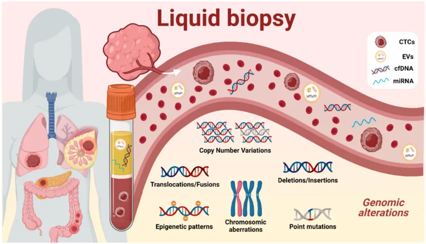
Figure 1: Schematic Representation of Liquid Biopsy Showing the Release of Tumour-Derived Biomarkers into Body Fluids, Sample Collection, and Molecular Analysis. [14]
3.1Advantages of Liquid Biopsy
Liquid biopsy offers benefits when compared to traditional tissue biopsy. It is less invasive and usually only needs a sample of body fluid, such as blood. This makes it possible to take repeated samples over time during the course of a disease or treatment. Tumors release different materials into body fluids, including circulating tumor cells (CTCs) cell-free DNA (cfDNA) or circulating tumor DNA (ctDNA) circulating RNA, extracellular vesicles (EVs) tumor-educated platelets and proteins. Studying these materials can reveal details about changes in the tumor tumor features, how well the treatment is working and signs of treatment resistance [15,16]
Liquid biopsy can also help track residual disease (MRD) and detect shifts in tumor behavior as time goes on. Since the tumor material in body fluids can come from parts of the tumor liquid biopsy can give insight into both the spatial and temporal differences, within the tumor. This is something that a single tissue biopsy might not fully capture [16].
3.2 Comparison of Traditional Tissue Biopsy and Liquid Biopsy
Liquid biopsy offers advantages over traditional tissue biopsy because it is minimally invasive and can be performed repeatedly during disease progression and treatment. Traditional tissue biopsy requires the removal of a tumor tissue sample through a needle-based or surgical procedure, which may cause discomfort and may not always provide a sample of the entire tumor. A single tissue biopsy may capture one region of a heterogeneous tumor and may therefore fail to reflect differences between the primary tumor and metastatic sites. In contrast liquid biopsy analyzes tumor-derived components present in body fluids. Can provide information about tumor characteristics, from multiple sites. Repeated liquid biopsy may help monitor tumor evolution, treatment response, treatment resistance, minimal residual disease (MRD) and cancer recurrence. However liquid biopsy does not completely replace tissue biopsy, which remains important for histopathological diagnosis and tumor characterization [17]
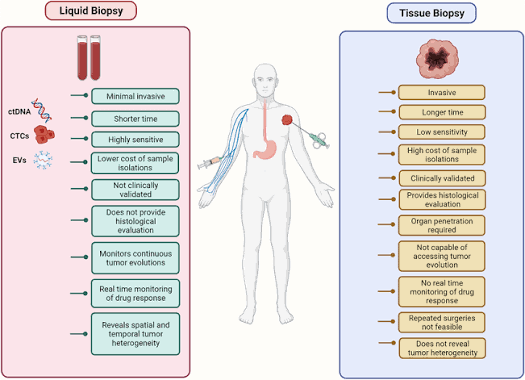
Comparison of Traditional Tissue Biopsy and Liquid Biopsy.[18]
3.3 Body Fluids Used for Liquid Biopsy
Plasma the liquid component of blood,is most commonly used body fluid for the liquid biopsy because cancer parts can get into the blood from the main cancer and from cancers that have spread. Other fluids like urine, saliva, fluid around the brain and spinal cord and fluid around the lungs can also be checked. Which fluid is used depends on the type of cancer and where it's the type of things from the cancer that are being looked at. [19]
3.4 Clinical Applications
Liquid biopsy has clinical applications across several areas of cancer care,including early detection, diagnosis, molecular profiling, prognosis, treatment selection, and monitoring of treatment response, detection of drug resistance, assessment of minimal residual disease , and early detection of recurrence.Despite this potential , liquid biopsy has limitations, as some cancer shed limited biomarker material into body fluids, making detection difficult.Tissue biopsy therefore remain essential for confirming diagnosis and guiding clinical decision,while liquid biopsy continues to be investigated and is increasing adopted as a complementary tool . [20]
4. Major Biomarkers
Liquid biopsy mainly looks for tumor-derived material, in body fluids in blood. The main biomarkers that liquid biopsy detects are circulating tumor DNA (ctDNA) cell- DNA (cfDNA) circulating tumor cells (CTCs) extracellular vesicles (EVs)/exosomes and circulating RNA. Each of these biomarkers gives an insight into tumor biology. Because each one provides information liquid biopsy can use any single biomarker alone or combine several of them to get a more complete picture. [21]
Major liquid-biopsy biomarkers and representative technologies:
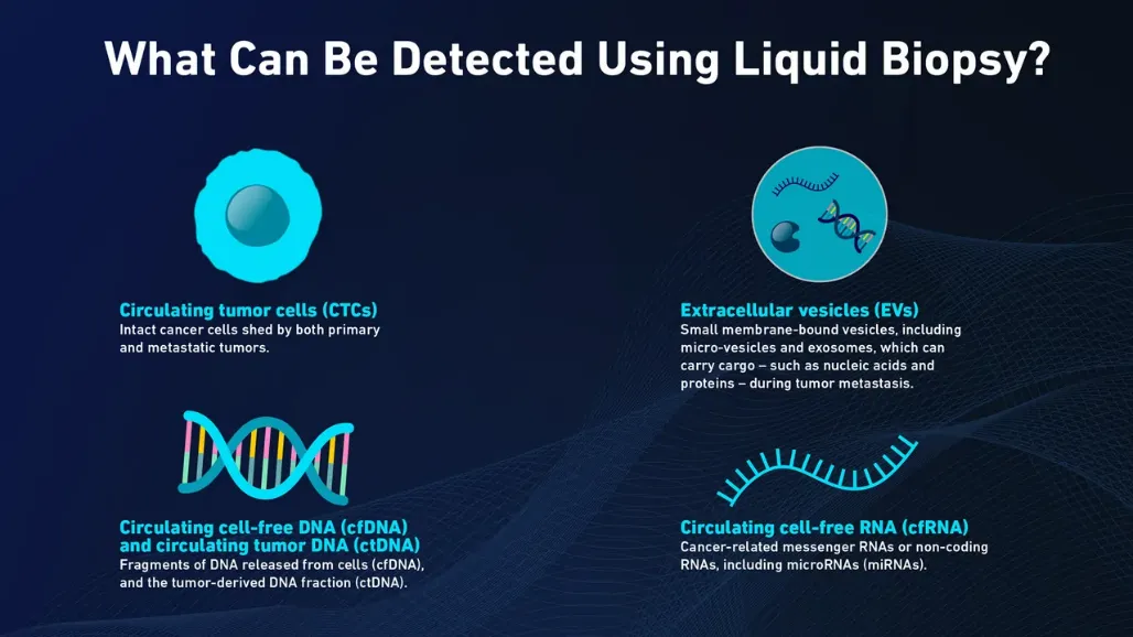
Figure 2: Major Liquid-Biopsy Biomarkers, Including Circulating Tumour DNA, Circulating Tumour Cells, Extracellular Vesicles, and Circulating RNA. [22]
| Biomarker | What it represents | What it represents | Main detection technologies |
|---|---|---|---|
| CtDNA/cfDNA | Tumor-derived and total cell-free DNA | Mutations, copy-number alterations, methylation and fragment patterns | PCR, ddPCR, NGS, methylation analysis, fragmentomics |
| CTCs | Intact tumor cells circulating in blood | Cell morphology, phenotype, DNA, RNA and proteins | Immunomagnetic capture, microfluidics, imaging |
| EVs/exosomes | Membrane-bound vesicles released by cells | DNA, RNA, proteins and signaling molecules | Ultracentrifugation, size-exclusion, affinity capture, biosensors |
| Circulating RNA | mRNA, miRNA and other non-coding RNA | Gene-expression and regulatory signatures | RT-PCR, RNA sequencing, biosensor |
| Other biomarkers | Proteins, tumor-educated platelets and combined biomarkers | Proteins, tumor-educated platelets and combined biomarkers | Immunoassays, proteomics, multi-omics |
4.1 Circulating Tumor DNA (ctDNA)
Circulating tumor DNA or ctDNA is a part of the cell-free DNA that comes from cancer cells and gets released into the bloodstream. It contains changes that're specific to the tumor, such as mutations, copy-number changes and patterns of DNA methylation. These pieces of DNA can be found using methods like PCR, next-generation sequencing (NGS) and methylation analysis. Because of this ctDNA helps in detecting cancer tracking how well treatment is working and spotting if the disease comes back later. . [21]
4.2 Circulating Tumour Cells (CTCs)
Circulating tumor cells, or CTCs, are cancer cells that break away from the original tumour or from areas where the cancer has spread. These cells travel through the blood. Unlike cells, CTCs carry full cellular information—like their shape, surface proteins, DNA changes, RNA activity and protein features. This means studying CTCs gives details about the tumour’s behaviour and how likely it is to grow or spread.
The big challenge with CTCs is that they are very rare in the blood. They also come in different types, and some do not have the surface markers that current detection tools depend on. Because of this, scientists are developing ways to find and study them. These include tools, microfluidic systems and label-free techniques that don’t need specific markers to work[23, 24]
4.3 Extracellular Vesicles and Exosomes
Extracellular vesicles or EVs are membrane-bound particles that cells release into their surroundings. These particles can carry molecules from one cell to another. Exosomes are a type of EV that are very small and can contain DNA, mRNA, miRNA, proteins and other substances. Since the contents inside these vesicles reflect the health and state of the cells that made them, researchers see EVs as signals for finding and watching cancer.
To study EVs, scientists first isolate them from body fluids. Then they analyse the molecules attached to the surface of the vesicles. Common isolation methods include ultracentrifugation, size-exclusion chromatography, precipitation, affinity-based capture and microfluidic approaches. Still, there are problems. EVs are highly diverse; they often mix with -EV materials, and different isolation methods can give different results. [21,25]
4.4 Circulating RNA and Other Biomarkers
Circulating RNA includes mRNA, microRNA (miRNA), long non-coding RNA and other RNA molecules found in body fluids like blood. These molecules can show how genes are being turned on or off and how cells are behaving when cancer is present. MiRNAs get a lot of attention because they can control genes at once and remain detectable in the blood even when protected inside protein complexes or small sacs known as extracellular vesicles.
Other circulating biomarkers, such as proteins and tumour-educated platelets, also give clues. When doctors combine information from different kinds of biomarkers—DNA, epigenetics, cells and proteins—they can get a clearer view of the cancer. Each type of signal shows a piece of the disease puzzle.[22]
5. Sample Collection and Isolation Technologies
The quality of results from biopsies depends a lot on how the samples are handled before analysis. Blood collection tubes, the time between when the blood is taken and when it is processed, how it is centrifuged, how it is stored, and the methods used to extract the DNA can all affect how well the biomarkers are recovered and how good the quality is. That is why it is important to follow procedures to get results that are reliable and repeatable.[26]
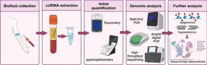
Figure 3: Workflow of Liquid Biopsy from Sample Collection and Biomarker Isolation to Molecular Detection and Clinical Interpretation. [36]
5.1 Plasma and cfDNA Isolation
Blood is usually processed to separate the plasma from the blood cells. Then the cell-free DNA is extracted using systems that're available on the market or automated tools. The goal is to get cell-free DNA while making sure there is not too much contamination from the DNA that comes from damaged blood cells. [21]
5.2 CTC Isolation
CTCs can be isolated using:
• Capture – this uses antibodies that target specific markers on the surface of cells.
• Size-based filtration – this separates cells based on their size and how flexible they are.
• Microfluidic systems – these use-controlled movement of fluids and the physical or molecular characteristics of the cells.
• Density-based separation – this takes advantage of differences in how dense the cells are
• Dielectrophoresis – this uses differences in the properties of the cells.
No one method can collect all types of CTCs efficiently because the cancer cells vary in size, appearance, and the markers they have on their surfaces.[23, 24]
5.3 EV/Exosome Isolation
EVs can be isolated by ultracentrifugation by size‑exclusion chromatography by precipitation by membrane‑based techniques and, by affinity capture. Newer microfluidic methods and nanotechnology‑based techniques aim to make the isolation of EVs faster, cleaner and easier to link with detection steps. [25]
6. Advanced technologies for detection The rapid development of liquid biopsy is driven by the advent of highly sensitive molecular and analytical technologies. Tumour-derived biomarkers are typically present at very low levels in body fluids, especially in early stages of cancer, making them difficult to detect using conventional methods. Emerging technologies such as digital PCR, next-generation sequencing, DNA methylation analysis, cfDNA fragmentomics, microfluidics and biosensors have improved detection and characterisation of circulating tumour DNA (ctDNA) , circulating tumour cells (CTCs) , extracellular vesicles (EVs), and other cancer-associated biomarkers. These technologies can provide information on tumour mutations, epigenetic changes, tumour burden, response to treatment and disease progression. [26,27]
6.1 Digital PCR and Droplet Digital PCR (ddPCR)
Digital PCR (dPCR) is a sensitive way to look at DNA. It helps. Count exact amounts of DNA that scientists are looking for. With dPCR, the sample is split into tiny reactions. This lets scientists see which reactions have the DNA they want and which do not. Then they can figure out how much of the DNA is there. Droplet digital PCR (ddPCR) is a type of dPCR. In ddPCR, the mix is split into thousands of drops before it goes through PCR.
In biopsy, ddPCR helps find small changes in DNA from tumours. It can spot changes in genes like EGFR, KRAS, BRAF and PIK3CA. These changes might be very small compared to all the DNA in the blood. Because it can find and count these amounts, ddPCR is used for finding cancer detection, checking how treatment is working, looking for remaining cancer after treatment and seeing if cancer comes back.
One important use is to watch for changes in DNA during treatment. For instance, ddPCR has been used to find signs of cancer returning in patients with cancer. In some cases, ddPCR found changes in ctDNA before regular tests found changes in CA-125.[28]
Benefits:
- Can find small changes in DNA
- Tells exactly how much DNA is there
- Needs only a little DNA
- Good for checking the same changes over time
- Works with blood and other body fluids
Drawbacks:
- Needs to know what change to look for
- Can’t check for many changes at once as well as some other methods
- If the DNA is not good or there is not much of it, it might not work well
So ddPCR is very useful when a certain change in DNA in a tumour needs to be found or checked many times.
6.2 Next-Generation Sequencing (NGS)
Next-Generation Sequencing (NGS) is a tool that allows the examination of DNA fragments at the same time. In biopsy, NGS is used to look at circulating tumour DNA and find changes that are linked to cancer. Depending on the method used, NGS can spot mutations and other changes in the DNA. Create a picture of the tumour.
The usual process involves taking plasma, extracting cfDNA, making a library, doing the sequencing, and then analysing the data with computers. NGS-based liquid biopsy is used in cancer screening studies to look at the details of the cancer, check how treatment is working, and find changes that might make treatment less effective. However, the low amount of ctDNA, especially when cancer is in early stages, is still a problem for analysis. [28]
6.3 DNA Methylation Analysis
DNA methylation changes how genes are controlled. It does not change the DNA sequence. DNA methylation controls whether a gene is working or not. When cancer starts, DNA methylation can become different from normal. These different DNA methylation patterns can be found in circulating cell-free DNA, which's DNA that moves around in the blood. Because of this, DNA methylation patterns in circulating cell-free DNA can help find cancer without tests.
Many ways exist to look at DNA methylation patterns. These ways include methylation-PCR, bisulfite-based techniques and sequencing-based methods. Some DNA methylation changes that are connected to cancer stay the same. These changes might happen early when a tumour starts to grow. Because of that, scientists are looking at DNA methylation analysis to find cancer, understand what type it is, guess how it might get worse, and keep track of it over time.
Many problems still exist. Different testing methods can have results. Differences in people's biology also affect the results. The amount of tumour DNA, in the blood can be small, making it hard to find. Large studies are also needed to prove that these methods work well. [30]
6.4 cfDNA Fragmentomics
cfDNA fragmentomics is about studying the features of pieces of cell DNA that are found in the blood. This includes looking at how big the pieces are, how they end and other ways they break apart. These features are connected to how DNA is organised in the genome, how chromatin is structured and what happens in cells when DNA is released.
This method is different from others that only look for mutations. Instead, fragmentomics looks at patterns across the genome. Changes in these patterns can help tell the difference between cfDNA from cancer cells and cfDNA from other cells. Scientists are now using computer tools and artificial intelligence to analyse features all at.
So fragmentomics is a way to detect cancer without tests. It could work well with methods like mutation or methylation analysis. We still need to standardise how we analyse the data and prove it works in real-world settings. [31]
6.5 Microfluidic Technologies
Microfluidic technologies deal with amounts of fluid moving through tiny channels. In the area of biopsy, microfluidic systems have been developed to isolate, concentrate and study circulating tumour cells (CTCs), extracellular vehicles (EVs), ctDNA and other signs of cancer.
These systems can use features like cell size or how flexible a cell is. They can also use techniques like attaching molecules to catch cancer-related signs from blood. Often these systems bring together sample preparation and detection in a device.
The main benefits of these systems include the need for small sample sizes, the ability to perform steps automatically, quick processing times and the chance to create devices that are small enough for use in portable or point-of-care settings. With these advantages, there are still problems. Variations in device designs, low recovery rates of biological signal impurities in the samples and the absence of standardisation make it difficult to use these systems widely in different settings. [32]
6.6 Biosensors
Biosensors are devices that can detect molecules and turn their interaction into a signal that can be measured. Biosensors have been studied for finding signs of cancer such as ctDNA, miRNA, proteins, exosomes and cells that are in the blood.
There are types of biosensor methods. These include systems that use electricity or light, as well as systems that use antibody pieces, DNA, special molecules called aptamers and tiny materials called nanomaterials to find the right molecules or make the signal stronger. These methods could be used for finding cancer, checking how treatment is working and testing at the place where care is given.
Making biosensors that're very good at finding small amounts of material and that can be carried around might help bring liquid biopsy to a place where it can be used quickly. There are still problems. These include ensuring biosensor accuracy, accounting for sample components that can confound results, ensuring consistent results, and proving they work in clinical settings. [33]
7. Clinical Application
The mature use of plasma-based assays is targeted molecular profiling in selected advanced cancers. This is especially true when tissue is not available or when a quick result is needed. Other important uses include checking how treatment is going over time, finding out if resistance has developed and looking for any remaining cancer cells after treatment that was meant to cure the disease. Screening a population is possible but needs very strong proof. This is because positive results can cause worry and lead to unneeded tests [34]
| Clinical question | Potential contribution of liquid biopsy | Evidence needed |
|---|---|---|
| Early detection | Multi-analyte signatures may flag an occult malignancy | Prospective population studies and diagnostic workup pathways [35] |
| Molecular profiling | Take actionable mutations when tissue is limited | Concordance with tissue and outcome-guided treatment benefit [34] |
| Treatment monitoring | Serial ctDNA changes may precede radiological progression | Thresholds and management action validation in trials [34] |
| MRD / recurrence | Molecular residual disease may identify her higher relapse risk | Proof that intervention based on result improves survival [34] |
8. Challenges and requirements for translation
• Pre-analytical factors: The type of collection tube, how quickly samples are processed, whether plasma or serum is used, storage conditions and the method of extraction can all affect the measured signal.
• Biological limitations: In early-stage cancers, little ctDNA may be released into the bloodstream. Also, the amount of signal can vary depending on the type of cancer, the tumour burden and where the tumour is located in the body. [34]
• Positive interpretation: Clonal haematopoiesis can cause genetic variants to appear in the blood that are not from the tumour but from blood cells. This can lead to misleading results.
• Clinical validation: Assays must have consistent limits of detection. They also need to be validated and linked to clear clinical actions that make a difference in patient care.
• Implementation: Cost, lab infrastructure, regulatory control, data privacy and access, for all patients must be. Addressed directly. [35]
Figure: pathway from Assay development to clinical use
[Figure could not be converted – please insert manually]
Biomarker discovery Analytical validation Clinical validation Clinical utility and equity Implantation and Monitoring
9. Future directions
Future liquid-biopsy research will likely combine several information layers, including mutations, methylation, fragment size, RNA, proteins, and imaging data. Multi-modal approaches may improve performance, but they must be compared against current standards of care in diverse populations. Priority areas include harmonised pre-analytical protocols, reference materials, prospective trials, transparent AI evaluation, and cost-effectiveness studies. [12]
Liquid biopsy offers a powerful route to serial, minimally invasive molecular assessment of cancer. Advanced technologies have expanded its ability to detect sparse tumour-derived signals and may support earlier detection, targeted treatment selection, and disease monitoring. At present, its strongest role is as an evidence-based complement to tissue pathology and imaging. Rigorous validation and equitable implementation are necessary before broad use in screening and routine early cancer detection [34]
CONCLUSION
Liquid biopsy offers a powerful route to serial, minimally invasive molecular assessment of cancer. Advanced technologies have expanded its ability to detect sparse tumour-derived signals and may support earlier detection, targeted treatment selection, and disease monitoring. At present, its strongest role is as an evidence-based complement to tissue pathology and imaging. Rigorous validation and equitable implementation are necessary before broad use in screening and routine early cancer detection.
REFERENCES


Madhuri Sable, Akshata Patil, Tushar Shelke, Advanced Technologies for Liquid Biopsy in Cancer Detection and Monitoring, Int. J. of Pharm. Sci., 2026, Vol 4, Issue 11, 1250-1262. https://doi.org/10.5281/zenodo.24002069
 10.5281/zenodo.24002069