

We use cookies to ensure our website works properly and to personalise your experience. Cookies policy
Department of Pharmaceutical Analysis, Bharat Institute of Pharmacy, Mangalpally, Ibrahimpatnam, Hyderabad, Telangana Pin code - 501510.
The present study was aimed at the formulation and evaluation of Gentamicin-loaded transferosomal gel for enhanced topical drug delivery. Transferosomes were developed to improve drug penetration and therapeutic efficacy. Preformulation studies including organoleptic evaluation, solubility studies, calibration curve preparation, and FTIR compatibility studies were carried out. Transferosomes were prepared by the thin-film hydration method using soya lecithin, cholesterol, and Span 80, followed by incorporation into Carbopol 940 gel. The prepared formulations were evaluated for particle size, pH, entrapment efficiency, SEM analysis, in vitro drug release, and stability studies.The FTIR studies confirmed the compatibility of Gentamicin with the selected excipients. The particle size of the prepared transferosomes ranged from 112 to 137 nm. SEM analysis revealed spherical vesicles with smooth surface morphology and no signs of aggregation. The pH of all formulations was found to be suitable for topical application. Entrapment efficiency ranged from 79.63% to 85.78%, with formulation F4 exhibiting the highest entrapment efficiency. In vitro drug release studies demonstrated sustained drug release, and formulation F4 showed the highest cumulative drug release of 98.84% after 8 hours. Stability studies indicated that the optimized formulation remained stable during the storage period without significant changes in its physicochemical properties. The results suggest that the developed Gentamicin-loaded transferosomal gel is a promising topical drug delivery system with enhanced drug entrapment, sustained release, and good stability. The optimized formulation may provide improved therapeutic efficacy for the management of topical bacterial infections.
Transdermal Drug Delivery System
Transdermal drug delivery systems are defined as self-contained, discrete dosage forms which, when applied to the intact skin, deliver the drug, through the skin, at a controlled rate to the systemic circulation. At present, the most common form of delivery of drugs is the oral route. While this has the notable advantage of easy administration, it also has significant drawbacks namely poor bioavailability due to hepatic first pass metabolism and the tendency to produce rapid blood level spikes (both high and low), leading to a need for high and/or frequent dosing, which can be both cost prohibitive and inconvenient. To overcome these difficulties there is a need for the development of new drug delivery system; which will improve the therapeutic efficacy and safety of drugs by more precise (i.e. site specific), spatial and temporal placement within the body thereby reducing both the size and number of doses. New drug delivery system are also essential for the delivery of novel, genetically engineered pharmaceuticals (i.e. peptides, proteins) to their site of action, without incurring significant immunogenicity or biological inactivation. Apart from these advantages the pharmaceutical companies recognize the possibility of repattening successful drugs by applying the concepts and techniques of controlled drug delivery system coupled with the increased expense in bringing new drug moiety to the market1. One of the methods most often utilized has been transdermal delivery - meaning transport of therapeutic substances through the skin for systemic effect. Closely related is percutaneous delivery, which is transport into target tissues, with an attempt to avoid systemic effects.
TRANSFEROSOMES
Transfersomes consist of a phospholipids component along with a surfactant mixture. The ratio of individual surfactants and total amount of surfactants control the flexibility of the vesicle. The uniqueness of this type of drug carrier system lies in the fact that it can accommodate hydrophilic, lipophilic as well as amphiphilic to drugs.20 Transfersomes are applied in a non- occluded method to the skin and have been shown permeate through the stratum corneum lipid lamellar regions as a result of the hydration or osmotic force in the skin. Transfersomes can deform and pass through narrow constriction (from 5 to 10 times less than their own diameter) without measurable loss. Transfersomes can pass through even tiny pores (100 mm) nearly as efficiently as water, which is 1500 times smaller.
MATERIALS
Gentamicin was procured from Synpharma Research Labs, Hyd, Cholesterol, Span 80, Carbopol 934, Chloroform and Methanol were obtained from Synpharma Research Labs, Hyderabad. Other chemicals and the reagents used were of analytical grade.
METHODOLOGY
Fourier Transform Infrared Spectroscopy:
Fourier transform IR spectra were obtained on Shimadzu FT-IR spectrometer. Samples were prepared in KBr disks (2mg sample in 200mg KBr). The scanning range was 450-4000 cm-1 and the resolution was 4 cm-1.
Formulation Development:
Table 1: Formulation Table Gentamicin
| Ingredients | F1 | F2 | F3 | F4 |
|---|---|---|---|---|
| Gentamicin | 5 | 5 | 5 | 5 |
| Soya lecithin | 100 | 125 | 150 | 175 |
| Cholesterol | 50 | 50 | 50 | 50 |
| Span 80 | 2 | 2 | 2 | 2 |
| Chloroform | 3 | 3 | 3 | 3 |
| Methanol | 1 | 1 | 1 | 1 |
Preparation Method Transferosomes
A thin-film hydration technique was employed to form transferosomes. Specific amounts of lipids were dissolved in a mixture of organic solvents consisting of methanol and chloroform (1:3) in a dry round-bottom flask. The evaporation of organic solvents was performed under vacuum using a rotary evaporator at 100 rpm, at 48-50 °C. The thin lipid film thus obtained was then hydrated using water containing surfactant and drug by rotation for 1 hour at 50 °C. Each formulation was placed in an ultrasonicator bath at 150 W for 20 seconds to reduce vesicle size. A dialysis bag was used to remove the free drug, and the final formulation was stored at 4 °C for further use. Blank transfersomes were prepared using the same method for each formulation.44
Incorporation into gel base (Carbopol gel)
Table 2: Formulation Development of Transferosomal Gel
| Ingredients | F1 | F2 | F3 | F4 |
|---|---|---|---|---|
| Transferosomes (ml) | 5 | 5 | 5 | 5 |
| Carbopol 940 (mg) | 100 | 200 | 300 | 400 |
| Triethanolamine (ml) | 0.01 | 0.01 | 0.01 | 0.01 |
| Glycerine (ml) | 1 | 1 | 1 | 1 |
| Water | q.s | q.s | q.s | q. s |
Preparation of topical Trasferosomes gel:
As a vehicle for incorporation of transfersomes for topical delivery, Carbopol gels were prepared. Transfersomes aqueous dispersion was utilized for the formulation of topical gel. Gel polymer such as Carbopol 940 was utilized to prepare transfersome gel. Various quantity of Carbopol- 940 powder was dispersed into vigorously stirred (stirred by magnetic stirrer Remi 5MLH) distilled water (taking care to avoid the formation of in dispersible lumps) and allowed to hydrate for 24 hrs. The dispersion was neutralized with tri ethanolamine to adjust the pH 7.4 by using pH meter (Lab India Sab 5000).
Characterization
Vesicle size
Vesicle size was determined using Dynamic Light Scattering (DLS) with a particle size analyzer after suitable dilution with distilled water.
SEM Analysis
The shape, surface characteristics, and size of the Transferosomal gel were observed by scanning electron microscopy. Once again, 0.2 g of the Transferosomal gel in a glass tube was diluted with 10 ml of pH 7.4 phosphate buffer. The Transferosomal gel were mounted on an aluminium stub using double-sided adhesive carbon tape. Then the vesicles were sputter-coated with gold palladium (Au/Pd) using a vacuum evaporator (Edwards) and examined using a scanning electron microscope (Hitachi 3700N, Germany) equipped with a digital camera, at 10 kV accelerating voltage.
Determination of pH:
The value of pH of topical Transferosomal gel was measured by using digital pH meter (Lab India Sab 5000 pH meter) at the room temperature.
Determination of entrapment efficiency percentage:
The amount of Gentamicin entrapped in Trasferosomes gel was estimated by centrifugation method. 1gm of Trasferosomes gel was taken and diluted with 10ml phosphate buffer (pH 7.4). This suspension was sonicated using bath sonicator for 20 minutes. Later this solution was placed in centrifugation tube and centrifuged at 14000 rpm for 30 minutes. 0.5ml of supernatant was withdrawn and diluted before going for absorbance measurement using UV spectrophotometer (UV-2960 Lab India). This gives us the total amount of unentrapped drug. Entrapment efficiency is expressed as the percent of drug trapped.
Total drug – Diffused drug
% Entrapment = _____________________ × 100
Total drug
In-Vitro Drug Release Studies
Modified Franz diffusion cell with a receiver compartment volume of 18ml and effective diffusion area of 2cm2 was used for this study. In-vitro drug study was performed by using Dialysis membrane in phosphate buffer solution (pH 7.4). To perform in-vitro drug release study, Dialysis membrane was mounted horizontally on the receptor compartment of Franz diffusion cell. The effective permeation area of donor compartment exposed to receptor compartment was 2cm2 and capacity of receptor compartment was 30ml. The receptor compartment was filled with 30ml of phosphate buffer (pH 7.4) maintained at 37± 0.50C and stirred by a magnetic bar at 100rpm. Trasferosomes gel formulation equivalent to 80mg drug was placed on Dialysis membrane and the top of the diffusion cell was covered. At appropriate time intervals 5 ml aliquots of the receptor medium were withdrawn and immediately replaced by an equal volume of fresh phosphate buffer (pH 7.4) to maintain sink conditions. The samples were analyzed spectrophotometrically at λ max 255 nm.
Stability Studies of Trasferosomes gel
After measuring the initial percentage entrapment of the drug in the optimized formulation, the three batches of the same formulation were stored in sealed glass ampoules (one each) using ICH like 250C/60%RH % Release, 300C/75% RH % Release, and 40 ± 2°C / 75 ± 5% RH for a period of at least 3 months. The percentage entrapment of the drug and % drug content was determined in the formulations after 90 days to know the amount of drug leaked out. The percent drug lost was calculated taking the initial entrapment of drug as 100%.
RESULTS & DISCUSSION
FT-IR Spectrum of Gentamicin
FT-IR Spectra of Gentamicin and F4 formulation were recorded. All these peaks have appeared in formulation and physical mixture, indicating no chemical interaction between drug and excipients.
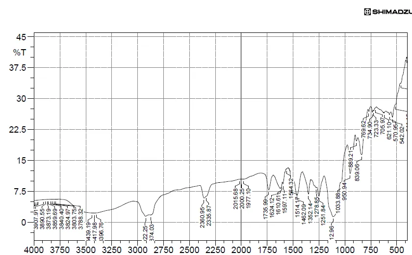
Figure 1: FTIR Studies of Gentamicin
Table 3: Characteristic Peaks for Gentamicin
| S.No. | Characteristic Peaks | Frequency range (cm-1) |
Frequency (cm-1) |
|---|---|---|---|
| 1 | OH stretching | 4000-3500 | 3890.55 |
| 2 | OH Bending | 3500-3000 | 3417.96 |
| 3 | C-H stretching | 2750-2250 | 2335.87 |
| 4 | C=O stretching | 1500-1000 | 1278.85 |
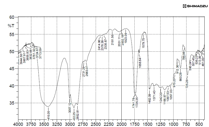
Figure 2: FTIR Studies of Physical Mixture of Drug and Excipients
Table 4: Characteristic Peaks for Drug and Excipients
| S.No. | Characteristic Peaks | Frequency range (cm-1) |
Frequency (cm-1) |
|---|---|---|---|
| 1 | OH stretching | 4000-3500 | 3907.91 |
| 2 | OH Bending | 3500-3000 | 3416.05 |
| 3 | C=O stretching | 1500-1000 | 1242.20 |
FTIR spectra of drug in KBr pellets at moderate scanning speed be 4000-400 cm-1 was made. Compatibility studies were performed using IR spectrophotometer. The IR spectrum of Pure drug and physical mixture of drug and excipients were studied. The characteristic absorption of peaks was obtained as above and as they were in official limits (±100 cm-1) the drug is compatible with excipients.
Particle Size of Transferosomes
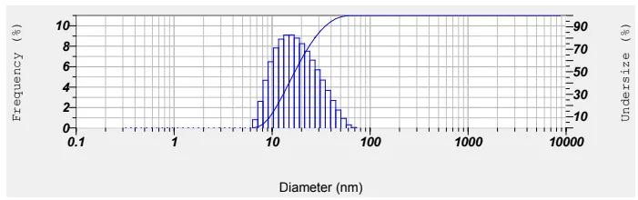
Figure 3: Particle Size of Optimized Formulation
The mean particle size of optimized transferosomes was found to be 112 nm
Table 5: Particle Size
| F. No | Particle size |
|---|---|
| F1 | 132 |
| F2 | 137 |
| F3 | 125 |
| F4 | 112 |
pH value of Transfersomal gel:
The value of pH of topical transfersome gels was measured by using digital pH meter (Labindia Sab 5000 pH meter) at the room temperature. The pH of all topical transfersomal gels were found to be in the range of 6.4 to 7.5.
Table 6: pH Value of Transfersome Gel
| F. No | pH |
|---|---|
| F1 | 6.5 |
| F2 | 7.2 |
| F3 | 6.6 |
| F4 | 7.5 |
The pH values of all formulations were within the acceptable range for topical application and are unlikely to cause skin irritation.
SEM Analysis
SEM revealed that the Gentamicin transferosomes were spherical in shape with a smooth surface and showed no signs of aggregation.
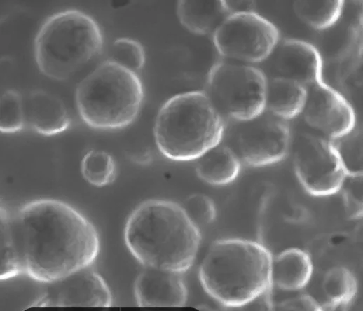
Figure 4: SEM Analysis of Transfersomal Gel
Entrapment efficiency
Table 7: Percentage Entrapment Efficiency
| F. No | % EE |
|---|---|
| F1 | 79.63 |
| F2 | 81.23 |
| F3 | 82.39 |
| F4 | 85.78 |
[Figure - see the formatted PDF]
Figure 5: Entrapment Efficiency
In Vitro Drug Release Studies
The diffusion medium used was phosphate buffer solution pH 7.4. In the Franz diffusion cell, the Dialysis membrane was mounted between donor and receptor compartment of diffusion cell. Dialysis membrane separated both the compartments. Area of membrane separating two compartments was 1.7662 cm2. Transfersomal gel (20 ml) volume was accurately pipetted into donor compartment which was then covered with aluminum foil to avoid any evaporation. Diffusion medium (20 ml) was maintained at 37 ±1 o C, so that the membrane just touches the receptor medium surface. A magnetic bar continuously stirred in the diffusion medium at 100 rpm. Each of 1 ml volume was withdrawn at required time intervals and replaced by 1 ml volume of receptor medium (phosphate buffer pH 7.4) to maintain the sink condition. These samples were analyzed by UV spectrophotometer at maximum wavelength 255 nm.
Table 8: Diffusion Studies of Transfersomal Gel
| Time | F1 | F2 | F3 | F4 |
|---|---|---|---|---|
| 0 | 0 | 0 | 0 | 0 |
| 1 | 17.19 | 16.42 | 18.12 | 21.63 |
| 2 | 27.10 | 24.60 | 26.39 | 30.25 |
| 3 | 35.13 | 32.99 | 38.56 | 42.16 |
| 4 | 45.32 | 55.37 | 56.83 | 49.80 |
| 5 | 56.35 | 65.10 | 67.89 | 59.86 |
| 6 | 78.10 | 77.52 | 78.15 | 79.54 |
| 7 | 83.69 | 84.56 | 85.93 | 88.96 |
| 8 | 94.50 | 92.38 | 95.69 | 98.84 |
[Figure - see the formatted PDF]
Figure 6: Diffusion Studies of Transferosomes (F1-F4)
Formulation F4 exhibited the highest cumulative drug release of 98.84% after 8 h and was selected as the optimized formulation.
Stability Studies:
There was no significant change in physical and chemical properties of the transferosomal gel formulation F4 after 3 months. Parameters quantified at various time intervals were shown.
Table 9: Results of Stability Studies of Optimized Formulation F-4
| F. Code | Parameters | Initial | 1st Month | 2ndMonth | 3rd Month | Limits as per Specifications |
|---|---|---|---|---|---|---|
| F-4 | 250C/60%RH % Release |
98.84 | 97.57 | 96.60 | 95.55 | Not less than 85 % |
| F-4 | 300C/75% RH % Release |
98.84 | 97.69 | 96.57 | 95.50 | Not less than 85 % |
| F-4 | 400C/75% RH % Release |
98.84 | 97.25 | 96.16 | 95.12 | Not less than 85 % |
SUMMARY & CONCLUSION
The present study was successfully carried out to formulate and evaluate Gentamicin-loaded transferosomal gel for enhanced topical drug delivery. Preformulation studies confirmed the suitability of Gentamicin for formulation development, and FTIR compatibility studies revealed no significant interaction between the drug and excipients used in the formulation.Transferosomes were successfully prepared by the thin-film hydration method using soya lecithin, cholesterol, and Span 80, and subsequently incorporated into a Carbopol 940 gel base. The formulated transferosomes exhibited nanosized vesicles with particle sizes ranging from 112 to 137 nm. SEM analysis confirmed the spherical shape and smooth surface morphology of the Transferosomes without aggregation. The pH of all transferosomal gel formulations was found to be within the acceptable range for topical application, indicating good skin compatibility. Entrapment efficiency studies demonstrated efficient drug incorporation within the vesicles, with values ranging from 79.63% to 85.78%. Among all formulations, F4 showed the highest entrapment efficiency (85.78%) and the smallest particle size (112 nm).In vitro drug release studies revealed sustained drug release from all formulations. Formulation F4 exhibited the highest cumulative drug release of 98.84% after 8 hours and was selected as the optimized formulation. Stability studies conducted for three months demonstrated that the optimized formulation remained physically and chemically stable with no significant changes in drug release characteristics.
Based on the results obtained, it can be concluded that the developed Gentamicin-loaded transferosomal gel is a promising vesicular drug delivery system for topical administration. The optimized formulation (F4) showed excellent physicochemical properties, high entrapment efficiency, enhanced drug release, and good stability, indicating its potential for improved therapeutic effectiveness in the treatment of topical bacterial infections.
REFERENCES

Dr. D. Usha Rani, S. Niharika, S. Shiva Kumar Reddy, S. Shiva Kumar, T. Keerthana, Formulation and Invitro Evaluation of Gentamicin Transfersomal Gel, Int. J. of Pharm. Sci., 2026, Vol 4, Issue 11, 1367-1376. https://doi.org/10.5281/zenodo.24003636
 10.5281/zenodo.24003636