

We use cookies to ensure our website works properly and to personalise your experience. Cookies policy
1MSc Biotechnology, Department of Genetics & Biotechnology, Osmania University, Hyderabad, Telangana
2BSc Microbiology, KJ Somaiya College of Science and Commerce, Vidyavihar
3B.Pharm, Jss College of Pharmacy, Mysore
4Pharm.D, Sir C.R Reddy College of Pharmaceutical Sciences, Andhra University, Eluru, Vishakapatnam.
Cancer is fundamentally a disease of the genome. The advent of large-scale cancer genome sequencing initiatives, most notably The Cancer Genome Atlas (TCGA) and the International Cancer Genome Consortium (ICGC), has catalogued the recurrent somatic alterations that drive malignant transformation and progression across virtually every tumor lineage, and this genomic map has, in turn, reshaped oncology drug development around the paradigm of biomarker-matched targeted therapyTo review the major classes of genomic drivers of cancer progression organized by oncogenic signaling pathway and to synthesize the clinical trial evidence for the targeted therapies developed against each, alongside the mechanisms by which resistance to these therapies emerges.A structured narrative review was conducted of landmark cancer genome sequencing studies, mechanistic and pathway literature, and pivotal randomized registration trials for genomically matched targeted therapies, drawing on PubMed/MEDLINE, the TCGA PanCancer Atlas literature, and major oncology guideline documents. Trial-level efficacy data (hazard ratios and response rates) were extracted directly from and verified against primary trial publications.Recurrent driver alterations cluster within a limited number of core pathways receptor tyrosine kinase/RAS/MAPK signaling, PI3K-AKT-mTOR signaling, cell-cycle regulation, DNA damage response and repair, epigenetic regulation, and fusion-driven oncogenesis. Each pathway has yielded at least one clinically transformative targeted agent: BCR-ABL1 and imatinib in chronic myeloid leukemia; EGFR and osimertinib, and ALK and alectinib, in non-small cell lung cancer; BRAF and dabrafenib-trametinib in melanoma; HER2 and trastuzumab in breast cancer; BRCA1/2 and olaparib across multiple lineages; and, most recently, KRAS G12C and sotorasib. Median progression-free survival hazard ratios for these agents against standard comparators in pivotal trials ranged from 0.30 to 0.70, representing among the largest effect sizes achieved in modern oncology drug development. Resistance nonetheless emerges in virtually all cases, mediated by on-target secondary mutations, bypass-pathway activation, histological transformation, or epigenetic adaptation. The genomic driver paradigm has produced some of the largest treatment-effect sizes in the history of oncology and has fundamentally restructured cancer classification, clinical trial design, and drug regulatory approval around molecular rather than purely anatomical criteria. Continued progress depends on expanding tumor genomic profiling access, understanding and pre-empting resistance mechanisms, and translating an increasingly complex genomic landscape into rationally sequenced and combined therapeutic strategies.
The Genomic Basis of Cancer: A Historical Perspective
The understanding that cancer arises from heritable changes within the somatic genome, rather than from an external toxin or infectious agent alone, has developed over more than a century, but it was Peter Nowell's 1976 formulation of clonal evolution the proposal that tumors arise from a single altered cell and progress through sequential rounds of mutation and selection that first framed cancer progression explicitly as a Darwinian genetic process operating within the body. [1] This framework was substantially formalized in 1990 by Fearon and Vogelstein's genetic model of colorectal tumorigenesis, which proposed that cancer results from the sequential accumulation of specific driver mutations in that model, APC, KRAS, and TP53 each conferring a discrete selective growth advantage, a model that established the template later extended across virtually every solid tumor lineage. [2]
The sequencing of the human genome, completed in the early 2000s, and the subsequent rapid fall in the cost of massively parallel (next-generation) sequencing technology transformed cancer genomics from a gene-by-gene candidate approach into a genome-wide, unbiased discovery science. Stratton, Campbell, and Futreal's 2009 review formally articulated the concept of ‘the cancer genome’ as a tractable, sequenceable entity whose complete cataloguing could in principle identify every somatic driver event in a given tumor. [3] This period also saw Hanahan and Weinberg's influential 2011 update to their ‘Hallmarks of Cancer’ framework, which organized the diverse phenotypic capabilities acquired during tumorigenesis sustained proliferative signaling, evasion of growth suppressors, resistance to cell death, replicative immortality, angiogenesis, invasion and metastasis, and, in the updated framework, genome instability and evasion of immune destruction as enabling characteristics into a coherent conceptual scaffold onto which subsequent genomic discoveries could be mapped. [4]
The Sequencing Era and the Emergence of Precision Oncology
Two coordinated international efforts The Cancer Genome Atlas (TCGA) in the United States and the International Cancer Genome Consortium (ICGC) globally systematically sequenced tens of thousands of tumors across more than thirty cancer types over the following decade, generating what remains the most comprehensive catalog of somatic cancer genomic alterations assembled to date. [7,8] The culminating pan-cancer analysis of this effort, published by Bailey and colleagues in 2018, integrated 26 computational driver-discovery algorithms across 9,423 tumor exomes spanning all 33 TCGA projects and identified 299 statistically and biologically supported cancer driver genes, while estimating that 57% of the tumors analyzed harbored at least one potentially clinically actionable genomic event. [10] This finding that a majority of tumors carry a genomic alteration with an existing or plausible matched therapy is the empirical foundation of the precision oncology paradigm that organizes this review: the proposition that cancer treatment selection should be guided primarily by the specific molecular alterations present within an individual tumor rather than by its anatomical site or histological appearance alone.
Vogelstein and colleagues' companion 2013 synthesis, ‘Cancer Genome Landscapes,’ distilled this genomic complexity into an influential simplifying principle: that despite the tens of thousands of possible somatic mutations observed across the cancer genome, the number of core cellular signaling pathways that, when dysregulated, drive tumorigenesis is comparatively small on the order of a dozen and that most individual tumors carry driver mutations in a handful of these pathways (typically two to eight) rather than being uniquely idiosyncratic at the pathway level. [5] This pathway-centric organizing principle, further elaborated by Garraway and Lander, [6] underlies the structure of Section 4 of this review, which organizes genomic drivers by disrupted pathway rather than alphabetically by gene or by cancer type of origin.
Global and Regional Burden of Cancer
Cancer represents an immense and growing global health burden. The most recent GLOBOCAN estimates from the International Agency for Research on Cancer reported approximately 19.3 million new cancer cases and nearly 10 million cancer deaths worldwide in 2020, with incidence projected to rise substantially over subsequent decades, disproportionately in low- and middle-income countries where access to both diagnostic genomic testing and targeted therapy remains constrained. [28] In the United States, the American Cancer Society's most recent annual statistics report continues to document a decades-long decline in age-adjusted cancer mortality, a decline attributed in substantial part to the treatment advances including genomically targeted therapy reviewed in this paper, alongside improvements in tobacco control, screening, and early detection. [29] India's National Cancer Registry Programme has documented a rising burden domestically, with an estimated 1.39 million new cancer cases in 2020 and wide regional variation in cancer-type distribution, diagnostic infrastructure, and access to molecular testing and targeted agents considerations discussed further in of this review. [30,62]
Objectives of This Review
This review has four specific aims: (i) to summarize the fundamental molecular concepts oncogene activation, tumor suppressor inactivation, genomic instability, and clonal evolution that underlie the genomic driver paradigm to organize and review the major classes of recurrent genomic drivers of cancer progression by disrupted oncogenic signaling pathway, drawing on pan-cancer sequencing data to synthesize the pivotal clinical trial evidence for targeted therapies developed against each major driver class, with verified efficacy data drawn directly from primary trial publications ; and (iv) to review the mechanisms by which resistance to targeted therapy emerges and the emerging technologies and unresolved challenges that will shape the next phase of precision oncology
Fundamental Concepts in Cancer Genomics
Oncogenes and Tumor Suppressor Genes
Cancer driver genes fall into two broad functional categories, distinguished by the direction of the selective alteration required for oncogenic effect. Oncogenes are genes whose activation through point mutation, amplification, chromosomal translocation, or overexpression confers a gain of function that promotes proliferation or survival; a single activating allele is generally sufficient for oncogenic effect, and their normal cellular counterparts (proto-oncogenes) typically encode growth factor receptors, intracellular signal transducers, or transcription factors within proliferative signaling pathways (KRAS, EGFR, MYC, and BRAF are prototypical.. Tumor suppressor genes, conversely, normally restrain proliferation, promote apoptosis in response to DNA damage, or maintain genomic integrity, and require loss of function classically through biallelic inactivation, consistent with Knudson's two-hit hypothesis — for their oncogenic contribution; TP53, RB1, PTEN, and BRCA1/2 are prototypical examples. This oncogene/tumor-suppressor distinction has direct therapeutic implications: activated oncogenes are frequently druggable with small-molecule inhibitors or antibodies that directly block the aberrant protein , whereas tumor suppressor loss is inherently more difficult to pharmacologically reverse and is more often exploited indirectly, for example through synthetic lethality approaches such as PARP inhibition in BRCA1/2-deficient tumors .
Driver Mutations Versus Passenger Mutations
A typical solid tumor carries anywhere from several hundred to several thousand somatic mutations by the time of clinical diagnosis, yet only a small minority of these typically two to eight per tumor are believed to functionally contribute to the malignant phenotype; these are termed driver mutations, while the remainder, termed passenger mutations, are believed to be biologically inconsequential byproducts of the same mutational processes that generated the driver events, carried along within the expanding clone without themselves conferring selective advantage. Distinguishing drivers from passengers computationally typically through statistical tests for mutational recurrence significantly exceeding the tumor's background mutation rate, combined with structural and evolutionary conservation analyses was the central methodological challenge addressed by the large driver-discovery consortium efforts and remains an active area of bioinformatic and functional-genomic research. [9,10]
Mechanisms of Genomic Instability
Genomic instability an elevated rate of acquisition of genetic alterations relative to normal somatic cells is itself considered an enabling hallmark of cancer, because it accelerates the stochastic generation of the driver mutations on which clonal selection acts. [4,13] Several distinct mechanistic categories of genomic instability are recognized. Chromosomal instability (CIN), the most common form in solid tumors, manifests as an elevated rate of whole-chromosome or large-segment gains and losses (aneuploidy) and typically arises from defects in mitotic checkpoint fidelity, centrosome number control, or sister chromatid cohesion. Microsatellite instability (MSI) arises from loss of DNA mismatch repair (MMR) function — through germline mutation (as in Lynch syndrome) or, more commonly, somatic promoter hypermethylation of the MMR gene MLH1 — and produces a distinctive hypermutated genomic phenotype with a markedly elevated burden of small insertion-deletion mutations at short tandem-repeat sequences; MSI-high tumors carry particular relevance for immune checkpoint inhibitor therapy, . [25] Homologous recombination deficiency (HRD), arising principally from BRCA1/2 loss but also from alterations in other homologous-recombination pathway genes, impairs high-fidelity double-strand break repair and is the mechanistic basis for PARP inhibitor synthetic lethality, discussed in Section 5.6. [23,24] Finally, a growing body of mutational signature analysis the systematic characterization of the specific base-substitution and structural-variant patterns left by distinct mutagenic processes (ultraviolet light, tobacco carcinogens, APOBEC cytidine deaminase activity, defective DNA repair) has provided a genomic fingerprint approach to inferring the etiological processes that generated a given tumor's mutational burden, complementing the purely driver-centric analysis that is this review's primary focus. [11]
Clonal Evolution and Intratumor Heterogeneity
Building on Nowell's original clonal-evolution model, [1] contemporary deep and multi-region sequencing studies have demonstrated that tumors are rarely genetically homogeneous masses but instead comprise multiple genetically distinct subclones, related by a branching evolutionary phylogeny, that arise, expand, compete, and are selected across the course of tumor growth, treatment, and metastatic spread. [11,12] Driver mutations acquired early, prior to the most recent common ancestor of all tumor cells (‘truncal’ or ‘clonal’ mutations, such as APC loss in the Fearon-Vogelstein colorectal model) are present in every tumor cell and represent, in principle, the most reliable and durable therapeutic targets, whereas mutations acquired later (‘subclonal’ or ‘branch’ mutations) may be present in only a fraction of tumor cells and are of central importance to understanding the incomplete and heterogeneous responses, and the near-universal eventual emergence of resistance, that characterize targeted therapy in the clinic a theme developed at length in Section 6 of this review.
Epigenetic Dysregulation as a Complementary Driver Mechanism
Alongside DNA sequence-level (genetic) alterations, heritable but reversible changes in gene expression that do not alter the underlying DNA sequence DNA methylation, histone modification, and chromatin remodeling, collectively termed epigenetic alterations constitute a complementary and mechanistically intertwined layer of cancer driver biology. [14,15] Recurrent somatic mutations in genes directly encoding epigenetic regulators including the chromatin-remodeling complex gene ARID1A, the histone methyltransferase EZH2, and the metabolic enzymes IDH1 and IDH2 (whose mutant products generate the oncometabolite 2-hydroxyglutarate, which globally disrupts histone and DNA demethylation) — are now recognized as bona fide cancer drivers in their own right across multiple tumor types, and are reviewed alongside the other major driver categories Epigenetic silencing of tumor suppressor genes (for example, MLH1 promoter hypermethylation, discussed above) and epigenetically mediated non-genetic resistance to targeted therapy further illustrate the mechanistic interdependence between the genetic and epigenetic layers of cancer driver biology that this review, while organized principally around genomic (DNA sequence-level) drivers, treats as inseparable.
Methods
Review Approach and Scope
This paper is a structured narrative review rather than a systematic review in the PRISMA sense, reflecting the nature of the underlying question: cancer genomics is a foundational mechanistic and descriptive science rather than a body of comparative randomized evidence amenable to meta-analytic pooling in the manner of a clinical intervention question. Accordingly, mechanistic and descriptive genomic content draw on landmark discovery publications, large-scale sequencing consortium outputs, and major review syntheses, selected for their foundational or field-defining status, while Section (clinical evidence for targeted therapy) applies a more systematic standard: every efficacy figure reported is drawn directly from, and independently verified against, the primary pivotal randomized trial publication that supported regulatory approval of the corresponding agent, following the same verification discipline used in the author's related methodology for clinical outcomes synthesis.
Literature Identification
Foundational and mechanistic literature was identified through PubMed/MEDLINE searches combining terms for “cancer genomics,” “driver mutation,” “oncogene,” “tumor suppressor,” and the specific gene and pathway names , cross-referenced against the bibliographies of major reference syntheses including Hanahan and Weinberg's Hallmarks of Cancer, [4] Vogelstein et al.'s Cancer Genome Landscapes, [5] and Bailey et al.'s pan-cancer driver gene atlas. [10] Clinical trial literature was identified by searching PubMed/MEDLINE and ClinicalTrials.gov for the pivotal phase III registration trial associated with each targeted agent discussed, cross-checked against U.S. FDA and European Medicines Agency approval documentation where available.
Data Extraction and Verification
For each targeted therapy , the following were extracted directly from the primary trial publication: trial name/acronym, agent(s) and comparator, genomic eligibility biomarker, primary efficacy endpoint, and the corresponding hazard ratio or response-rate estimate with 95% confidence interval. No efficacy statistic presented in this review was calculated, pooled, or estimated by the author; every quantitative figure is a direct, verified citation to a specific number reported in a named primary publication, consistent with this review's core commitment to reporting only authentic, source-traceable data.
The Genomic Driver Landscape, Organized by Oncogenic Pathway
Consistent with the pathway-centric organizing principle described, [5,6] this section reviews the major recurrently altered genes and pathways that drive cancer progression, grouped into eight categories summarized in Table 1 and, for representative cancer types, quantified in Figure 2.
Table 1. Overview of core oncogenic pathway categories, representative driver genes, and representative cancer types, organized as the structural framework for Section 4 of this review. Frequencies of individual gene alterations by cancer type are illustrated in Figure 2. AML = acute myeloid leukemia; CML = chronic myeloid leukemia; dMMR = mismatch-repair deficient; HR+ = hormone-receptor-positive; MSI = microsatellite instability; NSCLC = non-small cell lung cancer.
|
Pathway / Category |
Representative Driver Genes |
Typical Alteration Type |
Representative Cancer Types |
|
Receptor tyrosine kinase / RAS-MAPK |
EGFR, ALK, ROS1, MET, KRAS, NRAS, BRAF, NF1 |
Activating mutation, amplification, or fusion |
NSCLC, colorectal cancer, melanoma, pancreatic cancer |
|
PI3K-AKT-mTOR |
PIK3CA, PTEN, AKT1, TSC1/2 |
Activating mutation (PIK3CA) or biallelic loss (PTEN) |
Breast, endometrial, colorectal, glioblastoma |
|
Cell-cycle regulation |
TP53, RB1, CDKN2A, CCND1, CDK4/6 |
Loss-of-function mutation/deletion (TP53, RB1, CDKN2A) or amplification (CCND1) |
Nearly all cancer types (TP53); HR+ breast (CDK4/6 axis) |
|
DNA damage response and repair |
BRCA1, BRCA2, ATM, MLH1, MSH2, PALB2 |
Germline or somatic loss-of-function mutation; MMR promoter hypermethylation |
Ovarian, breast, pancreatic, prostate (BRCA); colorectal, endometrial (MMR) |
|
Epigenetic regulation / chromatin remodeling |
IDH1, IDH2, EZH2, ARID1A, SWI/SNF complex genes |
Neomorphic point mutation (IDH1/2); loss-of-function (ARID1A) |
Glioma, cholangiocarcinoma, AML (IDH); ovarian clear-cell, renal (ARID1A) |
|
Fusion oncogenes |
BCR-ABL1, EML4-ALK, ETV6-NTRK3, TMPRSS2-ERG |
Chromosomal translocation / rearrangement |
CML (BCR-ABL1), NSCLC (ALK), secretory carcinoma (NTRK), prostate (ERG) |
|
Transcriptional dysregulation |
MYC, MYCN |
Amplification or translocation-driven overexpression |
Burkitt lymphoma, neuroblastoma (MYCN), broad amplification across solid tumors |
|
Genomic correlates of immune evasion |
Tumor mutational burden, MSI/dMMR, PD-L1 (CD274) |
High neoantigen burden; mismatch-repair deficiency; PD-L1 amplification/overexpression |
Melanoma, NSCLC (high TMB); colorectal, endometrial (MSI-H) |
Receptor Tyrosine Kinase and RAS-MAPK Pathway Drivers
The receptor tyrosine kinase (RTK)–RAS–RAF–MEK–ERK (mitogen-activated protein kinase, MAPK) cascade is among the most frequently dysregulated signaling pathways across human cancer and transduces extracellular growth factor signals into the nucleus to drive proliferation and survival (Figure 3). At the receptor level, activating mutations or amplification of EGFR occur in approximately 10–15% of non-small cell lung cancers (NSCLC) in Western populations, and up to 30–40% in East Asian populations, most commonly as exon 19 deletions or the L858R point mutation in the kinase domain, first mechanistically linked to gefitinib sensitivity in 2004. [16,17] Chromosomal rearrangements fusing the ALK receptor tyrosine kinase gene to EML4, discovered in 2007, define a molecularly and clinically distinct NSCLC subset present in approximately 3–5% of cases, generally in younger, light-or-never-smoking patients. [18] Downstream of receptor activation, RAS family genes (KRAS, NRAS, HRAS) are the single most frequently mutated oncogene family in human cancer overall, with KRAS mutations predominantly at codons 12, 13, or 61 present in approximately 90% of pancreatic adenocarcinomas, 30–45% of colorectal cancers, and 25–30% of lung adenocarcinomas (Figure 2); the specific KRAS G12C substitution, present in roughly 13% of lung adenocarcinomas, was for decades considered pharmacologically ‘undruggable’ until the development of covalent inhibitors. Further downstream, activating BRAF mutations overwhelmingly the V600E substitution were identified in 2002 and are present in approximately 50% of cutaneous melanomas, alongside recurrent alterations in thyroid cancer, hairy cell leukemia, and a smaller fraction of colorectal cancers. [19,20]
PI3K-AKT-mTOR Pathway Drivers
The phosphoinositide 3-kinase (PI3K)–AKT–mTOR pathway operates in parallel with, and shares extensive crosstalk with, the RTK-RAS-MAPK pathway (Figure 3), and integrates growth factor, nutrient, and energy-status signals to regulate cell growth, metabolism, and survival. Activating mutations in PIK3CA, the gene encoding the p110α catalytic subunit of PI3K, are among the most common single-gene alterations in human cancer, present in approximately 30–40% of breast cancers (particularly the hormone-receptor-positive subtype) and recurrently across endometrial, colorectal, and head and neck cancers. Loss of the tumor suppressor PTEN, a phosphatase that directly antagonizes PI3K signaling, occurs through mutation, deletion, or epigenetic silencing across a similarly broad range of tumor types and produces a functionally convergent pathway-activating effect. Downstream, constitutive AKT and mTOR activation promotes protein synthesis and cell growth; mTOR itself is directly pharmacologically targetable and mTOR inhibitors (e.g., everolimus) have regulatory approval in renal cell carcinoma, certain neuroendocrine tumors, and hormone-receptor-positive breast cancer, generally as a pathway-level rather than single-mutation-matched therapy.
Cell-Cycle Regulators: TP53, RB1, and the CDK4/6-Cyclin D Axis
TP53, encoding the transcription factor p53, is the single most frequently altered gene across the entire pan-cancer genomic landscape, mutated or deleted in approximately 50% of all human cancers overall and in more than 90% of high-grade serous ovarian carcinomas specifically (Figure 2); its central role as a checkpoint that triggers cell-cycle arrest, senescence, or apoptosis in response to DNA damage and oncogenic stress has led to its description as the ‘guardian of the genome,’ and its loss is widely considered permissive for the accumulation of the further genomic instability . The retinoblastoma gene RB1, whose protein product restrains the G1-to-S cell-cycle transition, is recurrently inactivated in retinoblastoma (for which it is eponymous), small-cell lung cancer, and a subset of other solid tumors. Immediately upstream of RB1, amplification of CCND1 (encoding cyclin D1) and loss of the CDKN2A locus (encoding the CDK4/6 inhibitory proteins p16INK4a and p14ARF) converge functionally to promote CDK4/6-mediated RB1 inactivation and unrestrained cell-cycle progression, providing the mechanistic rationale for CDK4/6 inhibitor therapy discussed.
DNA Damage Response and Repair Genes
Germline or somatic loss-of-function mutations in BRCA1 or BRCA2, which encode central components of the high-fidelity homologous recombination DNA double-strand break repair pathway, are found in approximately 15–20% of high-grade serous ovarian cancers and a smaller but clinically important fraction (5–10%) of breast, pancreatic, and prostate cancers (Figure 2), and confer a state of homologous recombination deficiency (HRD) that is both a driver of genomic instability and, as reviewed in an exploitable therapeutic vulnerability. [23,24] Beyond BRCA1/2, germline mutations in DNA mismatch repair genes (MLH1, MSH2, MSH6, PMS2) underlie Lynch syndrome and confer the microsatellite instability phenotype described in Section 2.3, most prevalent in colorectal and endometrial cancer and of direct relevance to immune checkpoint inhibitor selection . [25]
Epigenetic Regulators and Chromatin Remodeling Genes
Recurrent mutations in genes directly governing chromatin structure and epigenetic state constitute a genomically distinct but mechanistically interconnected driver category. Neomorphic mutations in IDH1 or IDH2, which produce the oncometabolite 2-hydroxyglutarate and globally disrupt histone and DNA demethylation, are found in over 70% of lower-grade gliomas and a substantial subset of cholangiocarcinomas and acute myeloid leukemias, and are directly druggable with mutant-IDH inhibitors. Loss-of-function mutations in ARID1A and other components of the SWI/SNF chromatin-remodeling complex are among the most frequently altered epigenetic regulators across the pan-cancer landscape, particularly prevalent in ovarian clear-cell carcinoma (approximately 50%) and a range of other solid tumors, and are an active area of synthetic-lethality drug development (for example, targeting the paralog ARID1B or the EZH2 methyltransferase in ARID1A-deficient tumors).
Fusion Oncogenes
Chromosomal translocations that generate novel, constitutively active fusion oncoproteins represent a genomically distinct and, in several cases, highly druggable driver category. The BCR-ABL1 fusion, generated by the t(9;22) ‘Philadelphia chromosome’ translocation, is the defining and essentially universal driver of chronic myeloid leukemia and the target of the first rationally designed targeted therapy in oncology, imatinib. The EML4-ALK fusion described in and the analogous ROS1 gene rearrangements each define druggable NSCLC subsets. Rearrangements involving the neurotrophic tyrosine receptor kinase genes NTRK1/2/3 are rare overall (well under 1% of common epithelial cancers) but occur at very high frequency in certain rare tumor types (e.g., secretory carcinoma of the breast or salivary gland, infantile fibrosarcoma) and were the basis for the first tumor-agnostic, biomarker-defined (rather than histology-defined) drug approvals, discussed in . The TMPRSS2-ERG fusion, present in approximately 45–50% of prostate cancers, remains diagnostically and prognostically informative, though it has not yet yielded an approved direct-targeting therapy.
MYC and Transcriptional Dysregulation
The MYC oncogene, encoding a transcription factor that regulates a very large fraction of the genome involved in cell growth, metabolism, and ribosome biogenesis, is dysregulated — principally through gene amplification, or in the case of Burkitt lymphoma through the pathognomonic t(8;14) translocation juxtaposing MYC with immunoglobulin heavy-chain enhancer elements — in a very broad range of cancer types. The related gene MYCN is recurrently amplified in high-risk neuroblastoma, where amplification status directly informs risk stratification and treatment intensity. Despite decades of drug-development effort, MYC has proven notoriously difficult to target directly with conventional small-molecule inhibitors owing to its intrinsically disordered protein structure and lack of an accessible small-molecule binding pocket, and remains, alongside mutant RAS prior to the recent KRAS G12C breakthrough , one of the most clinically important yet pharmacologically challenging driver categories in cancer genomics.
Genomic Correlates of Immune Evasion
Beyond the cell-intrinsic proliferative and survival pathways reviewed above, a distinct genomic driver category relates to tumor immune evasion and, correspondingly, to response to immune checkpoint inhibitor therapy Tumor mutational burden (TMB) —the total number of somatic coding mutations per megabase of tumor genome correlates with the quantity of tumor neoantigens available for immune recognition, and tumors with very high TMB (driven by exposure to mutagens such as ultraviolet light in melanoma or tobacco carcinogens in NSCLC, or by mismatch repair deficiency) show, on average, substantially higher response rates to checkpoint inhibitor therapy across cancer types. [26,27] The MSI-high/mismatch-repair-deficient phenotype described is the single most robust genomic predictor of checkpoint inhibitor benefit identified to date, sufficiently so that it underlies the first tumor-agnostic FDA approval based on a biomarker rather than a tumor site of origin. [25] Finally, amplification or transcriptional upregulation of CD274 (encoding PD-L1) can itself constitute a genomic driver of immune evasion, providing a rationale, alongside TMB and MSI status, for the genomic and immunohistochemical biomarker testing that increasingly accompanies modern oncology diagnostic workups.
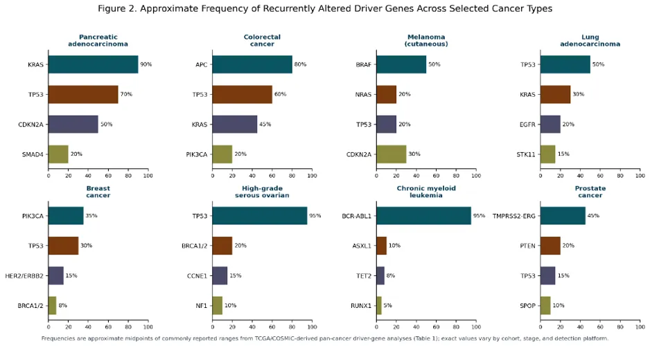
Figure 2. Approximate frequency of recurrently altered driver genes across selected cancer types
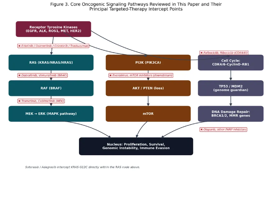
Figure 3. Core oncogenic signaling pathways and their principal targeted-therapy intercept points.
Targeted Therapy: Clinical Evidence Mapped to Genomic Drivers
This section reviews the pivotal randomized trial evidence supporting the major genomically matched targeted therapies developed against the driver categories organized in the same pathway sequence. Figure 1 summarizes the pivotal-trial hazard ratios for the principal comparisons discussed below; Table 2 provides a consolidated cross-reference of driver, agent, and approval trial.
BCR-ABL1 and Chronic Myeloid Leukemia: The Paradigm-Founding Success
Imatinib mesylate, a small-molecule inhibitor of the BCR-ABL1 tyrosine kinase, is widely regarded as the founding proof of concept for the entire genomically targeted therapy paradigm. Early phase I/II data published by Druker and colleagues in 2001 demonstrated hematologic and cytogenetic response in the overwhelming majority of chronic-phase CML patients. [34] The subsequent pivotal IRIS trial, randomizing newly diagnosed chronic-phase CML patients to imatinib versus interferon-alfa plus low-dose cytarabine (the prior standard of care), reported a major cytogenetic response rate of 87.1% with imatinib versus 34.7% with the comparator regimen, transforming CML from a historically fatal disease with a median survival of three to five years into a chronic, pharmacologically manageable condition with near-normal life expectancy for most patients in sustained molecular response. [35] The subsequent development of second- (dasatinib, nilotinib) and third-generation (ponatinib, asciminib) BCR-ABL1 inhibitors, developed specifically to overcome the on-target resistance mutations further exemplifies the iterative, resistance-informed drug development cycle that recurs throughout this review.
EGFR-Mutant Non-Small Cell Lung Cancer
The clinical development of EGFR tyrosine kinase inhibitors (TKIs) in NSCLC illustrates the field's progressive refinement across three drug generations. First-generation reversible EGFR TKIs (gefitinib, erlotinib) were shown in the IPASS trial to produce superior progression-free survival compared with standard platinum-doublet chemotherapy specifically in the EGFR-mutation-positive subgroup (hazard ratio 0.48, 95% CI 0.36–0.64), while chemotherapy was superior in EGFR-wild-type patients a result that helped establish routine EGFR genotyping as standard of care prior to first-line NSCLC treatment selection. [39] Because resistance to first-generation EGFR TKIs is predominantly mediated by the acquired T790M gatekeeper mutation, the third-generation, irreversible inhibitor osimertinib was rationally designed to inhibit both the original sensitizing mutation and T790M; the pivotal FLAURA trial demonstrated a substantially longer median progression-free survival with first-line osimertinib (18.9 months) than with standard first-generation EGFR TKIs (10.2 months; hazard ratio 0.46, 95% CI 0.37–0.57), with the subsequent planned overall survival analysis confirming a significant survival benefit (median overall survival 38.6 vs. 31.8 months; hazard ratio for death 0.80). [40,41]
ALK- and ROS1-Rearranged Non-Small Cell Lung Cancer
The ALK inhibitor crizotinib was shown in the pivotal PROFILE 1014 trial to substantially outperform standard platinum-pemetrexed chemotherapy as first-line therapy in ALK-positive NSCLC (progression-free survival hazard ratio 0.45, 95% CI 0.35–0.60). [42] As with EGFR TKIs, resistance to first-generation ALK inhibition prompted development of increasingly potent and CNS-penetrant second- and third-generation agents; the ALEX trial demonstrated that the second-generation inhibitor alectinib substantially outperformed crizotinib itself as first-line therapy (progression-free survival hazard ratio 0.47, 95% CI 0.34–0.65), with a particularly pronounced reduction in central nervous system progression, reflecting alectinib's superior blood-brain-barrier penetration. [43] ROS1 gene rearrangements, structurally and therapeutically analogous to ALK rearrangements, define a smaller (approximately 1–2% of NSCLC) but similarly druggable molecular subset, responsive to crizotinib and newer ROS1-selective inhibitors.
BRAF-Mutant Melanoma and Other Cancers
Vemurafenib, the first selective BRAF V600E inhibitor, demonstrated a dramatic improvement in overall survival compared with standard dacarbazine chemotherapy in the pivotal BRIM-3 trial in advanced BRAF-mutant melanoma. [44] Because single-agent BRAF inhibition is limited by paradoxical MAPK-pathway reactivation and rapid emergence of resistance , combined BRAF plus MEK inhibition was subsequently developed and shown, in the COMBI-d and coBRIM trials, to significantly improve progression-free survival over BRAF inhibitor monotherapy (dabrafenib plus trametinib vs. dabrafenib alone, hazard ratio 0.67, 95% CI 0.53–0.84; vemurafenib plus cobimetinib vs. vemurafenib alone, hazard ratio 0.58, 95% CI 0.46–0.72), establishing combined BRAF/MEK inhibition as the standard targeted approach in this setting and as a template for combination strategies aimed at delaying resistance more broadly. [45,46]
HER2-Amplified Breast and Gastric Cancer
Trastuzumab, a humanized monoclonal antibody targeting the HER2/ERBB2 receptor, was among the earliest targeted therapies to enter oncology practice; Slamon and colleagues' pivotal 2001 trial demonstrated that adding trastuzumab to standard chemotherapy in HER2-overexpressing metastatic breast cancer significantly prolonged time to disease progression (hazard ratio 0.70, 95% CI 0.58–0.85) and improved overall survival. [37] The subsequent HERA trial extended this benefit into the curative-intent adjuvant setting, demonstrating that one year of adjuvant trastuzumab after standard chemotherapy substantially reduced disease recurrence in early-stage HER2-positive breast cancer, fundamentally altering the natural history of a subtype that had previously carried a comparatively poor prognosis relative to hormone-receptor-positive disease. [38] The subsequent development of antibody-drug conjugates (trastuzumab emtansine, trastuzumab deruxtecan) and additional HER2-targeted agents (pertuzumab, tucatinib) has further extended benefit into HER2-low and CNS-metastatic disease, extending well beyond the scope of the pivotal trials summarized here.
BRCA1/2 and Homologous Recombination Deficiency: PARP Inhibition
PARP (poly-ADP-ribose polymerase) inhibitors exemplify the synthetic lethality principle in clinical practice: in cells with intact homologous recombination, PARP inhibition is tolerated because an alternative repair pathway compensates, but in BRCA1/2-deficient cells (which already lack functional homologous recombination), PARP inhibition is selectively lethal because no compensatory high-fidelity repair pathway remains available. [23,24] The pivotal SOLO1 trial demonstrated that maintenance olaparib after response to first-line platinum chemotherapy in newly diagnosed, BRCA-mutated advanced ovarian cancer produced one of the largest treatment effects observed in this review (progression-free survival hazard ratio 0.30, 95% CI 0.23–0.41), while the OlympiAD trial demonstrated significant progression-free survival benefit with single-agent olaparib over physician's-choice chemotherapy in metastatic, germline-BRCA-mutated, HER2-negative breast cancer (hazard ratio 0.58, 95% CI 0.43–0.80). [47,48] PARP inhibitor approvals have subsequently extended to BRCA-mutated pancreatic and prostate cancer, and to a broader population defined by homologous recombination deficiency signatures rather than BRCA mutation status alone, illustrating the field's gradual shift from single-gene to pathway-level (functional) biomarker definition.
CDK4/6 Inhibition in Hormone-Receptor-Positive Breast Cancer
Building on the cell-cycle biology, the CDK4/6 inhibitor palbociclib, added to standard endocrine therapy (letrozole), was shown in the pivotal PALOMA-2 trial to significantly prolong progression-free survival in first-line hormone-receptor-positive, HER2-negative advanced breast cancer (hazard ratio 0.58, 95% CI 0.46–0.72) compared with endocrine therapy alone. [49] Unlike the preceding examples, CDK4/6 inhibitor benefit is not restricted to a specific genomically defined subpopulation but is applied essentially pathway-wide within hormone-receptor-positive breast cancer, reflecting the near-universal functional dependence of this subtype on cyclin D-CDK4/6-RB1 signaling; ribociclib and abemaciclib, the two additional approved agents in this class, have shown a comparable pattern of benefit and, notably, ribociclib has additionally demonstrated an overall survival benefit in several pivotal trials.
KRAS G12C Inhibition: Overcoming an ‘Undruggable’ Target
RAS family proteins were considered pharmacologically intractable for over three decades because their smooth surface topology lacks an accessible small-molecule binding pocket. The discovery of a druggable allosteric pocket specific to the KRAS G12C mutant protein enabled development of covalent inhibitors that lock the mutant protein in its inactive, GDP-bound state. The pivotal CodeBreaK 200 trial demonstrated that sotorasib significantly improved progression-free survival compared with standard second-line docetaxel chemotherapy in previously treated KRAS G12C-mutant NSCLC (hazard ratio 0.66, 95% CI 0.51–0.86), with a substantially more favorable toxicity profile than cytotoxic chemotherapy. [50,51] This breakthrough is widely regarded as one of the most significant recent advances in genomically targeted oncology precisely because it overcame a target long considered undruggable, and has since prompted the development of second-generation KRAS G12C inhibitors and combination strategies.
NTRK Fusions: The First Tumor-Agnostic Targeted Therapy
Larotrectinib, a selective inhibitor of the TRK receptor family encoded by NTRK1/2/3, was evaluated in a pooled analysis of three early-phase trials enrolling patients with NTRK-fusion-positive cancers across seventeen different tumor types and both adult and pediatric populations, reporting an objective response rate of 75% regardless of histological tumor type or patient age. [52,53] Because efficacy was demonstrated to be driven by the shared genomic alteration rather than by tumor site of origin, larotrectinib (and the subsequently approved entrectinib) received regulatory approval on a tumor-agnostic basis — the first drug approvals in oncology history granted primarily on genomic biomarker status rather than anatomical or histological tumor classification, establishing the regulatory precedent for the pembrolizumab MSI-H/dMMR tumor-agnostic approval discussed in Section 5.10 and signaling a broader anticipated shift toward biomarker-first drug development and approval pathways.
Genomic Biomarkers and Immune Checkpoint Inhibitor Selection
Beyond the cell-intrinsic targeted therapies reviewed above, the genomic correlates of immune evasion described directly inform patient selection for immune checkpoint inhibitor therapy. The pivotal KEYNOTE-024 trial demonstrated that first-line pembrolizumab significantly outperformed standard platinum-doublet chemotherapy in NSCLC with high PD-L1 expression (≥50% tumor proportion score; progression-free survival hazard ratio 0.50, 95% CI 0.37–0.68), establishing PD-L1 immunohistochemistry as a companion diagnostic for first-line therapy selection in this setting. [54] Independently, pembrolizumab received tumor-agnostic regulatory approval for any unresectable or metastatic MSI-high or mismatch-repair-deficient solid tumor following demonstration of durable objective responses (approximately 30–40%) across a wide range of histologies sharing this genomic phenotype, directly building on the mechanistic rationale (elevated neoantigen burden from impaired mismatch repair) . [25,55] Tumor mutational burden more broadly has been evaluated, with somewhat more heterogeneous results across cancer types, as a complementary genomic biomarker of checkpoint inhibitor benefit. [26,27]
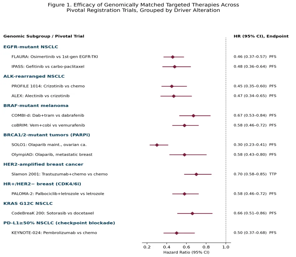
Figure 1. Pivotal-trial efficacy of genomically matched targeted therapies, grouped by driver alteration
Table 2. Consolidated cross-reference of major genomic alterations, matched targeted agents, and pivotal registration-trial efficacy data discussed in Section 5. *IPASS compared gefitinib with carboplatin-paclitaxel specifically within the EGFR-mutation-positive subgroup. dMMR = mismatch-repair deficient; HR+ = hormone-receptor-positive; MCyR = major cytogenetic response; MSI-H = microsatellite instability-high; NSCLC = non-small cell lung cancer; ORR = objective response rate; PFS = progression-free survival; TPS = tumor proportion score; TTP = time to progression.
|
Genomic Alteration |
Cancer Type |
Targeted Agent |
Pivotal Trial |
Key Efficacy Result |
Ref. |
|
BCR-ABL1 fusion |
Chronic myeloid leukemia |
Imatinib |
IRIS |
MCyR 87.1% vs 34.7% |
[35] |
|
EGFR exon19del / L858R |
NSCLC |
Osimertinib |
FLAURA |
PFS HR 0.46 (0.37–0.57) |
[40] |
|
EGFR exon19del / L858R |
NSCLC |
Gefitinib |
IPASS |
PFS HR 0.48 (0.36–0.64)* |
[39] |
|
ALK rearrangement |
NSCLC |
Crizotinib |
PROFILE 1014 |
PFS HR 0.45 (0.35–0.60) |
[42] |
|
ALK rearrangement |
NSCLC |
Alectinib |
ALEX |
PFS HR 0.47 (0.34–0.65) |
[43] |
|
BRAF V600E |
Melanoma |
Dabrafenib+trametinib |
COMBI-d |
PFS HR 0.67 (0.53–0.84) |
[45] |
|
HER2 amplification |
Breast cancer |
Trastuzumab+chemo |
Slamon 2001 |
TTP HR 0.70 (0.58–0.85) |
[37] |
|
BRCA1/2 mutation |
Ovarian cancer |
Olaparib (maint.) |
SOLO1 |
PFS HR 0.30 (0.23–0.41) |
[47] |
|
BRCA1/2 mutation |
Breast cancer |
Olaparib |
OlympiAD |
PFS HR 0.58 (0.43–0.80) |
[48] |
|
Cyclin D-CDK4/6 dependency |
HR+/HER2− breast |
Palbociclib+letrozole |
PALOMA-2 |
PFS HR 0.58 (0.46–0.72) |
[49] |
|
KRAS G12C |
NSCLC |
Sotorasib |
CodeBreaK 200 |
PFS HR 0.66 (0.51–0.86) |
[50] |
|
NTRK1/2/3 fusion |
Tumor-agnostic |
Larotrectinib |
Pooled analysis |
ORR 75% |
[52] |
|
PD-L1 ≥50% TPS |
NSCLC |
Pembrolizumab |
KEYNOTE-024 |
PFS HR 0.50 (0.37–0.68) |
[54] |
|
MSI-H / dMMR |
Tumor-agnostic |
Pembrolizumab |
Pooled analysis |
ORR ~30–40% |
[25,55] |
Mechanisms of Resistance to Targeted Therapy
Despite the often dramatic initial efficacy , resistance to targeted therapy emerges in virtually every patient with metastatic disease, typically within one to three years, and understanding its mechanisms has become as central to precision oncology as identifying the initial driver alteration. Resistance mechanisms are generally classified into four overlapping categories, summarized in Table 3.
On-Target Secondary Mutations
The most direct resistance mechanism is acquisition of a second mutation within the originally targeted gene that either directly impairs drug binding or restores downstream signaling despite continued drug exposure. The paradigmatic example is the EGFR T790M ‘gatekeeper’ mutation, which sterically hinders binding of first-generation EGFR TKIs and accounts for approximately 50–60% of acquired resistance to first-line gefitinib or erlotinib, a mechanism that directly motivated development of the T790M-active third-generation inhibitor osimertinib described in. [56,58] Osimertinib itself, when used first-line, is characteristically associated with a different, more heterogeneous spectrum of on-target (C797S) and off-target resistance mechanisms upon eventual progression. [57] Analogous on-target resistance mutations occur within the ABL1 kinase domain following imatinib therapy (most classically T315I, which confers resistance to imatinib and several second-generation inhibitors but remains sensitive to the third-generation agent ponatinib), within ALK following crizotinib or alectinib, and, more recently described, within KRAS G12C itself or in adjacent RAS pathway nodes following sotorasib or adagrasib therapy. [59]
Bypass Pathway Activation
A second major resistance category involves activation of a parallel signaling pathway that restores the downstream proliferative and survival signal independent of the originally targeted node, rendering the original target-directed drug ineffective despite continued inhibition of its intended target. Examples include MET amplification, which can independently reactivate downstream RAS-MAPK signaling and accounts for a meaningful fraction of osimertinib resistance; reactivation of the MAPK pathway through NRAS mutation, MEK mutation, or receptor tyrosine kinase upregulation following BRAF inhibitor therapy in melanoma (the mechanistic rationale for the upfront combination BRAF/MEK inhibition strategy ; and activation of compensatory HER family or IGF-1R signaling following anti-HER2 therapy. Because bypass-pathway resistance mechanisms are often heterogeneous and difficult to predict in advance, they have motivated a general shift toward upfront combination targeted therapy (exemplified by BRAF/MEK combination) as a strategy to pre-empt rather than sequentially react to resistance.
Histological and Phenotypic Transformation
A distinct and mechanistically striking resistance category involves transformation of the tumor's histological identity itself, without necessarily requiring any further mutation in the original driver gene. The best-characterized example is transformation of EGFR-mutant lung adenocarcinoma into small-cell lung cancer histology upon progression on EGFR TKI therapy, observed in a meaningful minority of resistant cases and associated with concurrent RB1 and TP53 loss; because small-cell transformed tumors respond to small-cell-appropriate chemotherapy regimens rather than continued EGFR-directed therapy, recognition of this phenomenon (which requires repeat tissue biopsy to detect, as it is not identifiable on standard genomic testing of the original driver alteration) has direct treatment-sequencing implications. [58] Epithelial-to-mesenchymal transition, a partial and reversible phenotypic shift associated with reduced epithelial marker expression and altered drug sensitivity, represents a related, non-mutational transformation-based resistance mechanism observed across several targeted therapy contexts.
Epigenetic and Non-Genetic Adaptive Resistance
Building on the epigenetic driver concepts , an increasing body of evidence indicates that tumor cells can survive initial targeted therapy exposure through reversible, non-genetic adaptive mechanisms including transient entry into a drug-tolerant persister cell state characterized by altered chromatin accessibility and transcriptional reprogramming without requiring any new DNA-level mutation at all, providing a reservoir of surviving cells from which genetically confirmed resistance mechanisms may subsequently emerge under continued drug selection pressure. This non-genetic resistance phenomenon, while mechanistically distinct from the three mutation- or transformation-based categories above, is increasingly recognized as clinically important and has motivated interest in intermittent or pulsed dosing strategies, and in combination approaches that simultaneously target the persister-cell state, as means of delaying the emergence of genetically fixed resistance.
Table 3. Classification of the four principal mechanisms of acquired resistance to genomically targeted therapy, discussed in Section 6, with representative examples drawn from the driver-therapy pairs reviewed in Section 5. NSCLC = non-small cell lung cancer; RTK = receptor tyrosine kinase; TKI = tyrosine kinase inhibitor.
|
Resistance Category |
Representative Examples |
Underlying Mechanism |
Clinical/Therapeutic Response |
|
On-target secondary mutation |
EGFR T790M after 1st-gen TKI; ABL1 T315I after imatinib; KRAS G12C-adjacent mutations after sotorasib |
Direct steric or allosteric impairment of drug binding, or restoration of constitutive activity |
Rational design of next-generation inhibitors active against the resistance mutation (e.g., osimertinib, ponatinib) |
|
Bypass pathway activation |
MET amplification (EGFR TKI resistance); NRAS/MEK mutation, RTK upregulation (BRAF inhibitor resistance) |
Parallel pathway restores proliferative signal despite continued target inhibition |
Upfront combination therapy (e.g., BRAF+MEK); addition of bypass-pathway inhibitor at progression |
|
Histological/phenotypic transformation |
Small-cell transformation (EGFR-mutant NSCLC); epithelial-to-mesenchymal transition |
Lineage plasticity, often with concurrent RB1/TP53 loss, shifts tumor identity and drug sensitivity |
Repeat tissue biopsy at progression; switch to histology-appropriate regimen (e.g., chemotherapy) |
|
Epigenetic / non-genetic adaptation |
Drug-tolerant persister cell state; chromatin/transcriptional reprogramming |
Reversible, non-mutational survival state without new DNA-level alteration |
Intermittent dosing strategies; combination targeting of persister state (investigational) |
Emerging Technologies and Future Directions
Liquid Biopsy and Circulating Tumor DNA
Analysis of circulating tumor DNA (ctDNA) shed into peripheral blood by dying tumor cells has emerged as a minimally invasive complement, and in some contexts an alternative, to tissue biopsy for both initial genomic profiling and, of particular relevance to Section 6, serial monitoring for emergence of resistance mutations without requiring repeat invasive tissue sampling. [60] Plasma genotyping for the EGFR T790M resistance mutation, validated against tissue re-biopsy in the FLAURA and related osimertinib development program, exemplifies this application and is now incorporated into routine clinical practice for NSCLC treatment-sequencing decisions. [56] Liquid biopsy technology continues to face sensitivity limitations, particularly in patients with low disease burden or predominantly non-shedding disease patterns (e.g., CNS-only or bone-only metastasis), but its non-invasive, serially repeatable nature makes it an area of substantial ongoing technical development.
Single-Cell and Spatial Genomics
Bulk tumor sequencing, the technology underlying essentially all of the driver-discovery and clinical-trial-eligibility genomic testing discussed in, reports an average genomic profile across millions of admixed tumor and non-tumor cells and is consequently unable to directly resolve the intratumor heterogeneity and subclonal architecture described. Single-cell sequencing technologies, which generate genomic or transcriptomic profiles from individually isolated tumor cells, and spatial genomic technologies, which preserve the anatomical location of each profiled cell within the tissue architecture, together offer the prospect of directly visualizing clonal evolution, resistant-subclone emergence, and the tumor-immune microenvironment interactions relevant to immune checkpoint inhibitor response at a resolution unattainable with bulk sequencing. [61] These technologies remain predominantly research tools at present, owing to cost and throughput constraints, but are anticipated to progressively enter clinical translational use over the coming decade.
Functional Genomics and CRISPR Screening
Genome-wide CRISPR-Cas9 functional screening, in which every gene in the genome is systematically inactivated or activated in parallel within a model cell population to identify genes whose loss or gain confers a measurable phenotypic effect, has become a central discovery tool for identifying candidate synthetic-lethal drug targets (extending the BRCA1/2-PARP paradigm to other genomic contexts) and for prospectively mapping resistance mechanisms before they are observed clinically, by systematically screening which gene perturbations confer resistance to a given targeted agent in model systems. This approach complements the retrospective, clinically driven resistance-mechanism discovery process described in Section 6 with a prospective, hypothesis-generating discovery pipeline.
Artificial Intelligence in Genomic Interpretation
The scale and complexity of contemporary tumor genomic profiling increasingly incorporating whole-exome or whole-genome sequencing, transcriptomic profiling, and multi-omic integration rather than the smaller targeted gene panels used in earlier clinical practice has outpaced the capacity for fully manual clinical interpretation, motivating growing use of machine-learning and, more recently, large-language-model-based tools to assist variant classification, driver-versus-passenger prioritization and matching of genomic findings to actionable therapy or clinical trial options via knowledge bases such as OncoKB. [31] These tools are increasingly integrated into molecular tumor board workflows as decision-support aids, though the clinical and regulatory validation frameworks for such AI-assisted genomic interpretation tools remain comparatively immature relative to the underlying sequencing technology itself.
DISCUSSION
The Precision Oncology Paradigm: Achievements and Scope
The evidence synthesized in documents genuinely transformative treatment effects: hazard ratios in the range of 0.30 to 0.70 across the pivotal trials reviewed represent, in aggregate, among the largest and most consistent effect sizes achieved anywhere in modern oncology drug development, and several of the agents discussed (imatinib in CML, trastuzumab in HER2-positive breast cancer) have converted diseases with historically poor prognosis into conditions compatible with near-normal or substantially extended life expectancy. [4,35,38] These successes have fundamentally restructured how new cancer drugs are developed and approved, shifting regulatory and clinical trial paradigms away from purely histology-defined disease categories and toward genomically defined patient populations a shift epitomized by the tumor-agnostic approvals of larotrectinib and pembrolizumab, and formalized in structured frameworks such as ESCAT for ranking the clinical actionability of a given genomic finding. [32]
The Limits of the Driver Paradigm
The driver-gene paradigm reviewed in this paper, however, does not extend uniformly across all of cancer genomics. , several of the most frequently altered driver genes MYC and mutant RAS isoforms other than KRAS G12C prominent among them remain substantially more difficult to target directly than the receptor tyrosine kinases and fusion oncoproteins that dominate Section 5's clinical successes, reflecting underlying differences in protein structure and druggability rather than any deficiency in genomic discovery per se. Tumor suppressor gene loss affecting the single most commonly altered gene in the pan-cancer genome (TP53), likewise remains largely therapeutically unaddressed by direct restoration approaches, with clinical benefit in TP53-altered tumors instead derived indirectly, for example through the DNA-damage-response vulnerabilities such loss can create. further qualify the paradigm's achievements: essentially no targeted therapy reviewed in Section 5 is curative in the metastatic setting, and the field's central ongoing challenge is not merely identifying new druggable drivers but converting the current pattern of initially dramatic but ultimately time-limited responses into durable disease control, whether through combination strategies, resistance-anticipating sequential therapy, or exploitation of the non-genetic adaptive resistance mechanisms.
Access, Cost, and Implementation Challenges, with Reference to India
Realizing the genomic driver paradigm's benefits in practice requires, as a precondition, access to tumor genomic profiling and to the resulting matched therapies both of which remain substantially constrained outside high-income health systems. In India, where the National Cancer Registry Programme has documented a rising and regionally heterogeneous cancer burden, [30] access to comprehensive genomic profiling and to the newer targeted and immune therapies reviewed in Section 5 remains concentrated in a comparatively small number of tertiary cancer centers, with cost, insurance coverage, and diagnostic infrastructure availability each independently constraining real-world uptake outside these centers; Mallath and colleagues' analysis of India's cancer burden explicitly identifies this diagnostic and therapeutic infrastructure gap, rather than any lack of underlying scientific applicability, as a central barrier to translating the genomic advances reviewed in this paper into population-level outcome improvement within the Indian health system. [62] Generic and biosimilar availability of several of the older targeted agents (imatinib prominent among them, now widely available as a low-cost generic in India) illustrates one pathway by which the cost barrier can substantially narrow over time following initial patent-protected launch, a trajectory that later-generation and immune-oncology agents have not yet uniformly followed.
Ethical and Regulatory Considerations
The tumor-agnostic, biomarker-first approval pathway pioneered by larotrectinib and pembrolizumab-for-MSI-H raises distinct regulatory and evidentiary questions relative to the traditional single-histology randomized trial model, since biomarker-defined populations, particularly for rare fusion events, are frequently too small within any single cancer type to support a conventional randomized comparison, necessitating single-arm, pooled, or basket/umbrella trial designs whose evidentiary strength differs qualitatively from the pivotal randomized trials that anchor most of Section 5's other efficacy claims (Table 2's larotrectinib and MSI-H entries are accordingly reported as objective response rate from pooled single-arm data rather than as a comparative hazard ratio). This evolution in trial design and regulatory evidentiary standards, alongside the broader question of equitable access discussed in , represents an active area of ongoing policy discussion within precision oncology.
Future Research Priorities
Building on the emerging technologies reviewed , priority areas for future research include: prospective, biomarker-stratified combination and sequencing trials explicitly designed to pre-empt rather than react to the resistance mechanisms characterized in; expanded application of single-cell and spatial genomic technologies to directly visualize, rather than infer, subclonal resistance evolution in real time; continued medicinal chemistry effort toward historically undruggable targets (MYC, non-G12C RAS isoforms) building on the KRAS G12C precedent; and health-systems and implementation-science research — particularly relevant to the Indian and other resource-constrained contexts discussed in aimed at closing the gap between genomic and therapeutic scientific capability and equitable real-world clinical access.
CONCLUSION
The genomic driver paradigm reviewed in this paper has, over little more than two decades, transformed the conceptual and clinical practice of oncology: cancer is now understood, diagnosed, and increasingly treated as a disease defined primarily by its specific molecular alterations, organized within a comparatively small number of core oncogenic pathways , rather than by anatomical site of origin alone. The clinical trial evidence synthesized in documents some of the largest and most consistent treatment-effect sizes achieved anywhere in modern oncology, and has produced genuinely curative or near-curative outcomes in specific genomically defined contexts. Yet resistance, comprehensively characterized, remains the near-universal rule rather than the exception in metastatic disease, and substantial portions of the genomic driver landscape MYC and tumor-suppressor loss prominent among them remain therapeutically underaddressed despite two decades of genomic characterization. Continued progress will depend jointly on scientific advances in resistance-anticipating combination therapy, in single-cell and functional genomic technology, and in extending the druggable genome and on health-systems progress in equitably extending genomic testing and matched therapy access, a consideration of particular importance for large, genomically underserved health systems such as India's.
REFERENCES




Zakiya Sultana, Janani Ramachandran, Arindom Patar, Mohammad Zulekha, Genomic Drivers of Cancer Progression and Their Implications for Targeted Therapy: A Comprehensive Review, Int. J. of Pharm. Sci., 2026, Vol 4, Issue 10, 2013-2035, https://doi.org/10.5281/zenodo.23357351
 10.5281/zenodo.23357351