

We use cookies to ensure our website works properly and to personalise your experience. Cookies policy
Department of Microbiology and Biotechnology, Deogiri College, Chhatrapati Sambhajinagar - 431005, Maharashtra, India
The present study aimed to isolate and characterize cholesterol-degrading probiotic bacteria isolated from homemade curd collected across different localities in Chhatrapati Sambhajinagar, Maharashtra, India. A total of 16 bacterial isolates were obtained using standard microbiological isolation techniques on De Man, Rogosa and Sharpe (MRS) Agar. Out of these, four isolates (S1 to S4) demonstrated the ability to utilize cholesterol as the sole carbon source when cultured on M9 minimal agar supplemented with 1% cholesterol. These isolates underwent detailed morphological and biochemical characterization, including IMViC profiling, catalase test and carbohydrate fermentation capabilities. Acid tolerance assays indicated that the isolates were able to grow under selected pH conditions, with optimal growth observed at pH 7.0 with moderate tolerance between pH 3.0 to 5.0. The cholesterol reduction assay, evaluated via spectrophotometric OD analysis at 560 nm, showed a significant reduction in cholesterol concentration over a four-day incubation period, with isolate S1 showing the highest cholesterol reduction. These findings highlight the probiotic potential of the selected isolates, especially in terms of their ability to reduce cholesterol levels, suggesting their potential application in developing functional foods aimed at promoting cardiovascular health.
Microbial fermentation is a biological process in which microorganisms such as bacteria, yeasts, and molds convert organic compounds, typically sugars and carbohydrates, into other products like acids, gases, or alcohol, under anaerobic (low or no oxygen) conditions. This natural transformation has been utilized for centuries in the preservation and enhancement of food, making it safer, more nutritious, and often more flavourful.
Probiotics are live microorganisms that, when consumed in adequate amounts, confer health benefits to the host. To qualify as a probiotic, a microorganism must meet several essential criteria: it must be able to survive exposure to bile salts and gastric acids, produce antimicrobial substances that inhibit the growth of harmful microbes, remain susceptible to antibiotics, and successfully colonize the gastrointestinal tract (GIT). Common probiotic strains include bifidobacteria, lactic acid bacteria (LAB), and certain yeasts, which are typically isolated from sources such as milk and milk products, different parts of the GIT, and various fermented food products [1].
Globally, fermented foods produced with lactic acid bacteria (LAB) are recognized for their positive impact on human nutrition. As a result, LAB are regarded as promising probiotic candidates and are being widely researched to better understand their beneficial properties [16].
Curd is a traditional fermented dairy product that is widely consumed in many parts of the world, especially in South Asia. It is formed through the fermentation of milk by LAB, primarily strains like Lactobacillus and Streptococcus species. These bacteria ferment the lactose (milk sugar) present in milk into lactic acid. The lactic acid lowers the pH of the milk, leading to the coagulation of casein proteins, which results in the thick, tangy product known as curd [5].
Cardiovascular diseases (CVDs) remain a major global health concern, accounting for nearly 19.8 million fatalities annually according to World Health Organization (WHO) estimates [22]. One of the major risk factors contributing to CVDs is hypercholesterolemia, or elevated blood cholesterol levels. Individuals with hypercholesterolemia are estimated to have a threefold increased risk of experiencing a heart attack compared to those with normal blood lipid profiles.
Clinical studies have shown that the normal range of serum cholesterol levels in humans typically falls between 2.8 to 6.0 mmol/L. When cholesterol levels exceed this range, they are often associated with an increased risk of developing various health conditions, particularly cardiovascular diseases such as atherosclerosis, coronary artery disease, and stroke. Elevated serum cholesterol contributes to the accumulation of lipid plaques in the arteries, which can impair blood flow and lead to serious complications. Therefore, maintaining cholesterol levels within the recommended range is considered critical for cardiovascular health and overall well-being [17].
Probiotics have been suggested to lower cholesterol levels through multiple mechanisms. These include the co-precipitation of cholesterol with deconjugated bile salts, binding of cholesterol to the bacterial cell walls, incorporation of cholesterol into bacterial cell membranes during growth, production of short-chain fatty acids (SCFAs) during fermentation, and the conversion of cholesterol may also be into less absorbable compounds such as coprostanol [10,18].
Cholesterol oxidase (cholesterol: oxygen oxidoreductase, EC 1.1.3.6) is a class of oxidoreductase, and it is a crucial enzyme involved in cholesterol metabolism. It catalyzes the oxidation of cholesterol into 4-cholesten-3-one while concurrently reducing molecular oxygen to form hydrogen peroxide [4].
This enzymatic activity is particularly significant when the enzyme is derived from bacterial sources, as many probiotic and non-pathogenic bacteria possess the ability to produce cholesterol oxidase. In the context of fermented foods, cholesterol oxidase contributes to the biodegradation and reduction of cholesterol content, thereby potentially enhancing the health benefits of such foods by lowering dietary cholesterol intake.
Its application has attracted increasing attention in the development of functional foods aimed at cardiovascular health and cholesterol management.
Therefore, the objectives of the present study were as follows:
MATERIALS AND METHODS
Collection of Curd Sample:
In the course of the study, homemade curd samples were aseptically collected from different localities of Chhatrapati Sambhajinagar, Maharashtra, India, to ensure a representative range of microbial diversity. The samples were collected in sterile containers to prevent external contamination. Following collection, the samples were carefully transported to the Department of Microbiology at Deogiri College, Chhatrapati Sambhajinagar. During transit, strict precautions were taken to maintain sample integrity and minimize the risk of microbial alteration. Upon arrival at the laboratory, all samples were promptly stored under refrigerated conditions to preserve their native microbial populations until further microbiological studies.
Isolation of Probiotic Microorganisms
Each curd sample was subjected to a tenfold serial dilution technique, extending up to a dilution factor of 10-6, using sterile distilled water as the diluent. For the isolation of probiotic bacteria, De Man, Rogosa and Sharpe (MRS) agar was used.
A 0.1 mL aliquot from each dilution of the curd samples was aseptically transferred onto individual MRS agar plates. The samples were evenly spreaded across the surface of the medium using a sterile L-shaped glass spreader to ensure uniform distribution of the inoculum. This step was performed under strict aseptic conditions to prevent contamination and to facilitate the isolation of discrete microbial colonies.
All inoculated plates were incubated at 37°C for a period of 2 to 3 days in an inverted position. Upon completion of the incubation period, the plates were observed using colony counter for the number and presence of isolated microbial colonies. Plates exhibiting 30 to 300 colonies were considered statistically significant and were selected for calculation of bioload. The microbial load (bioload) of each curd sample was calculated based on the colony count expressed as the number of colony-forming units (CFU) and corresponding dilution factor using the following formula;

Detailed records of the number and types of colonies observed were recorded. Morphological characteristics of the colonies were noted, and microscopic observations (such as Gram staining) were performed to assess the purity and identity of the isolates.
Gram Staining
Gram staining was performed to determine the Gram’s nature and cellular morphology of the bacterial isolates. A loopful of actively growing bacterial culture was smeared onto a clean and grease free glass slide, heat-fixed, and stained using the Gram staining procedures according to the published protocol outlined by Dubey and Maheshwari (2008). The results were observed under a light microscope at 1000× magnification using oil immersion. Gram-positive bacteria appeared purple, while Gram-negative bacteria, stained pink.
Primary Screening of Cholesterol-Metabolizing Bacterial Isolates:
To screen for cholesterol-metabolizing bacterial isolates, M9 Minimal Salt Agar supplemented with 1% (w/v) cholesterol (M9-cholesterol agar) was employed. In this selective medium, cholesterol functioned as the sole source of carbon, thereby enabling the identification of microorganisms capable of utilizing it for growth. The previously isolated pure bacterial cultures were aseptically streaked onto the surface of the sterile M9-cholesterol agar plates under laminar airflow to maintain aseptic condition.
Following inoculation, the plates were incubated in an inverted position at 37°C for a period of up to 7 days. This extended incubation period was necessary to support the growth of slow growing cholesterol degrading microorganisms. Post incubation, only those bacterial isolates that were capable of metabolizing cholesterol as their sole carbon source demonstrated visible colony formation on the agar surface.
These cholesterol-utilizing bacterial strains were subsequently selected and preserved for further characterization and studies.
Preliminary Identification of Cholesterol Reducing Probiotic Bacteria:
The identification of cholesterol reducing bacterial isolates were conducted using conventional microbiological techniques for determining the morphological and biochemical characteristics of the isolates.
Biochemical Characterization
To further characterize the isolates, a series of biochemical tests were performed:
IMViC Tests: The Indole, Methyl Red, Voges-Proskauer, and Citrate utilization tests were conducted to assess the metabolic capabilities of the isolates.
These tests were conducted using standard microbiological media and protocols. The results were recorded based on color changes and the presence or absence of specific reaction indicators.
Catalase Test
The catalase test was performed to detect the presence of the catalase enzyme, which catalyzes the breakdown of hydrogen peroxide (H₂O₂) into water and oxygen. A small amount of fresh bacterial culture was placed on a clean glass slide, followed by the addition of a drop of 3% hydrogen peroxide. The appearance of immediate bubble formation indicating a positive catalase reaction.
Carbohydrate Oxidation and Fermentation Tests
To determine the carbohydrate utilization profiles of the bacterial isolates, fermentation tests were carried out using various sugars, including glucose, fructose, sucrose, lactose, maltose, and mannitol. Each sugar was incorporated separately into M9 media as a carbon source and bromothymol blue as a pH indicator, and Durham tubes were used to detect gas production. The tubes were inoculated with bacterial cultures and incubated at 37°C up to 3 days. A color change from blue to yellow indicated acid production (positive fermentation), while gas accumulation in Durham tubes suggested gas production during fermentation.
These combined conventional methods provided preliminary insights into the identity and functional characteristics of the cholesterol degrading probiotic strains.
Assessment of Acid Tolerance Capacity of Probiotic Isolates:
To evaluate the acid tolerance capacity of the presumptive probiotic bacterial isolates, an in vitro simulation of the acidic environment of the human stomach was performed. This assay is critical to determine the ability of isolates to survive under varying acidic conditions, which is one of the key attributes of effective probiotic strains.
Preparation of Acidified MRS Broth
MRS broth was prepared and adjusted to different pH levels (1.0, 2.0, 3.0, 4.0, 5.0, 6.0, and 7.0) using 1N HCl and 1N NaOH. Each pH-adjusted broth was autoclaved at 121°C for 15 minutes and thereafter pre-incubated for 24 hrs to ensure sterility.
Inoculation and Incubation
A loopful of actively growing cholesterol utilising bacterial isolates were aseptically inoculated into 25 mL of MRS broth at each pH level and all flasks were incubated at 37°C up to 7 days.
Measurement of Bacterial Growth (OD Analysis)
To monitor bacterial survival and growth, optical density (OD) readings were taken at 600 nm using a UV-Visible spectrophotometer. The OD was recorded at 24-hour intervals (i.e., at Day 1, Day 2, Day 3, Day 4 and Day 5) for each pH condition. This provided a quantitative measure of the bacterial population over time.
Cholesterol Reduction Assay
The ability of selected probiotic strains to assimilate cholesterol was evaluated using a modified colorimetric method as described by Rudel and Morris (1973) and Gilliland et al. (1985). This method assesses the reduction in cholesterol concentration in the culture medium after bacterial growth under controlled conditions.
Preparation of Cholesterol-Enriched Medium
Cholesterol was first dissolved in ethanol (95%) to prepare a stock solution and then added to sterile MRS broth to achieve a final concentration of 0.1% (w/v). The medium was thoroughly mixed to ensure homogenous distribution of cholesterol and autoclaved to maintain sterility.
Inoculation and Incubation
Each probiotic strain was cultured overnight and then inoculated into the cholesterol-supplemented MRS broth at 0.1% (v/v) concentration. The inoculated broths were incubated at 37°C for 24 hours to facilitate optimal bacterial growth and cholesterol assimilation.
Separation of Supernatant
After incubation, the cultures were centrifuged at 10,000 RPM for 5 minutes. The supernatant was carefully collected and used for cholesterol quantification.
Quantitative Determination of Residual Cholesterol
The cholesterol concentration in the supernatant was estimated using a modified colorimetric method:
2 mL supernatant of each inoculated broth were taken in separate tubes and equal volumes (2 mL) of freshly prepared FeCl3-acetic acid reagent were mixed thoroughly in a test tube.
The mixture was allowed to stand at room temperature for 10 minutes to allow the initial reaction to proceed.
Then, 1 mL of concentrated sulfuric acid (H₂SO₄) was added carefully to each test tube. The samples were placed in the dark for 45 minutes to allow color development.
The optical density (OD) of each sample was then measured at 560 nm using a UV-Vis spectrophotometer.
Calculation of Cholesterol Reduction
The percentage of cholesterol removed from the medium by each probiotic strain was calculated using the formula:
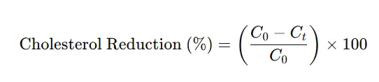
Where: C0 = Initial cholesterol concentration in uninoculated control broth.
Ct = Residual cholesterol concentration after 24 hours (Day 1) of incubation with probiotic strain.
The same procedure was followed for Day 2, Day 3 and Day 4 for determination of utilized cholesterol. This assay allowed for the comparative evaluation of the cholesterol-lowering ability of different bacterial isolates under simulated gut conditions.
Results and Discussion:
Isolation of Probiotic Microorganisms:
In the present study, a total of four homemade curd samples were used as sources for isolating potential probiotic bacteria. Each curd sample yielded four morphologically distinct bacterial colonies depending on their abundance, resulting in a total of sixteen (16) individual isolates. These isolates were obtained using standard serial dilution and spread plating techniques on MRS agar, which is selective for the growth of lactic acid bacteria (LAB).
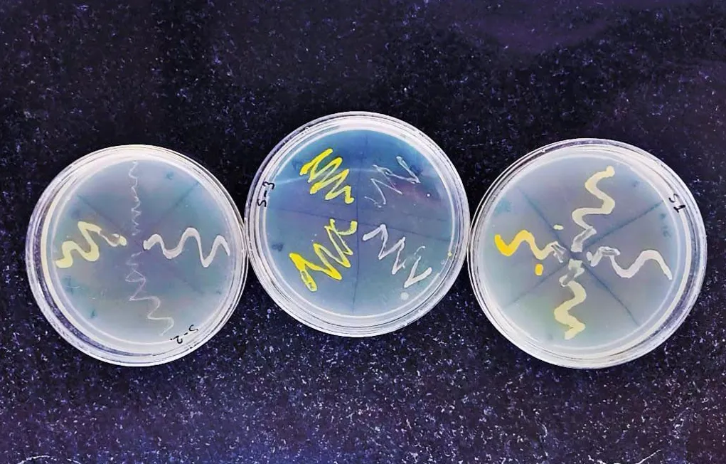
Figure 1: Representative Image of Bacterial Isolates Obtained from Curd Samples
The selection of colonies was based on morphological characteristics such as colony shape, size, margin, color, elevation, and texture and their abundance. Abundant colonies were selected for subculturing and purification.
This stage of the study demonstrated the microbial richness of homemade curd, particularly the diversity of lactic acid bacteria. The presence of multiple morphotypes within each sample suggests that homemade curd harbours a complex community of probiotics, influenced by factors such as geographical location, milk source, and fermentation practices.
Primary Screening of Cholesterol-Metabolizing Bacterial Isolates:
In the initial screening for cholesterol-reducing capability, sixteen bacterial isolates obtained from homemade curd were evaluated for their ability to utilize cholesterol as the sole carbon source. This was carried out using M9 minimal agar medium supplemented with 1% cholesterol, to assess microbial ability to metabolize cholesterol.
The plates were inoculated with each isolate and incubated at 37°C for up to 7 days. Growth on M9-cholesterol medium was considered a positive indication of cholesterol utilization, as the medium does not contain any alternative carbon sources.
Out of the sixteen isolates tested, four isolates exhibited (one from each curd sample) visible growth on the cholesterol-enriched M9 agar. These results suggest that these four isolates were able to utilize cholesterol as their primary carbon and energy source, highlighting their potential for cholesterol degradation. These isolates were designated as S1, S2, S3 and S4.
Table 1: Morphological and Microscopic Observation of Isolates
|
Colony characteristics |
S1 |
S2 |
S3 |
S4 |
|
Size |
Small |
Medium |
Medium |
Medium |
|
Shape |
Circular |
Circular |
Circular |
Circular |
|
Colour |
Off white |
Off white |
Yellow |
White |
|
Margin |
Entire |
Entire |
Entire |
Entire |
|
Elevation |
Raised |
Convex |
Convex |
Convex |
|
Consistency |
Smooth |
Glistening |
Smooth |
Smooth |
|
Opacity |
Opaque |
Opaque |
Opaque |
Opaque |
|
Gram’s nature |
Gram Positive Cocci |
Gram Positive Rods |
Gram Positive Rods |
Gram Positive Rods |
The observed growth suggests that the isolates may possess metabolic mechanisms enabling cholesterol utilization; however, the specific enzymatic mechanisms involved were not determined in the present study. These four isolates were thus selected for further biochemical and enzymatic characterization, including quantitative cholesterol assimilation assays and acid tolerance tests.
Preliminary Identification of Cholesterol Reducing Probiotic Bacteria:
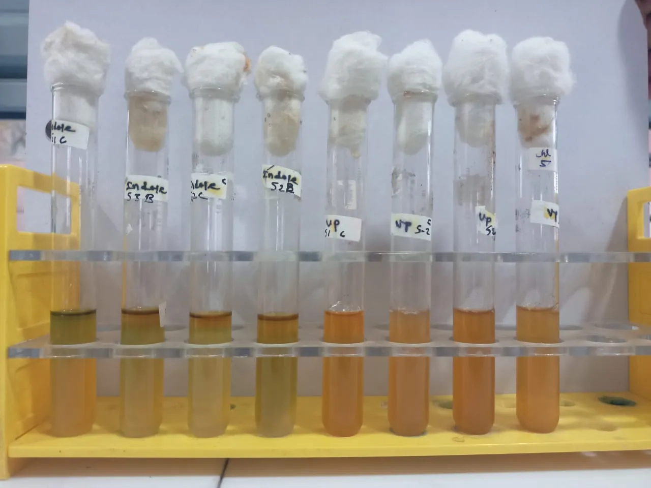
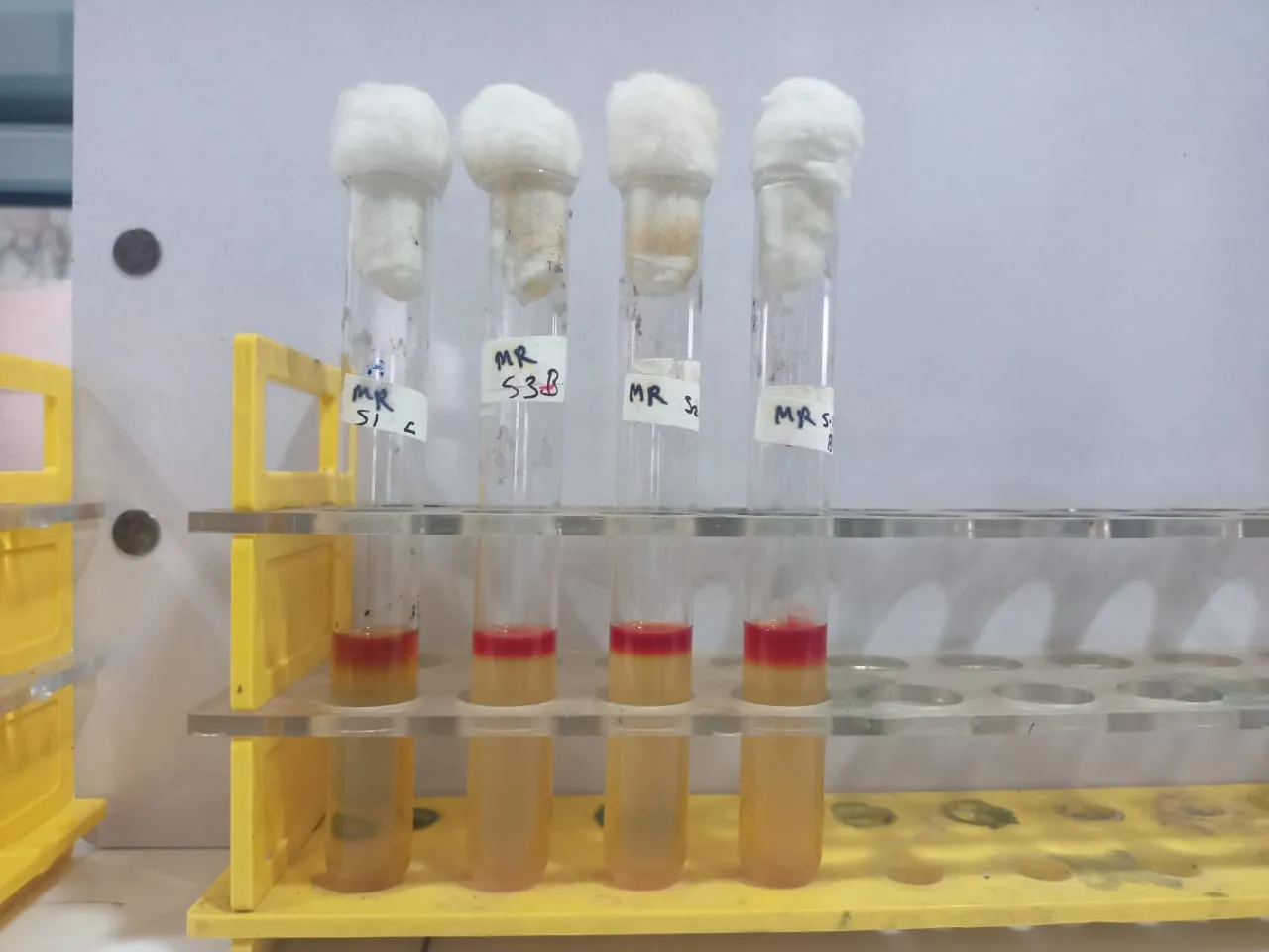
IMViC Test: All four isolates (S1 to S4) yielded almost identical outcomes in the IMViC series (Indole, Methyl Red, Voges–Proskauer and Citrate test) as summarized in table 2.
Table 2: IMViC Test Results of All Isolates
|
Isolate Number |
Indole Test |
Methyl Red Test |
VP test |
Citrate test |
|
S1 |
–ve |
+ve |
–ve |
+ve |
|
S2 |
–ve |
+ve |
–ve |
+ve |
|
S3 |
–ve |
+ve |
–ve |
–ve |
|
S4 |
–ve |
+ve |
–ve |
+ve |
Indole (–ve): No red ring formed with Kovács’ reagent, indicating absence of tryptophanase activity.
Methyl Red (+ve): Bright red color upon addition of methyl red indicator, signifying stable production of acids (pH < 4.4).
VP (–ve): No pink to red coloration after α‑naphthol/KOH addition, indicating that acetoin is not produced.
Citrate: Three isolates (S1, S2, and S4) showed positive citrate utilization, demonstrating their ability to use citrate as a sole carbon source, while isolate S3 was citrate-negative.
The IMViC test results (Indole–, MR+, VP–) across the isolate S1 to S4 shared fermentative metabolism: these isolates produce and maintain high levels of acidic end‑products from sugar fermentation but do not engage in indole formation or in the butanediol (acetoin) pathway.
|
|
|
|
Figure 2: Indole and VP Test Results |
Figure 3: Methyl Red Test Results |
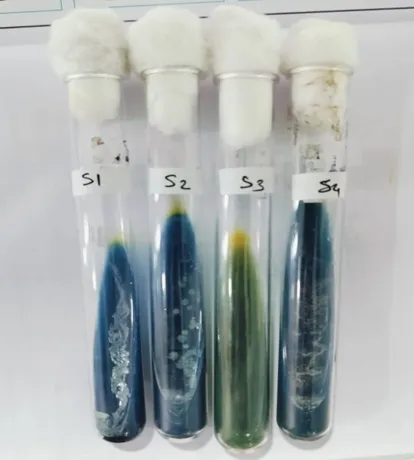
Figure 4: Citrate Test Results
Catalase Test:
The catalase activity of four probiotic candidate isolates (S1 to S4) was assayed by adding a drop of 3% H₂O₂ to a loopful of culture on a glass slide and observing immediate bubble formation. All four isolates did not produce rapid effervescence, indicating negative catalase activity.
Table 3: Catalase Test Results of All Isolates
|
Test Name |
Probiotic Isolate |
|||
|
S1 |
S2 |
S3 |
S4 |
|
|
Catalase Test |
-ve |
-ve |
-ve |
-ve |
Catalase is an enzyme that decomposes hydrogen peroxide (H₂O₂) into water and oxygen, protecting cells from oxidative damage. The catalase‑negative reaction observed among isolates S1 to S4.
Carbohydrate Fermentation Tests:
Carbohydrate fermentation tests were performed for all four isolates using different sugars as a sole carbon source and the results are as follows;
Table 4: Carbohydrate Fermentation Test Results of All Isolates
|
Type of Sugar |
Type of Test |
Isolate S1 |
Isolate S2 |
Isolate S3 |
Isolate S4 |
|
|
Glucose |
1. Oxidation |
Partial |
Positive |
Positive |
Positive |
|
|
2. Fermentation |
Acid |
Negative |
Positive |
Partial |
Positive |
|
|
Gas |
Negative |
Negative |
Negative |
Negative |
||
|
Fructose |
1. Oxidation |
Positive |
Positive |
Partial |
Positive |
|
|
2. Fermentation |
Acid |
Positive |
Positive |
Partial |
Positive |
|
|
Gas |
Negative |
Negative |
Negative |
Negative |
||
|
Sucrose |
1. Oxidation |
Positive |
Partial |
Positive |
Positive |
|
|
2. Fermentation |
Acid |
Positive |
Partial |
Partial |
Positive |
|
|
Gas |
Negative |
Negative |
Negative |
Negative |
||
|
Maltose |
1. Oxidation |
Partial |
Positive |
Positive |
Positive |
|
|
2. Fermentation |
Acid |
Positive |
Positive |
Positive |
Positive |
|
|
Gas |
Negative |
Negative |
Negative |
Negative |
||
|
Mannitol |
1. Oxidation |
Negative |
Negative |
Positive |
Positive |
|
|
2. Fermentation |
Acid |
Positive |
Negative |
Positive |
Positive |
|
|
Gas |
Negative |
Negative |
Negative |
Negative |
||
|
Lactose |
1. Oxidation |
Partial |
Partial |
Negative |
Positive |
|
Note: Positive = complete color change; Partial = weak or incomplete reaction; Negative = no reaction/no gas production.
|
|
|
|
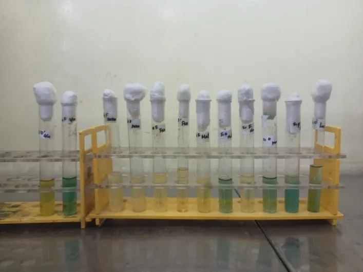
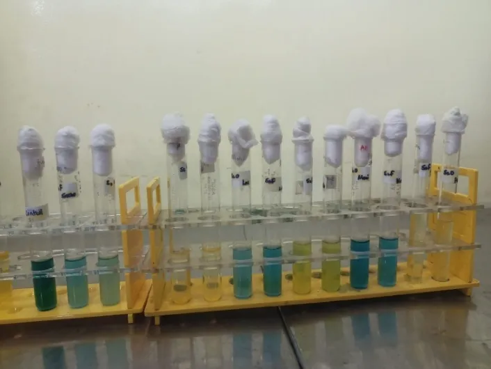
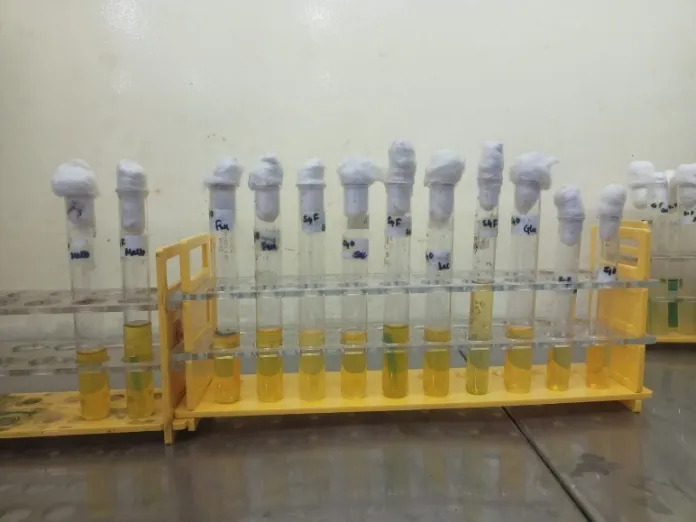
Figure 5: Carbohydrate Fermentation Test Results of S1 and S2 Isolate |
|
|
|
|
|
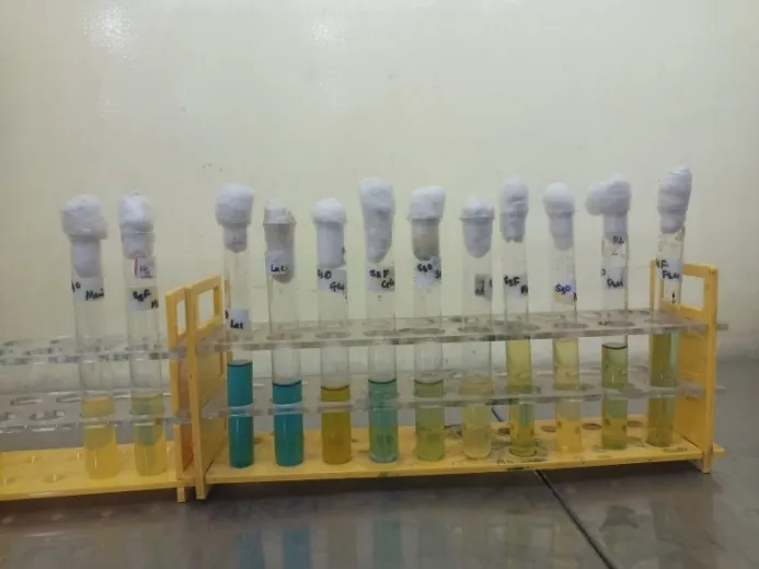
Figure 6: Carbohydrate Fermentation Test Results of S3 and S4 Isolate |
|
The carbohydrate fermentation test revealed significant metabolic diversity among the four bacterial isolates (S1 to S4) in their ability to oxidize and ferment various sugars.
Glucose oxidation was positive in isolates S2 to S4, with notable acid production in S2 and S4, confirming active fermentation pathways. In contrast, isolate S1 showed only partial oxidation and no acid production, indicating a limited ability to metabolize glucose.
For fructose, isolates S1, S2, and S4 demonstrated both oxidation and acid production, suggesting efficient utilization, while S3 showed only partial responses, indicating weaker metabolic adaptation to this sugar.
When sucrose was used as the carbon source, isolates S1, S3, and S4 showed positive oxidation, with strong acid production in S1 and S4, whereas isolate S2 exhibited only partial oxidation and mild acid production, suggesting a less effective metabolism of disaccharides.
All four isolates fermented Maltose, with S2 to S4 showing complete oxidation and fermentation, while S1 exhibited partial oxidation yet strong acid production, indicating effective fermentation despite incomplete oxidation.
Mannitol was oxidized and fermented exclusively by isolates S3 and S4, while S1 showed acid production without oxidation, and S2 was negative for both, suggesting the absence of the necessary enzymes in that isolate.
Lactose utilization was generally weak, with only isolate S4 demonstrating complete oxidation and fermentation. S1 and S2 showed partial activity, and S3 was entirely negative, likely due to the absence of β-galactosidase.
Across all sugar tests, gas production was absent indicating that the fermentation pathways followed by these isolates were primarily acidogenic rather than gas-producing. The differential sugar utilization patterns among the isolates provide valuable insights into their biochemical profiles and suggest potential for application-specific roles, including fermentation, probiotic use, or industrial enzyme production. These findings are critical for microbial identification and for understanding the metabolic versatility of the isolates.
Assessment of Acid Tolerance Capacity of Probiotic Isolates:
The acid tolerance of Isolate S1 to S4 were evaluated by measuring optical density (OD) at 600 nm over a 5-day period across a pH range of 1 to 7. The results show a clear pH-dependent growth pattern (Figure 7). Growth was minimal at extreme acidic conditions (pH 1 and pH 2), on Day 1 and increasing modestly over five days. Notably, growth increased progressively at pH 3 to pH 5, indicating moderate tolerance to acidity.
The highest OD values were consistently observed at pH 7, in all the isolates up to Day 5, indicating optimal growth under neutral conditions. This was followed by substantial growth at pH 4 and pH 5, suggesting the isolate’s moderate acid tolerance and potential adaptation to mildly acidic environments. The absence of sharp OD increases at lower pH levels indicates reduced metabolic activity and limited adaptability under strongly acidic conditions.
Overall, these findings suggest that all isolates exhibited optimal growth in neutral to mildly acidic environments, which could be important in tolerance to the acidic conditions of the stomach and potential applications in probiotic or fermentation industries.
Quantitative Determination of Residual Cholesterol
The cholesterol reduction assay conducted over a four-day incubation period revealed a progressive decrease in optical density (OD at 560 nm) across all four bacterial isolates (S1 to S4), indicating their potential to assimilate or degrade cholesterol (Figure 8).
Among them, isolate S1 exhibited the most efficient cholesterol-lowering activity, reducing OD from 0.75 on Day 0 to 0.07 by Day 4, followed by S4 (0.09), S2 (0.11), and S3 (0.09). This steady decline in OD suggests that the isolates possess mechanisms possibly involving bile salt hydrolase activity or direct assimilation capable of reducing cholesterol levels. These findings highlight the probiotic potential of the isolates, particularly S1, in contributing to cardiovascular health through cholesterol regulation.

Figure 7: Graphical Representation of Acid Tolerance Capacity of Probiotic Isolates

Figure 8: Graphical Representation of Cholesterol Assay
CONCLUSION:
The present study successfully isolated, screened, and characterized cholesterol-utilising probiotic bacteria from homemade curd samples collected from the Chhatrapati Sambhajinagar region. A total of sixteen isolates were initially obtained, out of which four (S1 to S4) demonstrated the ability to utilize cholesterol as the sole carbon source on M9 minimal media, indicating their potential in cholesterol metabolism. Morphological, biochemical, and enzymatic characterization revealed that these isolates possessed typical traits of probiotic lactic acid bacteria, including Gram-positive nature, catalase negative activity, and robust carbohydrate fermentation profiles.
IMViC test results affirmed the functional diversity of the isolates. Acid tolerance assays further indicated that all four strains could withstand and grow in pH ranges simulating gastrointestinal conditions, with optimal growth at pH 7 and moderate survival in mildly acidic environments. The cholesterol assimilation assay provided quantitative evidence of cholesterol degradation, with isolate S1 showing the most significant reduction in OD at 560 nm, followed by S4, S2, and S3, respectively. This suggests that the observed cholesterol reduction may be attributed to cholesterol assimilation and/or enzymatic activity, although further studies are required to confirm the mechanism.
These findings collectively affirm that bacterial strains isolated from traditional curd exhibit key probiotic features including acid tolerance, cholesterol-reducing ability, and carbohydrate versatility. Therefore, they hold promising applications in the development of functional fermented foods and dietary supplements aimed at promoting cardiovascular health. Further species level identification and in vivo evaluation of these strains are recommended to validate their efficacy and ensure safety in therapeutic or commercial formulations.
REFERENCES

Sushmita Pawar*, Shivaji Wankhede, Narayan Bajad, Isolation And Characterization Of Probiotic Bacteria From Homemade Curd With Cholesterol Reducing Activity., Int. J. of Pharm. Sci., 2026, Vol 4, Issue 8, 2245-2259. https://doi.org/10.5281/zenodo.21923782
 10.5281/zenodo.21923782