

We use cookies to ensure our website works properly and to personalise your experience. Cookies policy
Nalanda Institute of Pharmaceutical Sciences, kantepudi (v). Sattenapalli (m), Guntur (Dist.)- 522438.
Ischemic stroke refers to a serious condition causing both paralysis and death. This disorder is caused by interruption of blood flow to the parts of the brain, resulting in destruction of the nerve cells. Secondary injuries inflicted on the brain can occur as a result of neuroinflammation and oxidative stress, causing damage to neurons and threatening the function of the blood–brain barrier (BBB). Both ischemia and reperfusion promote the development of too much reactive oxygen species (ROS) and make mitochondria dysfunctional, which leads to damaging the cells. In addition, activation of microglia causes a release of proinflammatory cytokines, including interleukin-1?, interleukin-6, and tumor necrosis factor-alpha, that aggravate the situation. Together with the conditional processes mentioned above, a disturbance in such processes as nuclear factor kappa B (NF-?B), nuclear factor erythroid 2-related factor 2 (Nrf2), and the inflammasome signaling can help in understanding these intricate connections in order to develop drugs for overcoming ischemia. Using antioxidants, anti-inflammatory drugs, mitochondrial protective agents, and targeted pathways can help fight this disease.
Ischemic stroke is a serious neurological disease which occurs due to the interruption of oxygen supply to a certain part of the brain. The result of this is the loss of energy, timely dysfunction of neurons and tissue injury which may cause prolonged disability or even death to the patient. In spite of the fact that return of cerebral blood flow is crucial in order to minimize the impact of brain injury on the person, the process of reinstatement of blood flow can cause even more cellular damage. Neuroinflammation as well as oxidative stress are the major contributors of this secondary damage and therefore have become the most popular areas of research in ischemic stroke field.
Oxidative stress occurs when the generation of reactive oxygen species (ROS) surpasses the capability of endogenous and cellular antioxidant mechanisms. Ischemia and reperfusion of the brain cause mitochondrial dysfunction, excitotoxicity and altered cellular metabolism which results in excessive ROS production. Excess ROS may result in the damage of membranes, proteins and genetic material by causing lipid peroxidation and necrosis of neurons. The vulnerability of the brain to oxidative injury results from high oxygen consumption, presence of large numbers of polyunsaturated fatty acids and presence of limited amounts of antioxidant material in some cellular compartments. Various factors such as glutathione-dependent enzymes and antioxidant pathways regulated by Nrf2 play an important part in the maintenance of cellular redox status [1].
Neuroinflammation is one of the aforementioned mechanisms conducting ischemic brain damage. After cerebral ischemia, defective cells set free danger-associated molecular patterns and activate immune cells residing in the brain including microglia and astrocytes. Afterwards the immune cells can produce proinflammatory molecules, i.e. various cytokines and chemokines. Among the signaling molecules, interleukin-1β (IL-1β), interleukin-6 (IL-6), and tumor necrosis factor-alpha (TNF-α) have been mentioned as the ones which may lead to the injury of blood-brain barrier, to infiltration of leukocytes into the brain tissues, and to the damages of brain cells. Nevertheless, neuroinflammation is responsible not only for damaging effects, but also for inflammation helping to remove waste products from tissues, perform tissue repair, and support recovery of functioning of the brain [2].
The relationship between neuroinflammation and oxidative stress is inherent such that one can spur the other in the event of ischemic stroke. The reactive oxygen species (ROS) can activate a variety of signaling pathways that are responsible for inflammation, one of which is the nuclear factor kappa B (NF-κB) pathway. Likewise, inflammatory cells are capable of producing extra amounts of ROS and reactive nitrogen species. In addition, mitochondrial malfunction and activation of the NOD-like receptor pyrin domain containing protein 3 (NLRP3) inflammasome can interconnect the two processes triggering the release of inflammatory cytokines and cell damage. This mutual cooperation can create a vicious circle of oxidative damage and neuroinflammation, which will eventually lead to tissue damage that is greater than that caused by the initial ischemic event. Therefore, targeting only one mechanism may be impossible for the prevention of the complex process of secondary brain injury [3].
In order to identify potential therapeutic targets for ischemic stroke, it is necessary to explore the cellular and molecular mechanisms that connect neuroinflammation and oxidative stress. Some of the sought strategies are antioxidant therapies, anti-inflammatory therapies, modifications of Nrf2 and NF-κB pathways, inhibition of the inflammasomes, and treatments concerning mitochondria. Despite the above mechanisms being promising targets for the treatment of ischemic stroke, their transferability to clinical practice is sometimes difficult due to stroke severity, the time of treatment, specifics of drug delivery, and individual biological reactions. The review analyzes oxidative stress mechanisms and neuroinflammation processes in ischemic stroke and looks into their interplay in secondary brain damage as well as discusses the current trends of therapeutic target identification and barriers for the implementation of effective neuroprotective measures.
2. Pathophysiology of Ischemic Stroke
An ischemic stroke occurs when there is a blockage or a significant decrease of blood supply to the brain due to some kind of obstruction, like a thrombus or an embolus in one of the arteries of the brain. The lack of oxygen and glucose leads to alterations in energy metabolism of the cells in the affected part of the brain. The lack of ATP means that the cellular pumps work inefficiently that leads to disruption of ion concentration, swelling of cells as well as releasing of neurotransmitters in an irregular way. All these processes cause excitotoxicity, accumulation of calcium, failure of mitochondria to work, and initiation of the processes of dying of cells. The ischemic region experiences a great destruction, while the surrounding cortex can still be saved if circulation is restored in time. However, restoration of the blood flow can also generate the process of oxidative stress and damage of the brain tissue [4].
2.1 Cerebral Blood Flow Disruption and Energy Failure
Sufficient cerebral blood flow is crucial in ensuring that oxygen and glucose are fed into the neurons continuously. When an ischemic stroke occurs, the obstruction of the artery leads to the decreased supply of the substrates and reduces oxygen phosphorylation in mitochondria, which results in a failure of ATP production. Loss of ATP function in the sodium/potassium pump leads to sodium build-up inside cells, loss of potassium outside the cells, and swelling of the neurons. In order to function normally, neurons should produce ATP constantly and energy failure results in disruption of calcium handling, protein synthesis, and cell repair mechanisms. Dysfunction in mitochondria results in the increased production of free radicals and activation of apoptosis and regulated cell death processes. Degree of severity of impact upon cells depends on the duration of ischemia, degree of vascular obstruction, and collateral blood flow [5].
2.2 Excitotoxicity and Calcium Overload
Excitotoxicity is a neuron damage condition arising from the excessive activation of excitatory neurotransmitters. Ischemia leads to decreased ATP levels and reduced glutamate uptake which means that the concentration of extracellularly present glutamate is raised. Activation of NMDA and other receptors of glutamate causes the discharge of calcium and sodium into the cell. Increased levels of intracellular calcium led to the activation of different cytotoxic enzymes including proteases, phospholipases, and endonucleases. Calcium overload affects the mitochondria, reduces ATP synthesis, and creates free radicals. These phenomena lead to the aggravation of oxidative stress processes and the activation of the inflammatory processes. The process of lipid peroxidation affects integrity of neurons’ membranes and their sensitivity to ferroptosis [6].
2.3 Blood–Brain Barrier Disruption and Cerebral Edema
The blood-brain barrier (BBB) is responsible for maintaining the special environment of the central nervous system as it controls the transport of substances between blood and brain tissue. It is made up of the brain endothelial cells that are joined by tight junctions, besides pericytes, astrocytic end-feet, and supportive extracellular structures. During an ischemic stroke, a lack of energy, production of reactive oxygen species (ROS), and the release of inflammatory mediators result in a disruption of the endothelial function due to an impairment of the mechanisms of tight junction proteins, making the BBB more permeable. Once the BBB is damaged, the liquid and circulating components move into the brain tissue that leads to vasogenic edema and raises the intracranial pressure. Matrix metalloproteinases, especially the matrix metalloproteinase-9, canπplay a significant role in degrading the extracellular matrix and vascular barrier structure. Furthermore, the arrival of peripheral immune cells can enhance the local inflammation process. All of the above-mentioned processes can increase neuronal damage and impair the neurovascular unit. Thus, maintaining the integrity of the BBB is of great importance when it comes to treatment methods that are aimed at minimizing the secondary damage caused by ischemic stroke [7].
3. Molecular Mechanisms of Oxidative Stress
Oxidative stress is responsible for a huge part of ischemic brain damage; it is even more important during reperfusion. It is when the production of reactive oxygen species (ROS) and reactive nitrogen species (RNS) surpasses the capacity of the antixodants. Oxidative stress means that oxidative species take part in signaling but if their amount is excessive it results in damage to proteins, nucleic acids, lipids, and mitochondria. This type of stress affects the brain in particular due to its high metabolic needs, high polyunsaturated fatty acids level, and high dependence on oxidation-reduction Kinetics (redox) [8].
3.1 Reactive Oxygen and Nitrogen Species Generation
ROS are comprised of superoxide radical, hydrogen peroxide, and hydroxyl radical compounds, while RNS is represented by nitric oxide and the forms of peroxynitrite. In the event of cerebral ischemia, the mitochondria's electron transport deteriorates, causing an increased leakage of electrons which results in the production of superoxide. Reperfusion triggers the generation of ROS since the recovery of oxygen into disturbed tissue occurs. Furthermore, there exist other sources of ROS such as NADPH oxidase, xanthin oxidase, and activated inflammatory cells; in fact, all of them might act simultaneously to cause damage to tissue. Although nitric oxide plays normal physiological roles in regulation of blood flow and vessel function, its excessive production through pathological condition only induces nitrosative stress. The interaction between superoxide and nitric oxide gives birth to peroxynitrite compound capable of modifying proteins, disrupting mitochondria and blocking cellular functions. ROS and RNS also contribute towards activation of redox-sensitive inflammation; for example, ability of ROS and RNS to activate NF-κB inflammation pathway is rather important. In this regard, redox status might be responsible for the disturbance of the function’s cytokines, vessels and neurons [9].
3.2 Lipid Peroxidation and Oxidative Damage
Lipid peroxidation is the chain reaction in which radical species attack membrane phospholipids resulting in the formation of lipid hydroperoxides and their products, such as malondialdehyde (MDA) and 4-hydroxynonenal (HNE), that can change protein properties and modify cell signaling cascades. Following an ischemic stroke, the increased lipid peroxidation leads to changes in ion transport and the damage of cell membranes, mitochondria, and vascular endothelium. The amount of lipid peroxides accumulated in cells depends on iron levels, as redox-active iron generates reactive species and enhances lipid oxidation. When antioxidant defense mechanisms cannot sufficiently process lipid hydroperoxides (LHP), cells may become sensitive to iron-dependent regulated cell death, termed ferroptosis. Glutathione depletion and impaired GPX4 activity may increase susceptibility to ferroptosis. It should be noted that lipid peroxidation is a marker of oxidative injury as well as a mechanism of cell death. However, oxidative lipid products should not be regarded as specific for ferroptosis, and other markers of cellular processes also should be analyzed [10].
3.3 Antioxidant Defense Systems and Nrf2 Signaling
Antioxidants in the body help to keep cells safe from damage caused by active molecules called reactive oxygen species (ROS). Some important compounds in the oxidative stress cellular defense mechanism are glutathione, superoxide dismutase, catalase, and glutathione peroxidase. Superoxide dismutase is an enzyme that helps separate superoxide from hydrogen peroxide which is then destroyed by catalase and other similar substances. Vitamin E plays an essential role in the antioxidant processes to remove lipid hydroperoxide due to the activity of GPX4. However, during the ischemic stroke very much ROS are produced and the cells cannot eliminate them fast enough causing damage to neurons and vessels. The Nrf2 factor signal regulates cellular mechanisms associated with antioxidants, causing formation of compounds necessary for the cell to produce glutathione, detoxify, and maintain redox balance. The Nrf2 signal is also supposed to be linked to inflammation and susceptibility to ferroptosis. Still therapeutic activation of this mechanism depends on a lot of factors in the cell. Thus, methods which improve endogenous mechanisms of oxidative stress should be investigated more for the usage in clinical practice in ischemic stroke [11].
4. Neuroinflammation in Ischemic Stroke
The term neuroinflammation describes part of the body’s reaction to brain ischemia and plays a role in tissue recuperation and in the injury process. Ischemic nerves and glial cells produce DAMPs that stimulate immune system receptors and turn on signals of inflammation. Microglia and astrocytes react in a matter of minutes, while immune agents may pass through a damaged BBB afterwards as mentioned (Table 1). They enable release of cytokines, chemokines, ROS, and other elements causing inflammation. In spite of the fact that inflammation aids the process of getting rid of dead cells and prompt recovery, excessive inflammation may lead to more oxidative stress, destruction of vascular system and slow recovery [12].
4.1 Microglial Activation and Astrocyte Responses
The antioxidant defense systems are responsible for protecting cells against oxidative stress by eliminating the reactive oxygen intermediates. Furthermore, these systems keep oxidative balance within the cells. Some of the important players in this system are glutathione, superoxide dismutase, catalase, and glutathione peroxidases. The process of the creation of hydrogen peroxide from superoxide is carried out by superoxide dismutase; afterwards, the hydrogen peroxide is being metabolized by catalase and other detoxifying enzymes. Glutathione also plays a major role in eliminating lipid peroxides, where the process is performed by GPX4. Nrf2 is essential in regulating the response to oxidative stress in activating transcription of genes responsible for the production of glutathione and the detoxification process. Nrf2 pathway could help in the regulation of inflammation and susceptibility to ferroptosis. Nevertheless, the application of this pathway in the treatment is strongly dependent on the cellular environment including cell type and time of therapy [13].
4.2 Peripheral Immune Cell Infiltration
Ischemic strokes lead to systemic immune responses and a disruption of the blood-brain barrier (BBB), thus allowing circulating immune cells to enter the brain. Among the first cells to infiltrate the brain are neutrophils that can harm the tissues with the help of reactive oxygen species (ROS), protein-degrading enzymes, and inflammatory substances. Monocytes move into damaged tissues and transform into macrophages that are responsible for regulating inflammation and cleaning up cellular debris. The cells of lymphocyte lineage also play a significant role in post-stroke immune responses. The infiltration of immune cells can have both negative and positive effects. Overactivity can lead to damaging of the endothelium, decrease the perfusion of micro vessels, and increase the level of oxidative stress. On the other hand, certain immune responses are helpful for the clearance of debris. Thus, the timing, intensity, and structure of the immune cell infiltration matter [14].
4.3 Proinflammatory Cytokines and Chemokine Signaling
Cytokines and chemokines are responsible for the communication between cells of the central nervous system and cells of the immune system after ischemia. IL-1β, IL-6, and TNF-α are just some of the cytokines that are involved in the inflammatory process after stroke. The quantity and timing of the release of every particular cytokine influence its role in the modulation of BBB permeability, influx of immune cells to the injured area, functioning of neurons, and repair mechanisms. Overproduction of cytokines can augment the injury of the BBB as well as lead to the death of neurons. Chemokines guide the immune cells to the sites of damage during stroke, as they attract neutrophils and monocytes to the area. The expression of pro-inflammatory cytokines is controlled by NF-κB and activation of the inflammasome may result in the release of IL-1β and IL-18. Cytokine activity interacts with oxidative stress since the generation of ROS by the inflammatory cells can also lead to oxidative damage and intensification of inflammation. Such interrelation of two processes may provoke neuroinflammation and enhance tissue damage. Thus, selective modulation of cytokines and chemokines may serve the basis for the treatment of stroke [15].
Table 1: Inflammatory Mediators and Their Roles in Ischemic Stroke
| Mediator or cell | Major function | Role in ischemic stroke |
|---|---|---|
| Microglia | Immune surveillance | Regulate inflammation and debris clearance |
| Astrocytes | Maintain brain homeostasis | Influence BBB integrity and tissue repair |
| Neutrophils | Release ROS and proteases | Contribute to vascular and neuronal injury |
| Monocytes/macrophages | Phagocytosis and cytokine production | Participate in inflammation and tissue repair |
| TNF-α | Proinflammatory signaling | May exacerbate neuronal injury |
| IL-1β | Inflammatory activation | Contributes to secondary brain injury |
| IL-6 | Immune regulation | Influences inflammation and recovery |
| CCL2 | Immune cell recruitment | Promotes monocyte infiltration |
5. Molecular Crosstalk Between Neuroinflammation and Oxidative Stress
Neuroinflammation and oxidative stress are correlated occurrences that lead to secondary brain injury due to ischemic stroke. Increased production of ROS can lead to activation of inflammation signaling pathways, whereas immune cells will create further ROS, worsening the symptoms of oxidative injury as mentioned (Fig.1). This way the processes affect not only neurons but also glial cells and the vascular system thus causing damage not only because of ischemia. Nuclear factor kappa B, NLRP3 and mitochondrial stress are some of the most important processes involved in this connection between oxidative stress and neuroinflammation. Understanding these connections may give rise to new methods of treatment [16].
5.1 ROS-Mediated Inflammatory Signaling
The generation of reactive oxygen species (ROS) is central to cellular signaling pathways but elevated accumulation levels can lead to induction of inflammation and damage to cellular structures. In the processes associated with ischemia and reperfusion, mitochondria, NADPH oxidases, and the activated immune cells produce ROS and change the structure of proteins and membrane lipids. As a result of these events, redox-sensitive pathways (like NF-κB) are activated and the secretion of inflammatory cytokines occurs. Moreover, inflammatory substances activate microglia, astrocytes and immune cells which causes the increase in the ROS level. Thus, the cycle is created when oxidative stress leads to inflammation and inflammation intensifies the oxidative damage. Excess of the ROS can also cause the dysfunction of endothelial cells and violation of the blood–brain barrier (BBB) function allowing immune cells to penetrate the brain. So, regulation the influence of ROS at inflammatory signaling might help in mitigating secondary damage of neurons and maintaining physiological role of ROS in cell signaling [17].
5.2 NF-κB and NLRP3 Inflammasome Activation
NF-κB is an important player in the regulation of the genes responsible for inflammation, acting on cells under a variety of stress conditions, cytokines and danger-associated molecular patterns activated in response to ischemic injury. In its active form, it enables the transcription of those proinflammatory agents and sends the necessary signals for the NLRP3 inflammasome activation. Persistent activity of NF-κB can lead to prolonged neuroinflammation and increased tissue damage. NLRP3 is a multi-protein entity reacting to different disturbances in the cell such as ionic imbalance, mitochondrial malfunction, and oxidative stress. When activated, it promotes the activation of caspase-1 and leads to formation of interleukin-1β (IL-1β) and interleukin-18 (IL-18). The cytokines produced in this way contribute to the inflammation process and, thus, result in neuronal destruction and blood-brain barrier breakage. Even though NF-κB and NLRP3 are interrelated, they operate differently, with NF-κB being associated with the transcription of inflammatory genes and activating the inflammasome, while NLRP3 is responsible for activating the inflammatory cytokines [18].
5.3 Mitochondrial Dysfunction and Feed-Forward Inflammation
Mitochondrial dysfunction is one of the key elements that play an important role in oxidative stress caused by ischemic stroke. Low oxygen levels lead to decreased mitochondrial energy production and reperfusion leads to increased electron leakage and ROS production. Because of excessive ROS, the damage to various cellular structures occurs. Damaged mitochondria might as well release signals that trigger biological pathways of innate immunity and initiate inflammation. This process can develop into a circular process. The mitochondrial dysfunction leads to an increased ROS production, oxidative injury initiates the inflammatory signaling, and inflammation causes further impairment of mitochondria leading to changes associated with neuronal death and glial cell activation. Restricting the excessive ROS production might help to stop the above-mentioned process. However, the mitochondria-directed therapies can be effective only under certain conditions such as the time of intervention and severity of the injury [19].
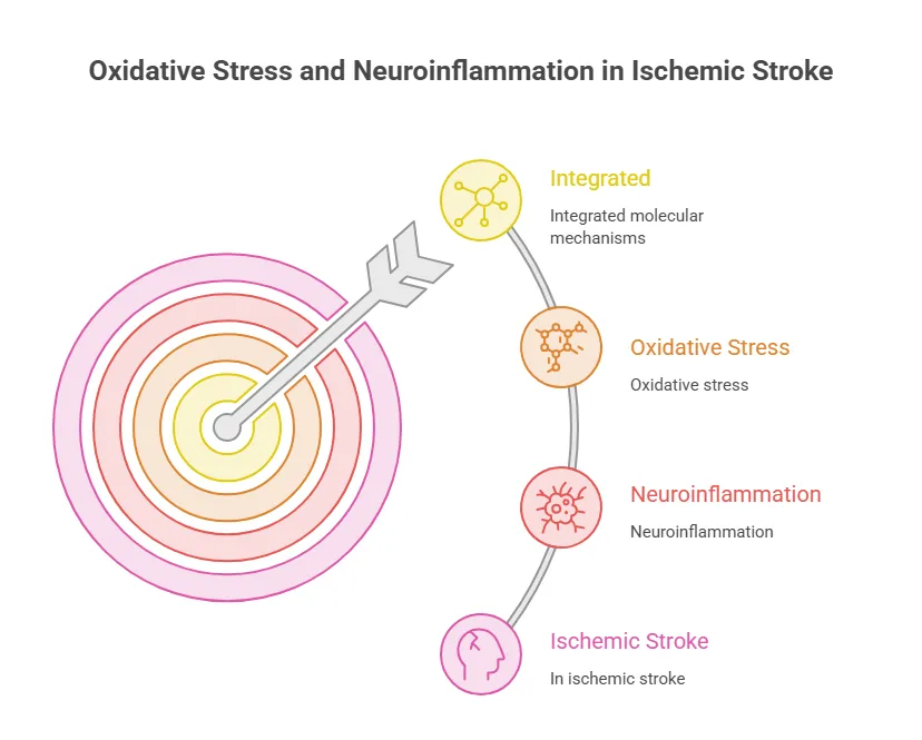
Figure 1: Integrated Molecular Mechanisms of Oxidative Stress and Neuroinflammation in Ischemic Stroke
6. Cellular and Molecular Consequences of Ischemic Injury
Ischemic injury impacts numerous cellular types and causes malfunctioning of brain functioning processes. Energy shortage, excitotoxic effect, oxidative trauma, and neuroinflammation may cause harm to nerves, change synapse transfer, and interfere with neurovascular complex work. The results depend on severity and duration of ischemic attack, amount of reperfusion, and ratio of damaging and reparative reactions. Some changes that occur in cells could be reversible, however, if the injury lasts too long there will be negative influence [20].
6.1 Neuronal Death and Synaptic Dysfunction
Neurons are especially susceptible to ischemia due to their need for energy and for maintaining the respective ionic and neurotransmitter concentrations. ATP depletion, glutamate excitotoxicity, calcium overload, and mitochondrial failure can serve as the triggers for multiple pathways of cellular death. Increased ROS production and lipid peroxidation cause additional destruction of the neuronal membranes and organelles, while inflammatory cytokines alter neuronal signaling and survival. Depending on the specifics of the cell context, the above-mentioned mechanisms can lead to apoptosis, necrosis, and other regulated forms of cell death. Synaptic dysfunction can take place long before about face, which is why the injury of neuronal cells could prove too late. The oxidative stress and inflammatory factors prevent normal neurotransmitter release, receptors’ functioning, and synaptic plasticity [21].
6.2 Blood–Brain Barrier and Neurovascular Unit Dysfunction
The blood-brain barrier (BBB) consists of endothelial cells, pericytes, astrocytes, and neurons as well as other supporting structures that work together to regulate blood supply and ensure that the brain functions properly. The process termed ischemia-reperfusion disrupts the normal interaction within this neurovascular unit by inducing the inflammatory process and oxidative stress conditions. Damage to the endothelial cells, which contain tight junction proteins, results in a loss of BBB integrity followed by the passage of fluid and blood components into the brain tissues. The increment in BBB permeability leads to edema of the brain and facilitates the infiltration of immune cells from the periphery. Inflammatory cytokines and growth factors released at the site of vascular injury produce further damages to the blood vessels. These can worsen microcirculation and consequently facilitate neuronal trauma. The treatments aimed at preserving the endothelial function and maintaining tight junctions need to be developed in order to prevent the dysfunction of the neurovascular unit after ischemic and traumatic injuries [22].
6.3 Neuroplasticity, Repair, and Recovery
Neuroplasticity is a characteristic of the brain that allows making new connections and altering neural pathways according to the necessity after an injury or during an experience. Following a stroke, remaining neurons can start to undergo changes in their synapses and structure, and their function might be restored. Various brain cells, and primary astrocytes, microglial cells, and the cells making up blood vessels help in the process of healing through the clearance of cellular debris and inflammation regulation. However, permanent oxidative stress and inflammation adversely affect the impact of the healing processes. If the level of cytokines produced is constantly high and mitochondria do not work properly, neurons can be damaged severely, and they will be unable to recover. Still, rehabilitation and some therapies aimed at maintaining cells alive can improve the recovery process, but the success of the healing process differs significantly for various people [23].
7. Therapeutic Targets and Emerging Treatment Strategies
Therapeutic approaches for ischemic stroke focus on the restoration of blood flow in the brain, keeping living brain tissue, and minimizing damage caused by reactions occurring in the brain due to oxidative stress and neuroinflammation. Reperfusion therapy is of great importance in the management of patients, although other means of protecting neurons and the neurovascular unit are also being studied as mentioned in (Fig.2). Among the targets of therapy are excessive production of reactive oxygen species (ROS) and inflammation, mitochondrial failure, and antioxidant defense mechanisms. The combination of treatment methods addressing interrelated mechanisms is promising but requires confirmation of safety, timing, and efficiency [24].
7.1 Antioxidants and ROS-Scavenging Agents
The general goal of antioxidant therapies is to decrease excessive accumulation of reactive oxygen species and prevent oxidative damage to cellular structures. Depending on endogenous defense mechanisms involving glutathione, superoxide dismutase, catalase, and glutathione peroxidase helps a lot in combating oxidative stress. Although promising results have been provided by studies on antioxidant therapy, its implementation into clinical practice encounters some challenges. During biological processes, reactive oxygen species play a significant role in normal cell signaling so that excessive suppression can interfere with the processes of the organism. Moreover, some of the compounds do not penetrate the blood-brain barrier or cannot have necessary concentrations in the ischemic tissue of the brain. Thus, future investigations should focus on finding out favorable moments for the use of such treatment options, improving the methods of brain delivery of such products as well as the choice of the type of impact [25].
7.2 Anti-Inflammatory and Immunomodulatory Therapies
Anti-inflammatory treatments focus on controlling the abnormal immune system and preventing the release of chemicals that cause secondary damage to the brain. Possible mechanisms for treatment are based on influencing the activity of the cytokines, interrupting inflammation mechanisms such as the NF-kb process, and blocking the function of processes linked to the inflammasome, e.g., NLRP3. Immunotherapy approaches can affect the activity of the microglia and the second immune cells. At the same time, neuroinflammation can have both positive and negative effects. The excessive inflammatory reactions cause the damage to the cells of the nervous system; at the same time, the immune response helps to clean debris, reorganize cut tissue, and heal it. Broad inflammatory reaction suppression can lead to the inability of recovery because of the lack of useful processes. Therefore, the ideal therapy should target specific inflammatory mechanisms during determined strokes [26].
7.3 Mitochondria-Targeted, Natural Compound, and Nanotechnology-Based Therapies
The main objective of mitochondrial therapies is to maintain the production of cellular energy while minimizing the generation of ROS by mitochondria. Some natural substances, namely resveratrol, curcumin, and sulforaphane have been evaluated as having antioxidant and anti-inflammatory properties that affect cellular signaling pathways. Unfortunately, the efficacy of these substances depends on their dose, as well as their availability and biological context, so their usefulness in the treatment of ischemic stroke is not well known. The use of nanotechnology-enabled delivery systems, e.g. liposomes, polymeric nanoparticles, and lipid-based carriers could improve drug stability, enable controlled drug delivery, and ensure drug delivery to the target area in the brain. This could result in increased delivery of antioxidants or anti-inflammatory agents with lesser systemic side effects. On the other hand, the penetration of nanoparticles through the BBB, targeted delivery, toxicity of nanoparticles, and consistency of their production remain areas of concern. The combination of mitochondrial protection with antioxidants and other anti-inflammatory approaches is a perspective area of research, however, the feasibility of routine clinical application will need thorough preclinical and clinical studies [27].
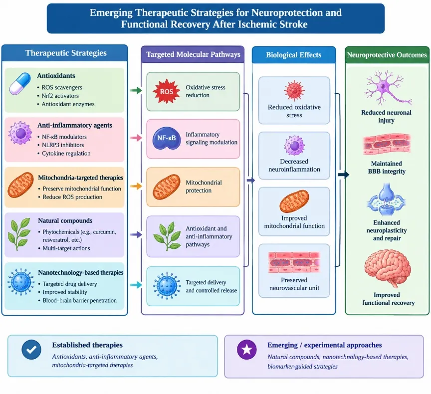
Figure 2: Emerging Therapeutic Strategies for Neuroprotection and Functional Recovery After Ischemic Stroke
8. Translational and Clinical Perspectives
In order to translate progress made in studies on oxidative stress and neuroinflammation into effective therapies for stroke, it is necessary to have trustworthy biomarkers, reproducible experiments, as well as clinical studies designed correctly. In particular, ischemic stroke differs regarding its causes, severity, brain regions, and response to reperfusion methodologies, which means that oxidative as well as inflammatory processes can have a different impact on different patients as mentioned in (Table 2). If these differences are defined, it can lead to more proper decision-making concerning patient selection, timing of treatment, and development of novel neuroprotection strategies [28].
8.1 Oxidative Stress and Inflammatory Biomarkers
Biomarkers for oxidative stress and neuroinflammation can help assess the severity of a certain illness, follow biological reactions and measure interventions' efficiency. Malondialdehyde, 4-hydroxynonenal and antioxidant capacity assessments are defined as oxidative stress biomarkers. In terms of inflammatory biomarkers, CRP, IL-6, TNF-α and other cytokines can be mentioned too. Nevertheless, the markers differ in their specificity and can be modified by the processes associated with systemic inflammation, comorbidities and complications not linked with ischemic stroke. At the moment, there is no one reasonable biomarker that could measure all parameters connected with oxidative stress and neuroinflammation in ischemic stroke. It should be acknowledged that combination of blood parameters with clinical data, neuroimaging and signs of vascular damage can help create a stronger scientific basis. Further research must be done on the reproducibility and accuracy of potential biomarkers, as only after validation of the mentioned studies [29].
8.2 Evidence from Preclinical and Clinical Studies
By making use of preclinical research with cellular systems and animal models, it has been possible to learn about the mechanisms involved in the production of reactive oxygen species, inflammation, dysfunction of mitochondrial systems, and pathology in neurons. In addition, the conclusions of the experiments were useful for the development of new antioxidant substances, inhibitors of inflammasomes, and other neuroprotective therapies. The results of preclinical studies consist of information on the volume of infarction, oxidative stress, levels of cytokines, neurons survived, and behavior of subjects. Nevertheless, the results obtained from animal experiments cannot be applied to humans without any reservations. There are significant differences between different species and different experimental models of stroke, conditions of treatment of various exposure to drugs. In addition, stroke in humans is a heterogeneous disease, and the treatment of stroke must be combined with the emergency treatment. Thus, the preclinical studies have to measure not only molecular changes but also the safety of treatments and functional outcomes [30].
8.3 Challenges and Future Directions in Precision Neuroprotection
To deliver oxidative stress and neuroinflammation therapies various risks pose restrictions. This includes narrow therapeutic opportunities during an acute stroke period, poor drug delivery to the affected brain area, unpredictability in terms of the inflammatory reaction, and the risk of side effects due to pathway inhibition. Moreover, the lack of accurate biometric systems that could signal the main injury process available only through the patient's body provides challenges for picking individual treatment approaches. Clinical practice requires the union of molecular profiling, neuroimaging, and clinical characteristics to elucidate patient groups with different reactions to the injury. Transcriptomics, proteomics, and metabolomics approaches may provide more information about the specific avenue under consideration. The other efficient future research direction is the combination of techniques aimed at mitigating oxidative injury and inflammation while securing the recovery process [31].
Table 2: Emerging Neuroprotective Therapies for Ischemic Stroke
| Therapeutic strategy | Mechanism | Major limitation |
|---|---|---|
| Antioxidants | Reduce ROS-mediated damage | Inconsistent clinical efficacy |
| Nrf2 activators | Enhance antioxidant defenses | Safety and optimal dosing |
| Anti-inflammatory agents | Modulate inflammatory signaling | May interfere with tissue repair |
| NLRP3 inhibitors | Suppress inflammasome activation | Limited clinical validation |
| Mitochondria-targeted therapies | Preserve mitochondrial function | Challenges in brain delivery |
| Natural compounds | Modulate oxidative stress and inflammation | Poor bioavailability |
| Nanotechnology-based therapies | Improve targeted drug delivery | Safety and regulatory challenges |
| Biomarker-guided approaches | Support patient stratification | Limited biomarker validation |
CONCLUSION
Neuroinflammation and oxidative stress are linked phenomena that aid in neuronal damage leading to secondary injuries after ischemic stroke. These mechanisms involve excessive oxidative stress production, mitochondrial disturbances, release of pro-inflammatory cytokines, and many more. Targeting pathways such as NF-κB, Nrf2, or the NLRP3 inflammasome could provide effective neuroprotective options for the future. However, validation of biomarkers, proving safety and efficacy of treatments, and development of targeted therapies to reduce the extent of damage caused by inflammation and oxidative stress must be continued.
REFERENCES

Kummari. Sathish, Dr. Thripuramallu J. Mohanrao, Neuroinflammation and Oxidative Stress in Ischemic Stroke: Molecular Mechanisms and Therapeutic Targets, Int. J. of Pharm. Sci., 2026, Vol 4, Issue 11, 387-399. https://doi.org/10.5281/zenodo.23923378
 10.5281/zenodo.23923378