

We use cookies to ensure our website works properly and to personalise your experience. Cookies policy
1,2,4,5,6Department of Pharmaceutics, Himalayan Pharmacy Institute, Majhitar, East Sikkim, 737136, India.
3Department of Pharmaceutics, Siliguri Institute of Pharmaceutical Sciences, Siliguri, West Bengal, 735134, India.
Promise for improved therapeutic results in the area of drug delivery to the eyes. Because drugs are quickly lost before they reach the cornea, standard ophthalmic formulations struggle to deliver drugs to the eye, an essential organ. With a focus on cutting-edge dosage forms, such as in situ gels, this review investigates novel drug delivery strategies for ocular administration. By extending the duration of drug interaction in the eyes, this technique seeks to address the problems with conventional delivery methods' bioavailability. The paper further details in situ gelation techniques, emphasising ion-activated, temperature-dependent, and pH-triggered systems. It investigates the creation of in situ gels using excipients such as polymers and solubilising agents. A full description of the commonly utilised polymers and their uses in ocular medication delivery is provided. Research on the use of nanoparticles in this gelling method to deliver drugs to the eyes. Techniques for assessing in situ gels are described, such as pH measurement, rheological investigations, drug content analysis, in vitro gelation, accelerated stability tests, and FTIR analysis. The benefits and uses of in situ gels are examined, as well as their drawbacks. The advantages of innovative ocular drug delivery systems—especially in situ ophthalmic systems, which provide regulated and prolonged drug release—as well as the prospects for in situ gels are highlighted in the review's conclusion.
The eye is a complex and unique part of the human organs that has been considered as the window to the human soul. Broadly, the human eye is divided into two segments that are anterior and posterior segments [1]. The specific disease conditions of the eye are associated with each of these broad segments. For instance, conjunctivitis, glaucoma, blepharitis, and cataract are some of the diseases that affect the anterior segment of the eye, while diabetic retinopathy and age-related macular degeneration are known to affect the posterior segment.[2]
Due to the unique structure of the eye, which inhibits the entry of drug molecules into the desired site, the ophthalmic delivery of the drug has been one of the most challenging tasks for a pharmaceutical scientist. Eye drops accounts for more than 90% of ophthalmic preparations on the markets. However, they are washed away from the eye and results in low ocular bioavailability (<5%) after topical administration by different elimination mechanisms.[3] This elimination process includes tear turnover, nasolacrimal drainage, protein binding, systemic absorption, enzymatic degradation and complex penetration barriers (Corneal Barrier, Blood Aqueous Barrier (BAB), and Blood Retinal Barrier (BRB). [4]
The eye serves as the body's most crucial organ. Currently, 90% of available ophthalmic formulations consist of typical dosage forms. The primary challenge faced is the rapid loss of drugs before reaching the cornea due to nasolacrimal drainage (Lynch et al.2020). To increase the availability of drugs in the eyes, significant efforts are being dedicated to the development of new drug delivery systems for ocular administration. Recent research in this field has created systems that prolong the contact time of the drug in the eye. Innovative dosage forms such as in situ gels, liposomes, niosomes, etc. are being explored (Shah et al., 2022). [5]
Furthermore, drugs that are administered systemically to exert their action in the ophthalmic system are also known to have poor access to the eye tissue. [6] Intravitreal and periocular routes are recommended in order to deliver drugs to the posterior part of the eye. However, there are disadvantages associated with these routes, such as the frequent intravitreal injections could be painful, thus affecting patient compliance. The periocular route is easy for administration, but the static and dynamic barriers constitute a problem. [7]
The low bioavailability of medications from the conventional delivery system results from a great extent of precorneal drug loss by nasolacrimal drainage. The rapid clearance of the topically applied drug into the eye often results in a short duration of pharmacological activity and, therefore, the need for a frequent dosing regimen. Moreover, 50%−100% of an instilled dose could undergo systemic absorption through drainage via the nasolacrimal duct. This could lead to a possible increased risk of unwanted systemic toxic effects.[8]
In last decades, various delivery systems such as using chemical permeability enhancers [6], prodrugs [9] stimuli-responsive in-situ gel, [10] and drug delivery carriers such as liposomes [11] and nano-or microparticles,[12] Niosomes,[13] dendrimers and microneedles [14] have been developed to increase ocular residence time, drug penetration across the ocular barriers and ophthalmic bioavailability. In-situ gelling system is one of the promising approaches to improve the retention time of drugs on the ocular surface. After instillation of the aqueous solution containing stimuli-responsive polymers such as pH-sensitive polymers, thermosensitive polymers, and ion-sensitive polymers, the viscous and mucoadhesive gels are formed on the eye surface,[15] subsequently, ocular retention time and ocular bioavailability of the ophthalmic drugs are improved. Therefore, in this review, we summarized and discussed the most recent research innovations in stimuli-responsive in-situ gelling system for ocular drug delivery system.
In the last decade, the development of in situ gelling drug delivery systems has gained an increasing attention in the scientific community. The majority of these systems presents the peculiarity to be in a soul-state before administration and to undergo gelation into the body. As a consequence, they are characterized by ease of administration, prolonged residence time and sustained drug release at the administration site, with a reduction in administration frequency and an improvement in patient compliance and comfort. Among the reasons for the great success of these formulations, there is the fact that they can be administered through various routes to achieve a local or a systemic etc of the drug loaded. Moreover, they can be successfully used as vehicles for nano- and micro-drug-delivery systems. [17,18]
OCULAR IN SITU GEL:
In situ gel-forming systems are drug delivery systems that are in solution form before administration in the body but, once administered, undergo gelation in situ to form a gel triggered by external stimuli such as temperature, pH, etc and release the drug in a sustained or controlled manner. This novel concept of producing in situ gel was suggested for the first time in the early 1980s. Gelation occurs via the cross linking of polymer chains that can be achieved by covalent bond formation (chemical cross-linking) or non-covalent bond formation (physical cross-linking). In situ gel-forming systems can be described as low-viscosity solutions that undergo phase transition in the conjunctival cul-de-sac to form viscoelastic gels due to conformational changes of polymers in response to the physiological environment. The rate of in situ gel formation is important because, between instillation into the eye and before a strong gel is formed, the solution or weak gel is produced by the fluid mechanics of the eye.[19]
Ophthalmic in-situ gelling comprises environmentally sensitive polymers that will be altered structurally by small changes in specific conditions like pH, temperature and ionic strength in the environment. In-situ forming gels are liquids during instillation into the eye and then undergo rapid gelation in the cul-de-sac of the eye to form viscoelastic gels in response to environmental changes; lastly release the drug slowly under physiological conditions.[20] Consequently, the residence time of the gel formed in situ will be extended, and the drug is released in a sustained manner, which leads to enhanced bioavailability, minimised systemic absorption and reduced dosing frequency, resulting in improved patient compliance.[21] Furthermore, some other potential advantages such as a simple manufacturing process, ease of administration, and delivery of an accurate dose have been exhibited by in situ gelling systems. [22]
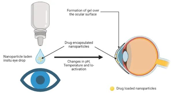
FIG: Ocular in situ gel formation from ocular in situ eye drop.[22]
MECHANISM OF OCULAR IN SITU GEL:
Temperature-dependent systems: Temperature-sensitive in situ gels are probably the most commonly studied class of environment-sensitive polymer systems in drug delivery research.[23] The use of biomaterials whose transitions from sol to gel are triggered by an increase in temperature is an attractive way to approach in situ gel formation for ophthalmic drug delivery. Three main strategies exist in the design of thermo-responsive sol-gel polymeric systems. For convenience, temperature-sensitive in situ gels are classified into negatively thermo-sensitive, positively thermo-sensitive, and thermally reversible gels. Negative temperature-sensitive in situ gels have a lower critical solution temperature (LCST) and contract upon heating above the LCST. Polymers with a low critical temperature (LCST) that transition between ambient and physiologic temperatures are used for this purpose. The formulation is liquid at room temperature (20-25°C), which undergoes gelation in contact with body fluid (35-37°C). Temperature increases the degradation of polymer chains, which leads to the formation of hydrophobic domains & transition of an aqueous liquid to an in-situ gel. [24-28]
pH-triggered in situ gelling system: pH-triggered in situ gelling systems are solutions that, upon exposure to the pH of the lachrymal fluid, convert into the gel phase, e.g. such as cellulose acetate phthalate and Carbopol. [29] The backbone of pH-sensitive polymers is made up of either weakly acidic or basic groups that, in reaction to a change in pH, either release or receive free protons. Electrostatic, hydrophobic, and hydrogen bonding interactions occur at a particular pH, which causes inter-diffusion and a conformational shift in the polymer that causes swelling. Therefore, pH triggers the sol-to-gel transition. [30] Polyacrylic acid (PAA, Carbopol 940), polycarbophil, and cellulose acetate phthalate (CAP) are the most widely utilised pH-responsive polymers in ophthalmic preparations. [31]
PHYSIOLOGY OF THE EYE: [16]
An eye is a spherical structure with a wall made up of three layers: the outer part, the sclera; the middle parts choroid layer, ciliary body and iris; and the inner section, the nervous tissue layer, retina. The sclera is a tough, fibrous coating that protects the inner tissues of the eye, which is white except for the transparent area at the front; that is, the cornea allows light to enter the eye. The choroid layer, situated in the sclera, contains many blood vessels that modify at the front of the eye as the pigmented iris, the coloured part of the eye (blue, green, brown, hazel, or grey). The clear, transparent bulge, the cornea situated at the front of the eye and conveys images to the back of the nervous system.
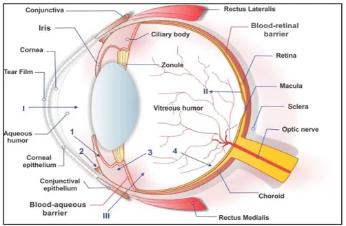
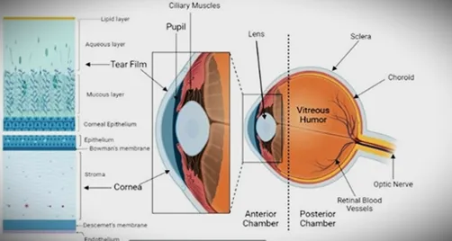
FIG: Schematic diagram and Anatomy of the eye.[16]
The adult cornea has a radius of approximately 7-8mm that is a vascular tissue to which nutrients and oxygen are supplied via lachrymal fluid and aqueous humour, as well as from blood vessels at the junction between the cornea and sclera. The cornea is made of five layers: epithelium, bowman’s layer, stroma, Descemet’s membrane and endothelium, which are the main pathways of drug permeation to the eye. The main barrier to drug absorption into the eye is the corneal epithelium, in comparison to many other epithelial tissues (intestinal, nasal, bronchial, and tracheal) that are relatively impermeable. The epithelium is stratified squamous (5-6 layers of cells) with a thickness of around 50-100 μm. The basal cells are packed with tight junctions, forming not only an effective barrier to dust particles and most microorganisms, but also limiting drug absorption. The transcellular or paracellular pathway is the main pathway for drugs to penetrate across the corneal epithelium. Lipophilic drugs choose the transcellular route, whereas the hydrophilic one chooses paracellular pathway for penetration (passive or altered diffusion through intercellular spaces of the cells). [36]
APPLICATIONS OF IN SITU GEL:
1. In situ gels are utilised in the eyes for a variety of treatments because of their capacity to form a gel upon contact with the ocular surface. Because they offer continuous release of medication, these gels can increase patient compliance, decrease the frequency of doses, and improve drug bioavailability. [39]
2. Oral Drug Delivery: The in-situ gel developed for oral delivery of metformin HCl provided a sustained release of the medicament. When these gels are exposed to gastrointestinal physiological circumstances, they can change from a solution or liquid state to a gel, which prolongs drug release and enhances therapeutic efficacy. In situ gel kept floating for 8 h, while slowly releasing the medicament with a cumulative % release of approximately 80% during 8 h. [40]
3. Ocular Drug Delivery System: In ocular delivery systems, natural polymers like gellan gum, alginic acid and xyloglucan are mostly used. Local ophthalmic drug delivery has been used for various compounds such as antimicrobial agents, anti-inflammatory agents and autonomic drugs used to relieve intraocular tension in glaucoma.4 Problems encountered with conventional drug delivery systems are poor bioavailability and low therapeutic response due to high tear fluid turnover and dynamics causing rapid elimination of the drug from the eye. Hence, to overcome this problem, in situ gels were developed. To further increase the bioavailability, viscosity enhancers like HPMC, CMC, Carbomers, and PVA are used, which prolong the precorneal residence time and hence improve bioavailability. Also, they are easy to manufacture. Penetration enhancers such as preservatives, chelating agents and surfactants are also used to enhance corneal drug penetration. Much of the significance in the pharmaceutical application of gellan gum has concentrated on its application on ophthalmic drug delivery. A drug used in an ophthalmic in situ gelling system is Indomethacin. [41]
4. Nasal Drug Delivery System: For nasal in situ gel system, gellan gum & xanthan gum are used as in situ gel forming polymers.2 An in-situ gel system for nasal delivery Mometasone furoate was furoate was developed and evaluated for its efficacy for the treatment of allergic rhinitis.[2] In-situ gel was found to inhibit the increase in nasal symptoms compared to marketed preparation Nasonex (Mometasone furoate suspension 0.05%) in situ gel drug delivery systems are suitable for protein and drug delivery. [42]
5. Wound Healing: By administering medicinal agents straight to the site of injury, in situ gels can aid in healing through the use of L-proline nanoparticles and oxygen nanobubbles. They are capable of preventing external infections while facilitating wound healing. [40]
LIMITATIONS:
i. Formulation viscosity plays a critical role; if it’s too high, the gel can cause ocular irritation, whereas excessively low viscosity prevents adequate gelation and adhesion to the ocular surface, impacting sustained release capabilities. [48]
ii. In situ gelation depends on specific triggers like temperature or pH; any deviation from these conditions can hinder gel formation and affect performance. The choice of polymers and additives influences the critical gelling temperature and, thus, the efficacy of the gel. [44]
iii. The choice of polymer is crucial; it should be both biocompatible and degrade properly (Chowhan & Giri, 2020). Biocompatibility concerns may arise with some materials used in in situ gels, potentially leading to irritation or toxicity. Hence, a careful evaluation is essential during the development phase to ensure safety, as different polymers can affect ocular tolerability differently.[49]
iv. Certain permeation enhancers used in formulations can potentially lead to ocular toxicity if not carefully controlled. The optimal concentration for the permeation enhancer was reported to be 20% when studied with other parameters like entrapment efficiency (Shukr et al.,2021). They may increase corneal hydration levels and affect blinking frequency, underscoring the need for cautious application. [60]
RECENT ADVANCEMENTS IN IN SITU GEL FORMULATION:
Ocular medication delivery could be revolutionised via in situ gelling. The problems with eye drops and ointments for the delivery of ocular medications are resolved by this technique. In situ gels are transformed into a gel on the ocular surface. They are sensitive to temperature. This increases the bioavailability and release of the medication. New preparations with even greater advantages, like better patient compliance and less frequent doses, have been made possible by recent developments in in situ gel technology. The creation of mucoadhesive in situ gels, which adhere to the surface of the eye for an extended period of time and enable prolonged drug release and enhanced therapeutic advantages, is one of the most significant developments. Using in situ gels, which release medication in reaction to physiological cues like pH or temperature, is another innovation. In general, in situ gel technology presents exciting new methods for administering eye medications and enhancing patient outcomes. [62]
One of the challenges in front of today's pharmaceutical industry centres on coming up with efficient treatment options that are readily tolerable to physicians and patients. Delivery systems should also have contribution in better therapeutic outcome is they are going to provide possible alternatives to pharmaceuticals currently delivered by other routes. In situ gelling formulations are one of the challenging drug delivery systems. [61] Various biodegradable polymers are used for formulation of in situ gels, but there are manufacturing problems, difficult processability, use of organic solvents for their preparation (particularly for synthetic polymer-based systems), burst effect and irreproducible drug release kinetics. Natural polymers suit the characteristics of an ideal polymer but batch-to-batch reproducibility is difficult; hence synthetic polymers are used. But all these problems are being overcome day by day, and these in situ forming gels are becoming a major tool for site-specific delivery of drugs.
Multiple stimuli driven in situ gelling system: Khan et al. (2015) developed and investigated a novel pH- and ion-triggered gelling technique for sustained release in ocular medication distribution employing sparfloxacin encapsulated in methylcellulose and sodium alginate. The preparation was in the sol phase at pH 4.7, but it quickly transitioned to the gel state at pH 7.4. In contrast to ocular droplets, the sparfloxacin release from ocular in situ preparations was sustained for 24 h. In comparison to traditional eye drops, the generated in situ gel's ex vivo corneal penetration test on goat eyes showed noticeably greater permeability. [57] Yu et al. (2017) used poloxamer and carboxymethyl chitosan to create a nepafenac in situ gel. The PEO-PPO PEO block copolymer experienced a changeable sol–gel transformation when the pH and temperature were changed at very low concentrations. The preparation was shown to be non-toxic to corneal epithelial cells at lower doses using the cell counting kit-8 method.[58]
In situ gelling system loaded with nanoparticles: Liu et al. (2016) used a temperature-sensitive gelling agent and cationic nanostructured phospholipid carriers to develop an ocular nano-gel loaded with curcumin. Researchers looked at corneal penetration, ophthalmic irritation, preocular retention, and in vitro release. Drug pharmacokinetics in aqueous humour were evaluated using microdialysis. Compared to curcumin ocular drops, curcumin nano-gel had a greater AUC, indicating better absorption. The non-irritating optimised in situ gelling ocular insert exhibited enhanced bioavailability, a higher Cmax, and a noticeably delayed Tmax. [60] A microsphere-encapsulated ofloxacin ion-triggered in situ gel was developed by Al-Khateb et al. (2016). Compared to commercial eye therapies, an in situ gel containing ofloxacin microspheres exhibited greater absorption in rabbit studies. The prolonged duration of action of ofloxacin-encapsulated microspheres in in situ gel formulation minimises the need for repeated administration and increases patient compliance. [59]
Additionally, an ion-activated in situ gel containing ofloxacin (OFX) loaded with microspheres was developed. When compared to commercial OFX eye drops, in vivo data in rabbits showed that OFX-loaded microspheres in-situ gel might increase the relative bioavailability by 11.7 times. Additionally, it is believed that the prolonged duration of action of OFX-loaded microspheres in-situ gel preparation prevents frequent administration, which enhances patient compliance. [61]
Compared to commercial eye drops, chitosan nanoparticulate in-situ gel performed better. [60] An enhanced ocular retention time was demonstrated by an in-situ gel loaded with levofloxacin nanoparticles. Levofloxacin-containing PLGA nanoparticles were created by Gupta et al. and added to chitosan in-situ gel. In rabbits, ocular retention was assessed using gamma scintigraphy. Compared to commercially available formulations, in situ gel, and nanosuspension alone, the new nanoparticle-laden in situ gel formulation showed a slower rate of clearance and was kept at the corneal surface for a longer period of time. [62]
Morsi and associates created a Mucoadhesive ion-sensitive in situ gel using an acetazolamide (AZA)-loaded nano-emulsion (NE). [61] Xanthan gum, hydroxypropyl methylcellulose (HPMC K4 M), or Carbopol 940 as a mucoadhesive agent was combined with 0.3% w/w GG, an ion-responsive anionic polysaccharide. Because of its poor aqueous solubility and low corneal permeability, which contribute to its low ocular bioavailability, loading AZA in a NE is the best method for topical delivery. In order to prepare PIL-loaded in situ gelling mucoadhesive formulations for ocular drug delivery, the usage of methacrylate GG derivatives was also studied; the methacrylation enhanced the mucoadhesive capabilities of GG. Even if the polysaccharide with the least amount of modification performed the best, in vivo investigations demonstrated that methacrylation increased the GG mucoadhesiveness. [62]
METHODS OF PREPARING IN SITU GEL:
The required quantity of polymers and co-polymers is dispersed in water to generate the polymeric solution. The final solution contained preservatives like benzalkonium chloride, buffering agents such as NaOH and citric acid to adjust the solution to the required PH, and osmolality agents such as NaCl to maintain the osmotic pressure according to the eye. The final volume of the resultant in -situ gel eye drop is maintained using the distilled water [45,46]. These formulations with stimuli-responsive polymers are sensitive to pH, temperature, and ion concentration form gel instantaneously upon ocular instillation. Combining multiple stimuli-responsive polymers in one formulation may improve gelation results.
Different methods have been reported for preparation of in situ gel. Some of these methods involves cross linking of co-monomers using cross linking agent or polymerization while some other methods involve cross linking of polymers by irradiation or by chemical means.
a) Solution polymerization or cross linking:
In this method, multifunctional cross-linking agents are mixed with ionic or neutral monomers. The polymerization is initiated thermally or by UV light or by redox initiator system. Solvent present minimizes the temperature control problem as well as serves as heat sink. The finished hydrogels require washing with distilled water for removal of the unreacted materials, cross linking agent and the initiator. One of the best examples of this method is poly (2-hydroxyethyl methacrylate) hydrogels from hydroxyethyl methacrylate, using ethylene glycol Di methacrylate as cross-linking agent. [47]
b) Suspension polymerization:
This method is widely used for preparation of spherical hydrogel microparticles with size ranging from 1µm to 1mm. In this method, the monomer solution is dispersed in the non-solvent forming fine droplets, which are stabilised by addition of stabilizer. [47] The initiation of the polymerization is by thermal decomposition of free radicals. The prepared microparticles require further washing to remove unreacted monomers, cross linking agent and initiator. [47] Hydrogel microparticles of poly (vinyl alcohol) and (hydroxyl ethyl methacrylate) have been prepared by suspension polymerization method.
c) Polymerization by irradiation:
High energy radiations such as gamma and electron beam are used to prepare the hydrogels of unsaturated compounds. The irradiation of aqueous polymer solution results in the formation of radicals on the polymer chains, which results in formation of macroradicals. [47] Recombination of the macroradicals on different chains results in the formation of covalent bonds, and finally a cross-linked structure is obtained. [47] Polymerization macroradicals may interact with oxygen during radiation, that's why radiation is performed in an inert atmosphere using nitrogen or argon gas. [47] Example of this method include poly (vinyl alcohol), poly (ethylene glycol) and poly (acrylic acid).
d) Chemically crossed linked hydrogels:
Polymers which contain functional groups like OH, -COOH, -NH2 are soluble in water. Due to presence of such functional groups on the polymer chain, it can be used to prepare hydrogels by forming covalent linkages between polymer chains and complimentary reactivity, such as amine-carboxylic acid, isocyanate -OH or -NH2 or by Schiff's base formation. Glutaraldehyde can be used as a cross-linking agent for preparation of hydrogels of polymers containing -OH groups such as poly (vinyl alcohol) and also polymers containing amine groups (albumin, gelatine, polysaccharides).[47] This cross -linking agent reacts with the functional groups present on the polymer via addition reaction. Since cross linking agents are highly toxic, unreacted agents have to be extracted. Also, the reaction has to carried out in organic solvents since water can react with the cross-linking agent. The drugs are loaded after the formation of hydrogel; hence the release is typically first order. [46]
e) Physically cross -linked hydrogel:
Almost all of the covalent cross -linking agents are known to be toxic, even in small traces. Hence to overcome this problem and to avoid a purification step, hydrogels are prepared by reversible ionic cross linking.[47] Chitosan, a polycationic polymer reacts with positively charged components, either ions or molecules forming a network through ionic bridges between the polymeric chains. In case of anionic molecules, phosphate containing groups, particularly sodium triphosphate is widely studied. Ionic cross linking is an effortless and easy-going procedure. Compared to covalent cross linking, no auxiliary molecules such as catalysts are required. [47] Chitosan is also known for forming polyelectrolyte complex with poly (acrylic acid).
EVALUATION OF IN SITU GEL:
a. Clarity: The clarity of the formulated solution is determined by visual inspection under black and white background. [47]
b. Texture analysis: The firmness, consistency and cohesiveness of hydrogels are examined using texture analyser which significantly indicates the syringe ability of sol so that the formulation can be easily administered in vivo. Higher values of adhesiveness of gels are required to maintain an intimate contact with surface. [44]
c. pH of gel: Formulation is taken in a beaker and 1ml NaOH added dropwise with continuous stirring, pH is checked by using pH meter. [41]
d. Sol-Gel transition temperature and gelling time: For in situ gelling systems with thermos-reversible polymers, the sol-gel transition temperature may be defined as the temperature at which the phase transition of sol meniscus is first noted when kept in a sample tube at a specific temperature and then heated at a specific rate. Gel formation is indicated by a lack of movement of meniscus on tilting the tube. [46]
e. . Depending on the mechanism of gelling of the gelling agent used, a defined amount of gel is prepared in a beaker form the sol form. This gel containing beaker is raised at a definite rate, so pushing a probe slowly through the gel. The changes in the load on the probe are measured as a function of depth of immersion of the probe below the gel surface. [40]
f. Rheological studies: This is one of the important parameters to be evaluated for in situ gels. Viscosity and rheological properties of in situ gelling drug delivery systems are assessed using Brookfield rheometer, or some other viscometers like Ostwald's viscometer. The viscosity of in situ gelling systems should be such that no difficulties are encountered during their administration by the patient, especially in parenteral and ocular administration. The formulation should have viscosity of 5-1000 mPas. [43]
g. High-performance liquid chromatography: The HPLC system if used in reversed phase mode.2 Analysis is performed on a Nova pack C18 packed column (150 mm length X 3.9 mm).[43]
h. Drug-polymer interaction study and thermal analysis: Interaction studies are performed with Fourier Transform Infra-Red (FTIR) spectroscopy. During gelation process, the nature of interacting forces can be determined using this technique by employing KBr pellet method. Thermo -gravimetric analysis (TGA) can be used for in situ gelling system to determine the percentage of water in hydrogel. Differential scanning calorimetry (DSC) used to observe if there are any changes in thermograms as compared to pure active ingredients used for gelation.[44]
i. In vitro drug release studies: For the in-situ gel formulations administered by oral, ocular or rectal routes, the drug release studies are done by using plastic dialysis cell. The cell is made up of 2 half cells, donor compartment and receptor compartment and both these compartments are separated with the help of cellulose membrane. The sol form of the formulation is sited in the donor compartment. The assembled cell is then shaken horizontally in an incubator. The total volume of the receptor solution is analysed for the drug release using analytical methods. For injectable in situ gels, the formulation is sited into vials containing receptor media and placed in a shaker water bath at required temperature and oscillation rate, samples are withdrawn periodically and analysed. [45]
j. Antimicrobial activity: Antimicrobial studies are carried out to determine the biological activity of sol-gel system against microorganisms. This is done using agar diffusion medium employing 'Cup Plate Techniques'. The microbial growth of bacteria is measured by concentration of antibiotic and compared with that produced with known concentrations of standard preparation of antibiotic and carried out the microbial assay serial dilution method is employed. [45,46]
k. Sterility Testing: Sterility testing is carried out as per IP 1996. The formulation is incubated for not less than 14 days at 30-35°C in the fluid thioglycolate medium to find the growth of bacteria and at 20-25°C in Soya casein digest medium to find the growth of fungi in formulations. [47]
l. Accelerated stability studies: Formulation is replaced in amber coloured vials and sealed with aluminium foil for the short term accelerated study at 40 ± 2°C and 75 ± 5% RH as per International Conference of Harmonization (ICH) Guidelines. Sample is analysed at every month for clarity, pH, gelling capacity, drug capacity, drug content, rheological evaluation and in vitro dissolution. [43]
FUTURE PROSPECTIVE:
In-situ gel technology is an innovative drug delivery system that employs sol to gel transitions upon administration, enabling controlled and sustained drug release. [48] These gels are made from crosslinked polymer networks and have numerous applications in drug delivery and disease treatment. However, further research is needed to address issues such as drug degradation. The technology can be customised to meet individual patient needs, enhancing patient compliance.
Additionally, it can carry multiple drugs simultaneously, potentially transforming the treatment of complex diseases. Future uses of in-situ gels include the delivery of proteins, peptides, and drug molecules, protecting them from degradation by providing targeted and localised release.[49]
The in-situ gelling system for ocular drug delivery is a well-studied strategy that can prolong precorneal residence time and provide sustained drug release, improving ocular bioavailability and therapeutic efficacy while reducing systemic absorption and toxicity. Exploring the integration of different drug delivery approaches, such as nanoparticles loaded into in-situ gels, is an attractive strategy for enhancing ocular drug delivery. [48]
Given the eye's sensitivity, safety is a crucial parameter for ocular formulations. Most studies reviewed indicate that in-situ gels do not cause significant cytotoxicity or irritation. However, further research is needed to assess the potential toxicity of repeated and long-term use, as well as the materials used in nanoparticle-based systems. Increased viscosity in in-situ gels can cause issues like blurred vision and discomfort, leading to faster elimination due to reflex tears and blinking. Therefore, careful control of viscosity is essential during formulation design and optimisation to mitigate these limitations. [48]
Future strategies should focus on developing formulas with multiple ingredients, such as traditional Chinese medicine, which uses a multi-target approach. Additionally, there is an expectation for new and more reliable in-situ forming polymers responsive to biochemical markers associated with eye diseases. Improved ocular permeation over longer durations would provide sufficient contact time for managing ocular diseases. The simple and economical preparation methods will facilitate industrial scale-up. These formulations can be further processed for large-scale batches and pharmacological evaluation through in-vivo studies. The future of in-situ gels is promising, with potential for innovative drug delivery solutions and applications across various industries, including cancer and ulcer treatment. [49]
In situ gels can also be utilised as templates for tissue engineering and cell transplantation by releasing growth factors and other bioactive molecules in a controlled manner to promote tissue regeneration. Combining in-situ gels with 3D printing technology could lead to patient-specific drug delivery systems. [50] Different types of stimuli-responsive gels, such as temperature, pH, and ionic, can be developed for remote drug release control. Incorporating in-situ gels into implantable devices like wound dressings can provide sustained drug release at target sites. The in-situ gel system is a dynamic field with ongoing research, and further technological advancements may lead to new applications beyond those currently envisioned. [51]
CONCLUSION
In conclusion, the drawbacks of existing drug delivery techniques may be mitigated using in situ gel technology for ocular medicine delivery. In situ gels decrease the frequency of doses, increase bioavailability, and extend drug release. Benefits include improved clinical results and patient acceptance. There are still issues in this field despite major advancements. A significant difficulty is in situ gelling techniques that sustain ideal drug concentration levels for extended periods of time. Another difficulty is optimizing formulation parameters for safety and stability of in situ gels. Stimuli-responsive devices that respond to changes in the ocular environment could be developed using in situ gel technology for the administration of ophthalmic medications. The effectiveness and safety of in situ gel can be enhanced by nanotechnology and other cutting-edge drug delivery techniques. In situ formulation technology has the potential to improve the quality of life for many individuals with ocular diseases and revolutionize the distribution of ophthalmic medications. In situ gels may emerge as the preferred ocular drug delivery technique as research develops.
Despite all of the difficulties with ocular drug delivery, numerous creative solutions have been created in recent years to address the issues with traditional ophthalmic preparations. One of the most promising and well researched methods for extending precorneal resident duration and providing sustained release drug administration is the in-situ gelling system. This could enhance ocular bioavailability and therapeutic efficacy while lowering systemic absorption and toxicity. Additionally, in-situ gel may increase patient compliance because of its capacity to sustain drug release and reduce the frequency of delivery. Different stimuli-responsive polymers with great sensitivity to changes in pH, temperature, and ion concentration are used in in-situ gel formulation. Nonetheless, it is well recognized that combining two or more stimuli-responsive polymers in one formulation results in better therapeutic efficacy and increased compliance. Additionally, investigating the integration of several drug delivery methods (such as nanoparticle-loaded in-situ gelling) to create in-situ gel has proven to be an appealing way to enhance the ocular drug delivery system. The safety of ophthalmic formulations is crucial since the eye is the most vital and delicate portion of the body. The majority of the cytotoxicity and irritability investigations that were part of this review demonstrated that the use of in-situ gel did not result in any appreciable changes or indications of toxicity. However, more research is needed to assess the potential toxicity resulting from long-term, repeated applications and materials used to create nanoparticles in nano-gel systems. Additionally, the increased viscosity of in-situ gel may result in certain limits, such as blurred vision and discomfort for the patient, which could accelerate removal owing to reflex tears and blinks. Therefore, in order to minimize the restrictions to a manageable level, important control of the viscosity should be taken into account throughout the design and optimization of the in-situ gel formulation. Currently, the majority of ophthalmic in-situ gels are only approved for formulations with a single active component. Future research should focus on developing more effective tactics for formulas with numerous constituents, such as Traditional Chinese Medicine, which uses a multi-target approach to create their activity. Finally, we anticipate the development of novel and more dependable in-situ forming polymers in the future that might be sensitive to certain biochemical indicators linked to eye diseases.
REFERENCES






Pragya Rani, Pragya Baghel, Debdatta Sarkar, Pempo Lhamu Bhutia, Priya Pandey, Rozila Rai, Ocular in Situ Gel-Based Drug Delivery System: Recent Development, Int. J. of Pharm. Sci., 2026, Vol 4, Issue 11, 132-145. https://doi.org/10.5281/zenodo.23876886
 10.5281/zenodo.23876886