

We use cookies to ensure our website works properly and to personalise your experience. Cookies policy
Department of Pharmaceutics, KMCH college of Pharmacy, Coimbatore, 641048, Affiliated to the Tamil Nadu Dr. M.G.R Medical University, Chennai, 600032, Tamil Nadu, India
Antimicrobial resistance (AMR) among Gram-negative pathogens has become a serious global health concern, driven by mechanisms such as reduced membrane permeability, efflux pump overexpression, enzymatic drug inactivation, biofilm formation, and quorum sensing–mediated virulence. Conventional antibiotics are increasingly ineffective against priority pathogens including Pseudomonas aeruginosa, Acinetobacter baumannii, Klebsiella pneumoniae, and Escherichia coli, highlighting the urgent need for innovative therapeutic strategies. Phyto-antibiotic synergistic nanoformulations offer a promising solution to this challenge. This approach integrates plant-derived bioactive compounds—such as flavonoids, polyphenols, and alkaloids—with antibiotics using advanced nanocarrier systems like polymeric nanoparticles, liposomes, and lipid-based carriers. These systems enhance drug stability, improve membrane penetration, inhibit efflux pumps, and disrupt biofilms, thereby restoring and amplifying antibiotic efficacy. Preclinical studies have demonstrated strong synergistic outcomes, including reduced minimum inhibitory concentrations and improved bacterial clearance. Additionally, this review outlines key preclinical evaluation methods and explores potential applications in treating infections such as pneumonia, sepsis, urinary tract infections, and diabetic wounds. However, challenges related to nanotoxicity, large-scale production, and regulatory approval remain significant barriers. Future research focusing on stimuli-responsive systems, CRISPR-based interventions, and AI-driven personalized therapies may further strengthen this emerging strategy against AMR.
Figure.1 Phytoactive Nano-Antibiotic Co-Delivery Systems for Disrupting AMR Mechanisms in Gram-Negative Bacteria
Antimicrobial resistance (AMR) has become one of the most significant global health crises, with the estimated number of deaths totaling to 4.95 million annually in 2019, with almost 1.27 million of them being directly related to the infection with the pathogen resistant to antimicrobials (AMR) ¹. Among them, multidrug-resistant (MDR) Gram-negative bacteria including Klebsiella pneumoniae, Escherichia coli, Acinetobacter baumannii, and Pseudomonas aeruginosa pose a particularly severe risk by virtue of limited treatment options and high predisposition to trigger serious healthcare-associated infections. As outlined in the 2024 revision of the WHO Bacterial Priority Pathogens List (BPPL), carbapenem-resistant Acinetobacter baumannii, Pseudomonas aeruginosa, and Enterobacterales were classified among the highest-priority pathogens due to their rapid transmission, high mortality rates, and increasing resistance to last-resort antibiotics, highlighting the severe AMR burden associated with Gram-negative bacteria.2 These also comprise outer membrane impermeability, active efflux pump systems, enzymatic degradation of antibiotics, biofilm formation, and regulated virulence by the quorum sensing which significantly diminish the effectiveness of the traditional antimicrobial treatment.3 Specifically, the growing number of carbapenem-resistant strain organisms have highly undermined treatment options and led to higher mortality rates up to approximately 30-50 percent in severe infections 4. In response to the growing threat of antimicrobial resistance, considerable research efforts have focused on the development of novel resistance-breaking therapeutic strategies that directly interfere with bacterial defense mechanisms while minimizing systemic toxicity and reducing the likelihood of treatment failure5. Such nanoengineered systems increase intracellular delivery of antibiotics, augment penetration of biofilms, inhibit efflux pumps, destabilize bacterial membranes, and prevent premature degradation of therapeutic agents5-6. So, the inclusion of antibiotics and resistance-modulating phytoactives into the nano-co-delivery systems can be considered a potential solution to recover the antibiotic susceptibility and enhance the treatment of the multidrug-resistant Gram-negative bacterial infections6.
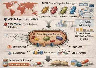
Figure.2 Overview of Antimicrobial Resistance in Gram-Negative Bacteria.
This review was done in line with the principles of the PRISMA guidelines to be able to have a systematic and transparent selection of literature. PubMed, Scopus, Web of Science, and Google Scholar were searched using the following keywords: nanoantibiotics, antimicrobial resistance, nanoparticles, drug delivery, biofilm inhibition, and stimuli-responsive nanocarriers. Articles describing nanotechnology-based antimicrobial systems against multidrug-resistant pathogens with in vitro or in vivo evidence were included and Non-English articles, conference abstracts, duplicates and studies lacking mechanistic insight were excluded. To conclude the current developments and define the research gaps in the nanoantibiotic strategies to combat AMR, the selected studies were critically analyzed and grouped into themes such as co-delivery system, stimuli-responsive nanocarriers, efflux pump targeted therapies, and personalized nanomedicine.
Table 1. Major Antimicrobial resistance mechanisms in multidrug-resistant gram-negative bacteria and their impact on antibiotic therapy
|
Resistance Mechanism |
Key Components |
Mechanism of Action |
Representative Pathogens |
Impact on Antibiotic Therapy |
|
Outer Membrane Barrier |
Lipopolysaccharide (LPS) outer membrane, porins, BepA, YcaL, DegP proteases, EnvZ–OmpR system, CpxR, micF, micA, micC |
Outer membrane remodeling alters permeability and reduces antibiotic influx. Downregulation or modification of porins decreases drug entry into bacterial cells. |
Escherichia coli, Pseudomonas aeruginosa, Klebsiella pneumoniae |
Reduced intracellular antibiotic accumulation limits the efficacy of β-lactams, fluoroquinolones, and other hydrophilic antibiotics. |
|
Efflux Pumps |
RND, MFS, ABC, MATE, SMR, and PACE transporter families; tripartite RND complexes |
Active extrusion of antibiotics from bacterial cells using ATP or proton motive force, thereby lowering intracellular drug concentration. |
Escherichia coli, Acinetobacter baumannii, Klebsiella pneumoniae, Pseudomonas aeruginosa |
Causes multidrug resistance against tetracyclines, aminoglycosides, fluoroquinolones, and β-lactam antibiotics. |
|
Enzymatic Degradation |
Carbapenemases, Extended-spectrum β-lactamases (ESBLs), plasmid-mediated resistance genes |
Enzymatic hydrolysis or chemical modification of antibiotics before reaching bacterial targets. |
Escherichia coli, Klebsiella pneumoniae |
Inactivates carbapenems and broad-spectrum β-lactams, severely restricting treatment options. |
|
Biofilm-Mediated Resistance |
Extracellular polymeric substance (EPS) matrix, persister cells, slow-growing bacterial populations |
EPS matrix prevents antibiotic penetration; reduced metabolic activity and persister cell formation enhance survival under antibiotic exposure. |
Pseudomonas aeruginosa, Acinetobacter baumannii, Klebsiella pneumoniae |
Biofilm-associated bacteria exhibit 10–1000-fold higher resistance, leading to chronic and recurrent infections. |
|
Quorum Sensing-Regulated Virulence |
Autoinducers, quorum sensing signaling pathways, virulence-associated genes |
Cell-to-cell communication regulates virulence factor production, motility, and biofilm formation according to bacterial population density. |
Pseudomonas aeruginosa, Acinetobacter baumannii |
Enhances bacterial survival, pathogenicity, and resistance persistence, reducing antibiotic effectiveness. |
Gram-negative bacteria have a variety of resistance mechanisms, including impermeability of outer membranes, active efflux pumps, enzymatic degradation of antibiotics, biofilm formation, and virulence regulation by quorum sensing. The combination of these mechanisms lowers penetration of antibiotics, decreases intracellular drug concentration, increases bacterial persistence and enhances antimicrobial tolerance. This concerted effort of these pathways is an important cause of multidrug resistance and of the necessity of advanced therapeutic strategies that have the capacity to simultaneously target biofilms, efflux activity, and bacterial virulence systems.
Phytoactives are plant-derived bioactive compounds widely explored as adjuncts in antimicrobial therapy due to their diverse pharmacological activities. These compounds can directly inhibit microbial growth and enhance antibiotic efficacy by targeting resistance mechanisms such as efflux pumps, biofilm formation, and membrane impermeability.
Phytoactives include a wide range of bioactive compounds derived from plants that have important antimicrobial potential. Major classes are polyphenols, flavonoids, alkaloids and terpenoids. Polyphenols disrupt the metabolism and cell wall integrity of microorganisms, whereas flavonoids disrupt microbial metabolism and cell wall integrity by affecting microbial signaling pathways. Alkaloids block DNA replication, protein synthesis and other important metabolic processes whilst the terpenoids disrupt the integrity of bacterial membranes which results in microbial cell death.
The other feature of bacterial resistance that extends to the efflux pumps is the pumping of the antibiotics in the cell by the efflux pumps therefore reducing the number of drugs in the cell³¹. These are the phytoactives among other phytoactives, the phytoactives are efflux pumps which make resistant bacteria susceptible to antibiotics³². The compounds may also act by the inhibition of transporter proteins which induce extrusion of inhibited drugs³³. Also, certain phytoactivities are able to repress the efflux pump gene expression and reduce the production of these resistance proteins³⁴. The other process is that it disrupts the functioning of ATPase that disrupts the energy provisioning of the effort of the efflux pump³⁵.
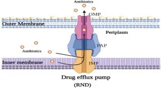
Figure.3 Efflux Pump Mechanism in Gram-Negative Bacteria
Incorporating antibiotics in bacterial biology to disturb biofilms and prevent infections caused by resistant microorganisms is a promising system to prevent infection³⁶. The biofilms are said to be one of the greatest dilemma in antimicrobial therapy since they possess protective extracellular matrix and high resistance against antibiotics³⁷. The phytoactives can interfere with the biofilm formation and integrity in many ways³⁸. Compounds that prefer the degradation of extracellular polymeric substance (EPS) are found and thus weaken the integrity of the biofilm matrix³⁹. The remaining ones inhibit quorum sensing, which is the bacterial communication which controls biofilm formation as well as expression of virulence factors⁴⁰. Additionally, certain phytoactives are disruptive to the primary contact between the bacterial cells and the host tissue or medical equipments by preventing its binding to surfaces⁴¹. These effects all lead to reduced biofilm and increased susceptibility of the embedded bacteria to the action of antimicrobial agents⁴².
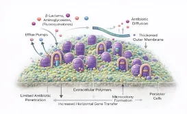
Figure.4 Biofilm-Mediated Resistance to Antimicrobial Agents in
Gram-Negative Bacteria
A high number of the phytoactives disturbs the bacterial cell membrane permeability and, thus, they possess antibacterial effects⁴³. The compounds help the antibiotics to be absorbed into the cells of the bacteria by disrupting the lipid bi-layers or altering the fluidity of the membranes⁴⁴. This is very high and may elevate the intracellular concentration of the drug and sensitize the resistant bacteria to the antibiotics even in instances that they were not as vulnerable⁴⁵.
Besides the direct antimicrobial effect, the phytoactives can possess the anti-inflammatory and immunomodulatory effect that can result in more favorable therapeutic outcomes⁴⁶. Most of the plant-derived products inhibit the synthesis of pro-inflammatory cytokine and oxidative stress at the site of the infection leading to the least amount of tissue damage⁴⁷. They can also induce innate immune reactions, as well as increase the ability of the host to destroy pathogens⁴⁸. Synergistic actions aid in infection control and tissue restoration and phytoactives are apt supplements to the combination of antimicrobial treatment⁴⁹.
Medicinal plants rich in phytoactives such as flavonoids, alkaloids, and terpenoids exhibit broad-spectrum antimicrobial activity with a lower likelihood of resistance development⁵⁰. However, their therapeutic application is limited by poor aqueous solubility, low oral bioavailability, rapid metabolism, and chemical instability⁵¹–⁵⁵. Many phytoactives, including curcumin, quercetin, and resveratrol, show limited membrane permeability and short biological half-lives, resulting in inadequate drug concentrations at infection sites, particularly against intracellular pathogens such as Mycobacterium tuberculosis and Salmonella species⁵²–⁵⁶.
Nano-co-delivery systems such as liposomes, polymeric nanoparticles, and solid lipid nanoparticles (SLNs) effectively overcome the limitations of free phytoactives by enhancing stability, bioavailability, and targeted delivery⁵⁷. Encapsulation protects phytoactives from hydrolytic, oxidative, and enzymatic degradation, significantly prolonging circulation time; for example, chitosan-coated nanoparticles can extend curcumin plasma half-life from minutes to nearly 24 hours⁵⁸. Their nanoscale size and surface modifications improve membrane interaction, intracellular uptake, and biofilm penetration, leading to higher accumulation at infection sites⁵⁹˒⁶⁰. Additionally, these systems enable sustained and stimuli-responsive drug release, such as pH-triggered release in acidic intracellular environments, thereby improving therapeutic efficacy and helping combat multidrug-resistant pathogens⁶¹–⁶³.
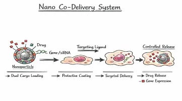
Figure:5. Nanoparticle-Based Co-Delivery System
The best thing in nano-co-delivery systems is that they have sophisticated cellular trafficking and retention⁶⁴. Nanoparticles can be targeted to evade reticuloendothelial system (RES) clearance by stealth coatings where surface ligands, e.g., mannose, interact with the macrophage receptors and facilitate phagocytic uptake⁶⁵. Once they are internalized, they may avoid lysosomal degradation by various mechanisms including; membrane fusion by cationic lipids or proton sponge effect caused by fusogenic peptides, which release the drugs into the cytosol⁶⁶. The phytoactive-antibiotic synergy also increases intracellular retention⁶⁷. Some of the phytoactives block efflux pumps decreasing drug extrusion and nanoparticle carriers delay exocytosis and preserve intracellular localization⁶⁸. Moreover, controlled release kinetics sustain drug concentrations beyond minimum inhibitory concentration (MIC) over a long time⁶⁹. All these effects allow improved eradication of intracellular pathogens, and much less bacterial burden in vivo and the reduction of systemic toxicity⁷⁰.
This has been enhanced by the appearance of multidrug-resistant (MDR) pathogens that has increased the speed at which effective nanoformulation systems are being formulated to reinstate the antimicrobial properties⁷¹. The nanosystems will improve the stability of the drug, the intracellular delivery, overcome biological barriers and interfere with the defence mechanisms resistance mechanisms like efflux pumps and biofilm defence. The categories of nanoformulations which are resistant-breaking antibacterial therapy are varied⁷¹.
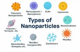
Figure:6. Inside Nanotechnology: A Closer Look at Nanoparticle Types
Table 2. Major Nanoformulation strategies and their advantages in combating multidrug-resistant gram-negative bacterial infections
|
Nanoformulation Type |
Description |
Advantages in Antimicrobial Therapy |
Representative Features |
|
Polymeric and Lipid-Based Nanocarriers |
This category includes polymeric nanoparticles, liposomes, solid lipid nanoparticles (SLNs), and nanostructured lipid carriers (NLCs). These carriers can efficiently encapsulate antibiotics and phytoactives, improving their stability and controlled release. |
Improve drug penetration into bacterial cells and biofilms, enhance intracellular antibiotic retention, and protect drugs from premature degradation. |
Chitosan nanoparticles, PLGA nanoparticles, liposomes, SLNs, NLCs |
|
Metallic, Hybrid, and Green-Synthesized Nanoparticles |
Metallic and hybrid nanoparticles possess inherent antibacterial properties. Green-synthesized nanoparticles are produced using plant extracts, offering a more eco-friendly and biocompatible alternative to conventional synthesis methods. |
Induce membrane damage, generate reactive oxygen species (ROS), inhibit bacterial resistance pathways, and reduce toxicity through sustainable fabrication methods. |
Silver nanoparticles, gold nanoparticles, zinc oxide nanoparticles, plant-mediated nanoparticles |
|
Nanoemulsions, Nanogels, and Hybrid Systems |
Nanoemulsions and nanogels are nanosized delivery systems capable of improving the solubility and localized delivery of antimicrobial agents. Hybrid systems combine multiple materials to enhance therapeutic performance. |
Enhance drug bioavailability, provide sustained and targeted release, improve antibiofilm activity, and increase therapeutic effectiveness at infection sites. |
Essential oil nanoemulsions, thermosensitive nanogels, multifunctional hybrid systems |
Overexpression of efflux pump systems is one of the major forms of antimicrobial resistance in bacteria, since the efflux systems actively pump antibiotics out of the cell, and eliminate the concentration of drugs within the cell⁸⁰. Phytoactives that are used in the nano-co-delivery system have the capability of being used as efflux pump inhibitors that prevent the movement of proteins like multidrug resistance pumps. Inhibition of these systems would severely amplify intracellular accumulation of antibiotics as the effect of effectively resensitizing resistant bacterial strains to traditional antibiotics. This approach reverses the resistance of pathogens otherwise demonstrated against antibiotics⁸¹.
Bacterial biofilms are a significant burden to therapy since they have thick extracellular polymeric matrix that impedes diffusion of drugs and shields populations of microbes against antimicrobial agents. The nano-co-delivery systems enhance the therapeutic effects as they penetrate the biofilm structures at a deep level⁸². They can be readily crossed into the biofilm matrix because of their nanoscale size and surface modifications and targeted active agents right to the bacterial cells embedded within the biofilm. Additionally, phytoactives have the capability of interfering with the integrity of biofilms, and quorum sensing, undermining the protective scaffold and exposing bacterial population to the effects of antibiotics⁸³.
This multi-target approach to antibacterial that is provided by the integration of phytoactives and synthetic antibiotics makes a significant impact on reducing the risk of the development of resistance⁸⁴. Phytoactives add complementary activity, e.g. membrane disruption, which leads to increased bacterial permeability and increased antibiotic uptake; enzyme inhibition, which disrupts critical cellular metabolic or resistance-linked enzymes; and generation of reactive oxygen species (ROS), which causes oxidative stress and damages cellular macromolecules. All of these simultaneous processes enhance antibacterial effectiveness⁸⁵.
The synergistic effect of the phytoactives and antibiotics in the nano-delivery systems usually leads to a considerable lowering of the minimum inhibitory concentration (MIC) to suppress bacterial development⁸⁶. Enhanced cellular uptake, enhanced intracellular retention, and multi-modal antibacterial effects improve the potency of the drug and permit the use of reduced therapeutic levels of these drugs. Therefore, this is not only an effective method of enhancing the efficacy of antimicrobials but also the reduction of toxicity, as well as, the delay of resistance, which makes nano-co-delivery systems a promising method of fighting multidrug-resistant infections⁸⁷.
The process of transforming the laboratory-based resistance-breaking nanoformulations to the clinical setting is a complicated venture that requires a proper preclinical evaluation to be completed effectively. The objectives of these findings are to assess the efficacy of anti-bacterial agents, namely antibiotics, synergy, safety, and therapeutic benefit of antibiotics in controlled in vitro models and applicable infection in vivo models⁸⁸.
The initial screening of the nanoformulations will involve a test of their bacteria killing capacity against the susceptible and multidrug resistant strains of bacteria89. The traditional method of determining the minimum concentration that will inhibit the apparent growth of bacteria is called MIC assay⁹⁰. The further investigation of the synergistic effects between phytoactives and antibiotics is generally conducted in the checkerboard assays. The results are interpreted with the help of the fractional inhibitory concentration index (FICI) which quantitatively investigates the possibility of interaction between combined agents being synergistic, additive, indifferent, or antagonistic. The assays are very essential to achieve the knowledge of the enhanced antimicrobial features of nano-co-delivery systems⁹¹.
This will require the development of the capacity of the nanoformulations to prevent and eliminate the establishment and annihilation of biofilms as they are amongst the leading causes of chronic and apparatus-related infections⁹². The identification of the reduction in the biomass and metabolic activity of biofilms following the treatment is done using the standard biofilm tests. This more invasive effect of biofilm matrix is also another characteristic of nanocarriers that, in its turn, are capable of killing the community of bacteria that are considered to be generally resistant to traditional antibiotics⁹³.
Time-kill kinetic studies are dynamic studies that give details of how much and how fast a bacterial killing occurs⁹⁴. These experiments determine the nature of nanoformulations as bacteriostatic or bactericidal by determining the viable bacterial counts at various time points following treatment. They are also used to establish antimicrobial action time and synergistic effect of antimicrobial action as is seen using MIC based assays⁹⁵.
Some of the factors that should be established before the animal research is the cytotoxicity and biocompatibility of the nanoformulations⁹⁶. The toxicity of the compound is commonly found using cell-based assays in which mammalian cell lines are used such as epithelial or macrophage cell lines. The parameters here include cell viability, membrane integrity and inflammatory reactions to give the safety of the nanocarriers to therapeutic use and protect the antimicrobial effect⁹⁷.
The use of animal models of infection is a problematic aspect and hard to study biological systems because they are complex and provide helpful information of therapeutic efficacy, pharmacokinetic and safety⁹⁸. By means of such models, the bacterial clearance, tissue distribution, immunological response and post-therapy survival can be studied with the help of this kind of models. The lack in the literature regarding the correlation of the in vitro findings with clinical practice is addressed in vivo experiments, which also demonstrate the prospects of nano-based resistance-breaking methodologies in clinical practice⁹⁹-100.
Multidrug-resistant Gram-negative bacteria are increasingly associated with severe clinical infections including hospital-acquired pneumonia (HAP), ventilator-associated pneumonia (VAP), urinary tract infections (UTIs), diabetic wound infections, sepsis, and bloodstream infections¹⁰¹–¹⁰⁹. Among these, Pseudomonas aeruginosa, Acinetobacter baumannii, and resistant Escherichia coli are major pathogens responsible for persistent infections and high mortality rates¹⁰¹–¹⁰⁴. Nanoformulation-based antimicrobial systems have shown considerable potential in improving therapeutic outcomes across these disease conditions by enhancing drug stability, targeted delivery, tissue penetration, and intracellular antibiotic retention¹⁰²,¹⁰⁴. In pulmonary infections such as HAP and VAP, nano-carriers improve lung deposition and penetration into infected tissues while minimizing systemic toxicity¹⁰². In UTIs, nano-delivery systems facilitate sustained antibiotic release and improved bacterial clearance, thereby reducing recurrent infections associated with biofilm formation¹⁰³,¹⁰⁴. For diabetic wound and skin infections, nanogels and antimicrobial wound dressings enable localized and prolonged drug release, while phytoactives provide additional anti-inflammatory and antioxidant effects that support tissue healing¹⁰⁵. Similarly, in sepsis and bloodstream infections, nano-co-delivery systems enhance circulation time, targeted biodistribution, and intracellular pathogen eradication, thereby reducing bacterial burden and inflammatory damage. Collectively, these findings highlight the significant therapeutic potential of nanoantibiotic strategies for managing multidrug-resistant infections106-107.

Figure.7 Clinical images of diabetic foot ulcers
The most preferable type of antimicrobial resistance is the nano-based co-delivery systems, however, the safety, pharmacokinetics, production capacity and marketing of the system after the translation of the idea to the clinics should be overemphasised. These are the issues which are meant to be resolved so that nano- therapeutics can be successful and safe when used in clinical practice in the long term108.
The nanoparticles that are specific with their therapeutic advantage may be toxic to the body depending on their composition, size, surface charge, dosage and sizes. The oxidative stress, inflammation or destruction of cells can be caused by the deposition of the nanoparticles into some of the major organs of the body such as the liver, spleen or kidney109. Even more so, some Nanomaterials are also unpredictable with the biological membranes and proteins. To furnish the information on safety with respect to nanoformulations to the therapy environment thus entire scope of toxicity test to be done should involve cellular viability test, hemocompatibility test, and long term toxicity test110.
Of interest to improve the clinical factors of nanoformulated therapeutics is the biodistribution and pharmacokinetics of such drugs. The absorption, distribution, metabolism and elimination of nanoparticle is usually varied compared to the traditional drug preparation. Some of the determinants of the circulation time, tissue targeting and uptake in the cells include particle size, surface modification and carrier composition. The pharmacokinetic works are executed in a thorough way to enable it in the determination of optimal doses of the drugs to be used in addition to ensuring the pharmacokinetic levels are established in the location of infections, as well as the minimization of systemic exposures111.
Physical and chemical stability of nanoformulations in a storage and transportation system is required to be effectively commercialized. The aggregation of the particles, leaking and degradation of the delicate Phytoactives, etc. may jeopardize the quality of the products and the therapeutic effect of the fragile phytotactives112. On top of this, reproducible, economically viable process, and consistent process of nanoparticles production must exist at least to be able to scale-up the nanoparticle in the laboratory scale to the large scale industrial scale. In order to attain uniformity in the sizes distribution of the particles, food loading efficacy and batch to batch uniformity is one of the key concerns of the development of the nano-based therapeutics113.
Nano-therapeutics is associated with special regulatory concerns in the case of a combination of antibiotics and Phytoactives. It takes a fair number of data on safety, efficacy, pharmacokinetics and quality of manufacturing to allow new drugs to be allowed by the regulatory authorities114. This may make its regulation to be difficult in the nanoformulations particularly those, which include over two active agents since there is no information on the mechanism of action, chronic toxicity, and interaction with the biological systems. The general assessment protocols and the administrative provisions will consequently be necessary to as much as commercialization and clinical translation of resistance-breaking nano-based antimicrobial therapies can be achieved115.
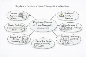
Figure.8 Regulatory Barriers in the Development of Nano-Therapeutic Combination Systems
Nevertheless, with its great potential, a major challenge is still evident in nanoantibiotic platforms, limiting their successful clinical translation against antimicrobial resistance (AMR). Key gaps involve the unavailability of effective co-encapsulation systems to coordinate the release of multiple therapeutics, limited in vivo and clinical studies because of scalability, biocompatibility, and regulatory issues116-117. In addition, insufficient development of stimuli-responsive nanocarriers targeting infection-specific microenvironments and underexplored efflux pump-targeted nanotherapies restrict therapeutic efficacy118. Moreover, individualized antimicrobial nanomedicine incorporating fast diagnostics, artificial intelligence, microbiome profiling, 3D bioprinting, and wearable biosensors are not well-developed yet. To tackle these interrelated problems with multidisciplinary innovation is needed to advance nanoantibiotics to the exact and effective management of AMR 119.
The use of Phytoactives in conjunction with nano-co-delivery system will be a revolution in the treatment of the MDR Gram-negative infections, and make a turnaround on the mechanisms of resistance i.e. efflux overexpression, biofilm fortification and enzymatic inactivation, to become a weakness that can be exploited. These platforms re-establish the action of antibiotics, minimal intracellular retention and development of resistance in preclinical sepsis, pneumonia and chronic wound models by the achievement of the desirable penetration, greater intracellular retention, and multi- modal synergy. Since polymeric nanoparticles (e.g., chitosan-PLGA) and lipid hybrids are not only a lot less soluble (less than 1mg/mL) but also highly degradable (half-life <1 h), an example of biocompatibility and controlled release would be in order.
Nevertheless, they continue to suffer certain translational bottlenecks which include organ-accumulation-related nanotoxicity, failure to scale the production and paucity of Phase II data. In order to fulfill these requirements, the combination of resistome sequencing with 3D-bioprinting should be used to co-encapsulate resistance to polymicrobial synergy, 3D-bioprinted organoids, efflux-selective CRISPR payloads, and customizable platforms. Interdisciplinary inventions will have transformed AMR, via AI-optimally designed, GMP-scalable and regulationally harmonized designs, that, by the year 2050, AMR inventions will be available in clinics in 10 million fewer deaths annually, and inventions of antimicrobial stewardship.
ACKNOWLEDGEMENTS
The authors acknowledge their guide and the management of KMCH College of Pharmacy, Coimbatore, for their guidance and institutional support.
AUTHORS' CONTRIBUTION
COMPETING INTERESTS
The authors declare no conflicts of interest.
ETHICAL APPROVAL
Not applicable.
REFERENCES

Srimathi Raj, Gayathri Rajaram* Pavinesh Kumar Krishnaswamy, Bharath Vishnu Sankar, Thirumurgan Periyayya , Phyto-Antibiotic Synergistic Nanoformulations As Resistance-Breaking Strategies Against Gram-Negative Pathogens, Int. J. of Pharm. Sci., 2026, Vol 4, Issue 9, 2448-2466. https://doi.org/10.5281/zenodo.22872428
 10.5281/zenodo.22872428