

We use cookies to ensure our website works properly and to personalise your experience. Cookies policy
Konkan Gyanpeeth Rahul Dharkar College of Pharmacy and Research Institute, Karjat, Maharashtra, India.
Diabetic wounds, particularly diabetic foot ulcers, remain among the most burdensome complications of diabetes mellitus because persistent hyperglycaemia, oxidative stress, chronic inflammation, impaired angiogenesis and recurrent infection together stall the normal sequence of repair. Conventional dressings and systemic antibiotics seldom address these overlapping defects at once. Hydrogels are attractive topical carriers because their hydrated three-dimensional networks maintain a moist wound environment, absorb exudate and permit controlled release of loaded agents. Silver, in ionic and nanoparticulate form, provides broad-spectrum antimicrobial and anti-biofilm activity, whereas curcumin, the principal polyphenol of Curcuma longa, offers antioxidant, anti-inflammatory, pro-angiogenic and collagen-promoting actions but is limited by poor aqueous solubility and instability. Combining the two within a single hydrogel is therefore a rational multi-target strategy. This review summarises the pathophysiology of impaired diabetic healing, the properties of silver and curcumin relevant to wound care, polymer matrices, formulation and crosslinking strategies, proposed mechanisms of complementary action, and the physicochemical and biological methods used for evaluation. Safety, silver accumulation, regulatory and scale-up challenges are discussed, and future directions such as stimuli-responsive, injectable and sensor-integrated systems are outlined. Silver–curcumin hydrogels show considerable preclinical promise, but standardised testing and well-designed clinical trials are required before clinical translation.
Diabetes mellitus is a chronic metabolic disorder whose long-term complications place a heavy clinical and economic burden on health systems. Among these, diabetic foot ulceration is one of the most serious. The lifetime risk of a person with diabetes developing a foot ulcer has been estimated at 19–34%, and ulcers frequently recur after apparent healing.1 A meta-analysis of studies from many regions reported a global prevalence of diabetic foot ulcer of roughly 6% among people with diabetes.2 Non-healing ulcers are a major precursor of infection, hospitalisation and lower-limb amputation, and they are associated with reduced quality of life and increased mortality.1,3
Normal cutaneous repair proceeds through overlapping phases of haemostasis, inflammation, proliferation and remodelling, coordinated by platelets, immune cells, keratinocytes, fibroblasts and endothelial cells.4,5 In diabetes this orchestrated sequence is disturbed at several levels. Hyperglycaemia drives the accumulation of advanced glycation end-products and excess reactive oxygen species (ROS), inflammation fails to resolve, angiogenesis and neurovascular supply are impaired, proteolytic enzymes degrade the extracellular matrix (ECM) and growth factors, and microbial colonisation, often organised as biofilm, sustains the cycle.6-8 Current standard care combines glycaemic control, offloading, debridement, infection management, revascularisation where indicated and moisture-balanced dressings.3,9 Yet a considerable proportion of wounds remain unhealed, which reflects the fact that most single-mode interventions address only one of several concurrent defects.10
Silver has been used in wound care for a long time, and silver ions and silver nanoparticles (AgNPs) retain broad-spectrum antimicrobial activity against bacteria, including strains resistant to common antibiotics.11-13 Concerns remain regarding cytotoxicity to host cells, silver accumulation and the potential for bacterial resistance, which make controlled and localised delivery desirable.14,15 Curcumin, the principal curcuminoid of Curcuma longa L. (turmeric), is a polyphenol with antioxidant, anti-inflammatory and antimicrobial properties and a broad spectrum of reported pharmacological effects.16,17 Its benefits for cutaneous repair have been documented in several animal models, including diabetic rats.18,19 However, curcumin is poorly soluble in water, chemically unstable at neutral to alkaline pH, photosensitive and rapidly metabolised, so that its practical use requires formulation support.20,21
Hydrogels are three-dimensional, hydrophilic polymer networks that can hold large amounts of water. They maintain a moist wound environment, absorb exudate, are generally soft and conformable, and can serve as depots for sustained local release of therapeutic agents.22-24 A hydrogel is therefore a natural vehicle in which a metal-based antimicrobial and a hydrophobic phytochemical can be combined. The resulting silver–curcumin (Ag–Cur) hydrogel aims to control infection and oxidative inflammation at the same time as supporting the proliferative and remodelling phases of healing.25
Studies on Ag–Cur systems are scattered across materials science, pharmaceutics and wound biology, and use diverse polymers, preparation routes and test models. The purpose of this review is to bring this information together. It describes the pathophysiology of impaired diabetic healing, the relevant properties of silver and curcumin, the polymer matrices and formulation strategies used to prepare Ag–Cur hydrogels, the proposed mechanisms of action, and the methods used for physicochemical and biological evaluation. It also discusses safety, translational challenges and future therapeutic perspectives.
MATERIALS AND METHODS
This narrative review was prepared by searching PubMed/MEDLINE, Scopus, Web of Science and Google Scholar for English-language publications, using combinations of the terms “diabetic wound”, “diabetic foot ulcer”, “silver nanoparticles”, “silver”, “curcumin”, “hydrogel”, “wound dressing”, “antibacterial”, “drug release” and “nanocomposite”. Original research articles, reviews, standards and guidelines were considered. Priority was given to studies that described the preparation and characterisation of hydrogels containing silver, curcumin or both, and to studies that evaluated them in vitro or in diabetic wound models. Reference lists of key articles were screened manually for further relevant work. Information was grouped thematically as pathophysiology, active components, polymer matrices, formulation, mechanisms, evaluation and safety. No quantitative meta-analysis was performed because of the heterogeneity of materials, doses and models.
RESULTS
3.1 Pathophysiology of impaired healing in diabetes
Understanding the defects of diabetic wounds is essential for rational design of a multi-component dressing. Table 1 summarises the normal phases of repair and the corresponding abnormalities in diabetes.
Table 1: Phases of Wound Healing and Abnormalities in Diabetes
| Phase | Normal events | Abnormalities in diabetic wounds |
|---|---|---|
| Haemostasis | Platelet aggregation, fibrin clot, release of PDGF and TGF-β | Altered platelet function and coagulation; microvascular disease limits perfusion7,26 |
| Inflammation | Neutrophil and macrophage infiltration, clearance of debris and microbes, then resolution | Prolonged neutrophil and M1 macrophage activity, high TNF-α, IL-1β and IL-6, impaired phagocytosis, delayed resolution6,27 |
| Proliferation | Angiogenesis, granulation tissue, fibroblast migration, re-epithelialisation | Reduced VEGF signalling and angiogenesis, impaired fibroblast and keratinocyte migration, high MMP activity26,28 |
| Remodelling | Collagen deposition and cross-linking, ECM maturation, scar formation | Disordered collagen, weak tensile strength, excessive protease degradation of ECM28,29 |
Chronic hyperglycaemia increases flux through polyol and hexosamine pathways and promotes mitochondrial superoxide production, which together with advanced glycation end-products activates inflammatory signalling and damages cells.30 The antioxidant transcription factor NRF2 is important for protection against oxidative stress in wounds, and its activity is compromised in diabetic tissue.31 Matrix metalloproteinase (MMP) activity is raised in diabetic ulcers relative to acute wounds, with a high MMP-to-inhibitor ratio, so that newly formed matrix and growth factors are degraded.28 Impaired angiogenesis and neuropathy further reduce oxygen and nutrient supply.26
Infection is another critical factor. Chronic wounds are colonised by mixed aerobic and anaerobic flora, and polymicrobial biofilms embedded in an extracellular polymeric matrix show tolerance to antibiotics and host defences.32,33 Because microbial burden, oxidative stress, protease excess and poor perfusion interact, a dressing that influences more than one of these factors has a theoretical advantage over single-target approaches.8 The wound microenvironment is also relevant to formulation: chronic wound fluid tends to have an alkaline pH, which affects the stability of pH-sensitive actives such as curcumin and the behaviour of pH-responsive polymers.34
3.2 Silver in wound care
3.2.1 Forms of silver and antimicrobial mechanisms
Silver is used as metallic silver, silver salts (for example silver nitrate and silver sulfadiazine), silver complexes and nanoparticles. Antimicrobial activity depends largely on the release of Ag⁺ ions, so that the dissolution rate, particle size, shape, surface coating and local chemistry of the wound all influence efficacy.11,12 Several complementary mechanisms have been proposed. Ag⁺ binds to thiol, amino and phosphate groups of proteins and enzymes and thereby inhibits respiratory and other metabolic enzymes. Silver interacts with and perturbs the bacterial cell envelope, causing increased permeability and leakage, and nanoparticles have been observed to accumulate in and damage bacterial membranes.35,36 Silver also promotes intracellular ROS generation and can interact with nucleic acids, which interferes with replication.37,38 Because several targets are affected simultaneously, the emergence of resistance is considered less likely than with single-target antibiotics, although silver-resistant strains have been reported and stewardship remains necessary.12,15
3.2.2 Effects on host cells and healing
Besides antimicrobial action, AgNPs have been reported to influence healing in animal models. In a thermal injury model, topical AgNPs were associated with faster re-epithelialisation and a modified cytokine profile.39 Other work indicated that AgNPs can influence the differentiation and migration of keratinocytes and fibroblasts, promoting the transformation of fibroblasts to myofibroblasts and thereby wound contraction.40 At the same time, dose-dependent cytotoxicity and genotoxicity toward human cells have been described, which sets a limit on the amount of silver that can be safely applied.14 The clinical evidence for silver-containing dressings is mixed. A Cochrane review found no clear evidence that topical silver prevents wound infection or promotes healing across wound types, which emphasises the need for targeted, well-designed products and trials.41
3.2.3 Synthesis of silver nanoparticles
Silver nanoparticles are commonly prepared by chemical reduction of silver nitrate using borohydride, citrate or other reducing agents, with stabilisers or capping agents to prevent aggregation.42 Physical methods and photochemical routes are also available. “Green” synthesis uses plant extracts, polysaccharides or other biomolecules as both reducing and capping agents, avoiding toxic reagents; this is attractive for biomedical use, although batch-to-batch reproducibility of extract-based syntheses can be difficult to control.13,42 Particle size generally ranges from a few nanometres to about 100 nm, and the optical surface plasmon resonance band, typically observed in the 400–450 nm region for spherical AgNPs, is widely used to monitor formation.42
3.3 Curcumin as a wound-healing agent
Curcumin [(1E,6E)-1,7-bis (4-hydroxy-3-methoxyphenyl)hepta-1,6-diene-3,5-dione] has a molar mass of 368.38 g mol⁻¹ and exists in equilibrium between diketo and keto-enol tautomers.17 The phenolic hydroxyl groups and the conjugated β-diketone system underlie its radical-scavenging ability and its metal-chelating and reducing properties.17,43
In wound models, curcumin has been reported to accelerate closure, enhance granulation tissue formation, increase collagen and hydroxyproline content, and reduce ROS and lipid peroxidation.44,45 Curcumin-incorporated collagen films improved dermal wound healing in rats,46 and in streptozotocin-induced diabetic rats topical curcumin accelerated closure while modulating antioxidant enzymes and inflammatory mediators.19 Mechanistically, curcumin inhibits NF-κB signalling and thereby reduces TNF-α, IL-1β and IL-6, activates NRF2-dependent antioxidant defences, and has been associated with increased TGF-β1 and VEGF expression, fibroblast proliferation and angiogenesis.16,18,47 Curcumin also has intrinsic antibacterial activity, though it is moderate compared with that of silver.17,18
The principal obstacle to clinical use is physicochemical. Curcumin is practically insoluble in water at acidic and neutral pH, degrades rapidly in neutral to alkaline solution and is sensitive to light, and its systemic bioavailability is poor because of limited absorption and rapid metabolism.20,21 Topical application avoids first-pass metabolism, but solubility, stability and skin retention still require formulation strategies.48,49 Moreover, curcumin has been classified as a compound that can give false-positive results in assays, owing to its colour, aggregation and reactivity, which is relevant to biological evaluation and is discussed further below.43 Although numerous clinical trials of curcumin for other conditions have been conducted, evidence for topical diabetic wound treatment in humans is limited.50,51
3.4 Hydrogel matrices for diabetic wound dressings
An ideal wound hydrogel is biocompatible, non-adherent, able to absorb exudate while keeping the wound bed moist, permeable to oxygen, mechanically adequate and easily removable. The concept of moist wound healing dates back to the observation that wounds covered by an occlusive layer re-epithelialise faster than dry wounds.52 Hydrogels may be derived from natural polymers, synthetic polymers or combinations; their properties are tuned through polymer concentration, crosslink density and chemistry.53,54 Table 2 lists polymers commonly used as matrices for silver and curcumin.
Table 2: Polymer Matrices Used for Silver and Curcumin Hydrogels
| Polymer | Key features for wound use | Limitations |
|---|---|---|
| Chitosan | Cationic, intrinsic antibacterial and haemostatic activity, promotes granulation; reduces and stabilises AgNPs55 | Soluble only in dilute acid; mechanical weakness; variable source and deacetylation degree |
| Sodium alginate | High exudate absorption, gentle gelation with Ca²⁺ ions, widely used in commercial dressings56 | Low intrinsic bioactivity; ion-dependent stability |
| Poly(vinyl alcohol) (PVA) | Good mechanical strength and film-forming ability, physical crosslinking by freeze–thaw cycles57 | Lacks bioactivity; may need blending for cell adhesion |
| Gelatin / collagen | Cell-adhesive sequences, biodegradable, supports fibroblast attachment46 | Thermally unstable; enzymatic degradation; needs crosslinking |
| Hyaluronic acid | Hydrating, supports migration and angiogenesis, native to skin ECM58 | Rapid enzymatic degradation; cost |
| Carbomer / cellulose derivatives | Easy to prepare, good spreadability, suited to topical gels53 | Limited biological activity; acidic pre-gel requires neutralisation |
| Poly(ethylene glycol) and synthetic copolymers | Tunable network, reproducible synthesis, injectable and self-healing designs possible59,60 | Bio-inert; scale-up and regulatory cost for complex designs |
Natural polysaccharides such as chitosan and alginate are especially popular for Ag–Cur systems. They are inexpensive, biodegradable and bioactive, and they provide functional groups that can bind or reduce silver and interact with curcumin.55,56 Synthetic polymers provide reproducibility and mechanical control, and hybrid networks combine the advantages of both.54,61
3.5 Formulation strategies for silver–curcumin hydrogels
The preparation of an Ag–Cur hydrogel involves selecting the matrix, introducing silver and curcumin in a suitable form, and fixing the network by crosslinking (Fig. 1). The order and method of addition determine particle size, curcumin state, release profile and stability.
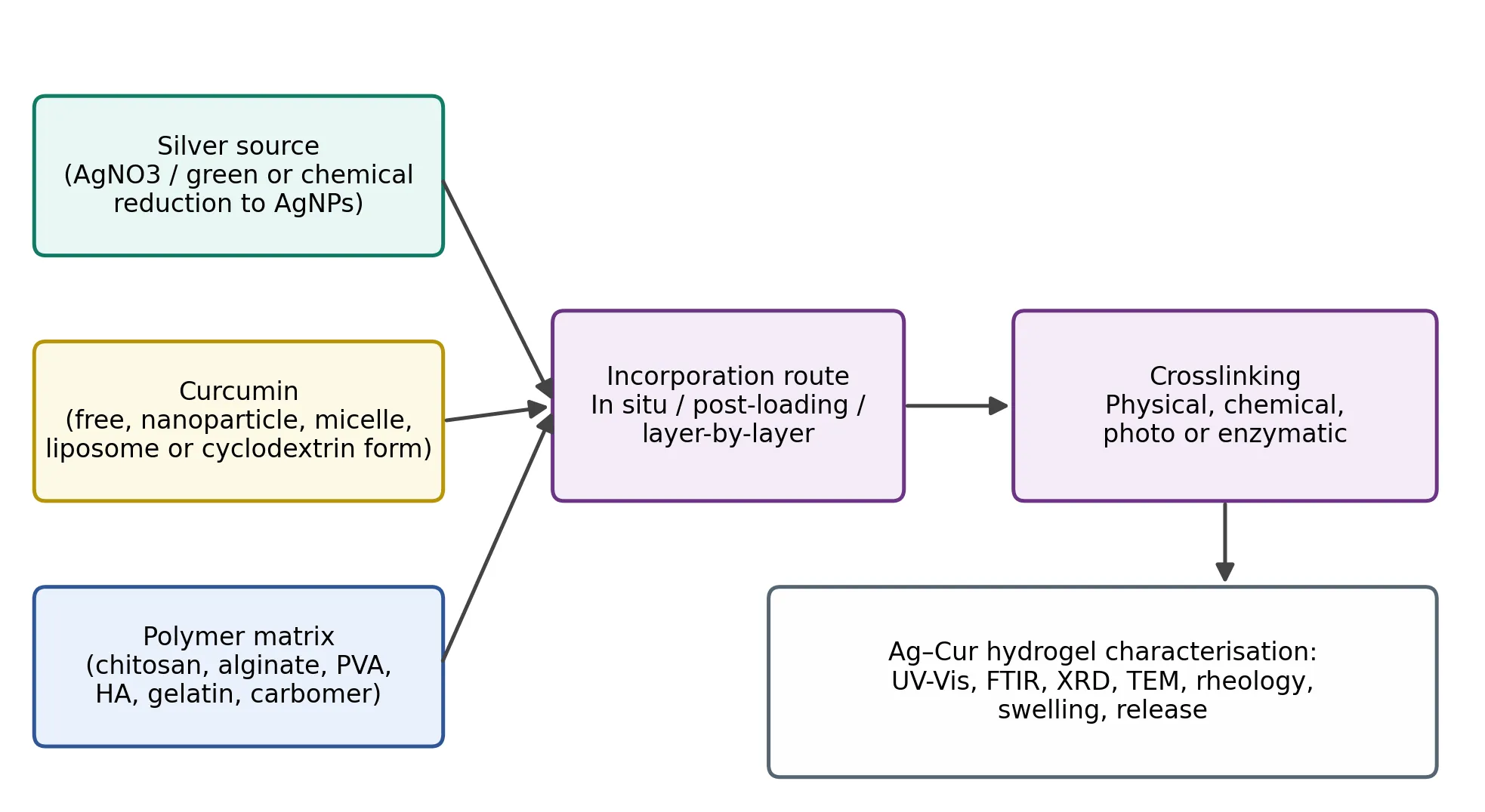
Figure 1: General Workflow for the Preparation of Silver–Curcumin Hydrogels.
3.5.1 Incorporation of silver
Silver can be introduced in three main ways. In in situ synthesis, AgNO₃ is dissolved within the polymer solution or pre-formed gel and reduced inside the network by the polymer itself (for example chitosan or polysaccharides with reducing end groups), by an added reductant, by heat or by UV irradiation. This yields well-dispersed nanoparticles stabilised by the matrix and avoids aggregation.25,42 In the second approach, pre-formed AgNPs prepared and characterised separately are mixed into the polymer solution, allowing better control of size and dose but requiring colloidal stability in the pre-gel medium. The third approach employs silver salts or complexes, such as silver sulfadiazine, which release Ag⁺ directly. Green routes using plant-derived reductants are widely explored in the literature on silver nanocomposite hydrogels.13,25 Because curcumin itself possesses reducing and chelating functionality, it can in principle participate in the reduction and capping of silver ions, so that in some preparations a single step produces curcumin-capped AgNPs; this can simplify processing but complicates the independent control of the two components.17,25
3.5.2 Incorporation of curcumin
Direct dispersion of crystalline curcumin in an aqueous hydrogel gives poor uniformity and limited bioavailability at the wound surface. Strategies to improve solubilisation and stability are summarised in Table 3.
Table 3: Approaches for Incorporating Curcumin into Hydrogels
| Approach | Principle | Consideration |
|---|---|---|
| Co-solvent or surfactant solubilisation | Dissolution in ethanol, propylene glycol or micellar surfactant before dispersion in the gel | Simple; solvent or surfactant levels must be compatible with skin and the polymer network |
| Cyclodextrin inclusion complexes | Host–guest encapsulation increases aqueous solubility and protects from light and hydrolysis49 | Requires complexation efficiency optimisation; adds mass to the formulation |
| Polymeric nanoparticles or micelles | Entrapment in biodegradable particles that are then dispersed in the gel48,49 | Improved stability and sustained release; adds a preparation step |
| Liposomes, niosomes, nanoemulsions | Lipid-based carriers solubilise the hydrophobic drug and promote skin retention49 | Physical stability of vesicles within the gel network |
| Covalent or ionic conjugation to polymer | Attachment to the polymer backbone gives slow, degradation-dependent release62 | Chemical modification must preserve activity; regulatory complexity |
Encapsulation within polymeric or lipid nanocarriers before incorporation into the hydrogel produces a dual-compartment system in which the gel controls the macroscopic behaviour and the nanocarrier controls the curcumin microenvironment, which is a common design in the recent literature.49,62
3.5.3 Crosslinking and gelation
Physical crosslinking relies on reversible interactions, including ionic gelation (alginate with Ca²⁺), polyelectrolyte complexation, hydrogen bonding and freeze–thaw induced crystallite formation in PVA. These methods avoid potentially toxic crosslinkers and often permit self-healing or injectability but yield weaker networks.53,57 Chemical crosslinking using glutaraldehyde, genipin, carbodiimide chemistry or free-radical polymerisation produces stronger networks with controllable swelling and degradation, but residual crosslinker and initiator must be removed or kept below acceptable limits.23,53 Photocrosslinking and enzymatic crosslinking offer spatial and temporal control and are compatible with in situ gelation on irregular wounds. For Ag–Cur hydrogels, the crosslinking method must be compatible with both active components: strong oxidants and high temperatures may degrade curcumin, whereas strong reducing conditions or halide-rich media may alter or precipitate silver species.
3.5.4 Advanced hydrogel designs
Injectable and self-healing hydrogels based on dynamic covalent or supramolecular bonds can conform to deep or irregular ulcers and recover after mechanical disruption.59,60 Stimuli-responsive systems are of particular interest for diabetic wounds: pH-responsive gels may release more payload in the alkaline, infected wound environment, and ROS- or enzyme-responsive networks can release curcumin or silver in proportion to oxidative stress or protease activity.8,34 Multilayer or bilayer constructs can place a silver-rich antimicrobial layer at the wound contact surface and a curcumin-rich layer for sustained anti-inflammatory delivery. Combination with nanofibrous or foam substrates and the use of microneedle arrays for deeper penetration are emerging variations, although reports specific to Ag–Cur combinations remain limited.63
3.6 Mechanisms of action of silver–curcumin hydrogels
The rationale for combining silver and curcumin lies in the complementary biological effects of the two agents and in the supportive role of the hydrogel itself (Fig. 2). The hydrogel provides moisture, exudate management and a physical barrier, and acts as a depot for both actives.54,58
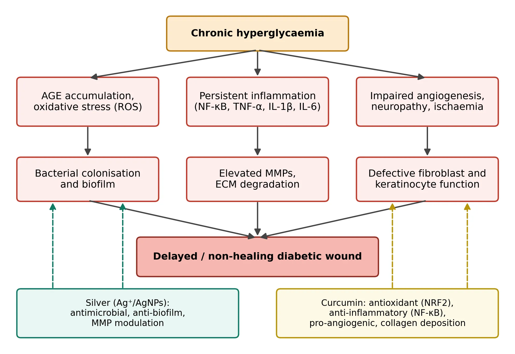
Figure 2: Principal Pathological Mechanisms in the Diabetic Wound and Proposed Points of Action of Silver and Curcumin.
Silver mainly addresses the microbial and biofilm component. Ag⁺ and AgNPs reduce planktonic and surface-associated bacteria, thereby decreasing the continuous stimulus for inflammation and protease production.12,37 Curcumin addresses the host response. By suppressing NF-κB signalling and reducing pro-inflammatory cytokines it may allow progression from the inflammatory to the proliferative phase, while its antioxidant properties and activation of NRF2-dependent pathways counter the oxidative stress that damages cells in hyperglycaemic tissue.19,31,47 Increased VEGF and TGF-β1 expression, fibroblast proliferation and collagen deposition support granulation and remodelling.18,45
Several types of interaction between the two agents have been proposed. First, curcumin has some antibacterial activity of its own, so that the two agents may contribute additively to microbial control. Second, by reducing the inflammatory and oxidative load, curcumin may counteract part of the oxidative and cytotoxic stress that silver can induce in host cells, so that a lower silver dose might be sufficient.14,19 Third, the polyphenolic structure of curcumin may stabilise AgNPs and modulate the rate of Ag⁺ release.17,25 Fourth, the hydrogel network can co-localise and co-release both agents at the wound interface, which helps to achieve simultaneous exposure. Most of these interactions are, however, supported by indirect or in vitro observations, and well-controlled studies that separate additive from truly synergistic effects (for example by isobologram or checkerboard analysis) are uncommon; this point is revisited in the Discussion.
3.7 Physicochemical characterisation
Comprehensive characterisation is necessary to link composition to performance. Table 4 lists the techniques most frequently applied to Ag–Cur hydrogels.
Table 4: Techniques for Physicochemical Characterisation of Silver–Curcumin Hydrogels
| Parameter | Technique | Information obtained |
|---|---|---|
| Nanoparticle formation | UV–visible spectroscopy | Surface plasmon band of AgNPs (typically 400–450 nm); curcumin absorption in the visible region; changes indicate interaction or degradation17,42 |
| Size and morphology | TEM, SEM, DLS, zeta potential | Particle size and distribution, shape, dispersion within the network, colloidal stability42 |
| Crystallinity | XRD | Face-centred cubic silver reflections; amorphous or crystalline state of curcumin25 |
| Chemical interactions | FTIR, Raman, NMR | Hydrogen bonding, complexation, crosslinking chemistry, drug–polymer compatibility53 |
| Thermal behaviour | DSC, TGA | Thermal stability, miscibility, water content |
| Swelling and water handling | Gravimetric swelling, water vapour transmission, moisture retention | Exudate absorption capacity and moist environment maintenance57,58 |
| Mechanical and rheological | Rheometry, tensile or compression testing, adhesion tests | Viscoelasticity, injectability, strength, self-healing, tack59,60 |
| Drug and silver release | Franz diffusion cell or dialysis with UV–Vis/HPLC (curcumin) and AAS or ICP-MS (silver) | Release kinetics, Higuchi or Korsmeyer–Peppas modelling64,65 |
| Degradation and stability | Enzymatic or hydrolytic degradation, accelerated and photostability studies | Shelf-life, curcumin retention, silver speciation21,66 |
Release studies deserve particular attention in a dual-component system. Curcumin and silver have different physicochemical properties, so their release profiles differ and should be quantified separately: curcumin by UV–Vis or HPLC under conditions that maintain sink conditions and curcumin stability (for example, medium containing a solubilising agent and protection from light), and silver by atomic absorption spectroscopy or ICP-MS. Release data are often fitted to the Korsmeyer–Peppas model to distinguish Fickian diffusion, anomalous transport and polymer-relaxation-controlled release.64,65 Because the aim is to maintain therapeutic concentrations of both agents while avoiding silver overexposure, the quantity of silver released over the dressing wear period should be reported explicitly.
3.8 Biological evaluation
3.8.1 In vitro studies
Antimicrobial activity is generally assessed against Gram-positive and Gram-negative wound pathogens such as Staphylococcus aureus, Pseudomonas aeruginosa and Escherichia coli using agar diffusion, minimum inhibitory and bactericidal concentration determinations and time–kill assays; anti-biofilm assays, for example crystal violet or viability staining of biofilms grown on the material, are more clinically relevant and should be included.33,67 Cytocompatibility is evaluated on fibroblasts, keratinocytes or endothelial cells, with extract-based or direct-contact tests following ISO 10993-5.68 Metabolic assays such as MTT are widely used,69 but the intrinsic colour of curcumin and the optical properties of AgNPs can interfere with colorimetric readouts, so orthogonal methods (for example live/dead staining or ATP-based assays) and appropriate material-only controls are advisable.43 Functional assays include scratch (migration) assays, collagen synthesis, tube formation assays for angiogenesis, and measurement of inflammatory mediators and ROS in stimulated macrophages or in cells cultured under high-glucose conditions to mimic diabetic hyperglycaemia. Haemocompatibility and antioxidant capacity (for example DPPH or ABTS radical scavenging) are commonly reported.
3.8.2 In vivo studies
Diabetic wound efficacy is commonly tested in rodents made diabetic by streptozotocin (STZ), sometimes combined with a high-fat diet, or in genetically diabetic strains, using full-thickness excisional wounds, often stented to limit contraction.70 Typical outcome measures are percentage wound closure over time, time to complete re-epithelialisation, histology of granulation tissue and epidermal thickness, collagen deposition (Masson’s trichrome, hydroxyproline), angiogenesis markers such as CD31 and VEGF, inflammatory cytokines, oxidative stress markers and bacterial load in infected wound models. The rat and mouse models used in studies of curcumin and of AgNPs have reported faster closure and improved histological quality compared with untreated or vehicle controls.19,39,48 Table 5 summarises recommended parameters and controls.
Table 5: Recommended Parameters for Biological Evaluation
| Level | Parameters | Suggested controls |
|---|---|---|
| Antimicrobial | MIC/MBC, zone of inhibition, time–kill, anti-biofilm activity on relevant wound isolates67 | Blank hydrogel, silver-only, curcumin-only, and reference antibiotic or commercial silver dressing |
| Cytocompatibility | Cell viability by two orthogonal assays, live/dead imaging, haemolysis68 | Material-only readout controls, negative and positive cytotoxicity controls |
| Cell function | Scratch closure, collagen synthesis, tube formation, cytokine and ROS levels under high glucose | Normal-glucose and high-glucose conditions |
| Diabetic animal model | Wound closure kinetics, histology, hydroxyproline, CD31/VEGF, cytokines, bacterial load70 | Untreated diabetic, vehicle gel, single-component gels, marketed dressing, non-diabetic reference |
| Safety | Skin irritation and sensitisation, silver tissue and blood levels, organ histology | Dose-ranging and repeated-application groups |
3.9 Safety, stability and regulatory aspects
Silver exposure is the main safety consideration. Although absorption through intact skin is low, absorption through large or chronic wounds can occur, and silver may accumulate in tissues over long-term use; argyria is the best-known consequence of high exposure, and cytotoxic and genotoxic effects have been demonstrated in cell cultures at sufficiently high concentrations.11,14,15 Dose-finding studies should therefore aim for the lowest effective silver content and quantify local and systemic silver levels. Curcumin is generally regarded as safe in oral use, but allergic contact dermatitis and staining of skin and dressings can occur.50,51
Stability testing under ICH conditions should evaluate curcumin content, silver speciation and particle aggregation, pH, viscosity, microbial quality and appearance over the intended shelf-life, with protection from light.66 Sterilisation is a particular challenge: gamma irradiation and autoclaving may degrade curcumin, alter polymer networks or change AgNP size, so sterilisation methods must be validated for each formulation. From a regulatory perspective, a dressing combining a medicinal component and a nanomaterial may be regulated as a drug, a device or a combination product depending on the jurisdiction and the principal mode of action, and the nanomaterial status will usually require additional characterisation and toxicological data.61,63
DISCUSSION
4.1 Therapeutic perspectives
The rationale for Ag–Cur hydrogels is conceptually strong. Diabetic wounds are multifactorial and a topical product that simultaneously controls bioburden, modulates inflammation and oxidative stress, and sustains a moist healing environment addresses more than one contributor to chronicity.8,10 Preclinical studies with curcumin or AgNPs individually, and with composite hydrogels, consistently report accelerated closure and improved histological parameters in rodent models.19,25,39,48 A further potential advantage is dose sparing: if curcumin mitigates oxidative and inflammatory stress, the silver concentration required for antimicrobial efficacy may be kept low, which would improve the benefit–risk balance.
Patient groups that could benefit most include those with infected or critically colonised neuropathic ulcers, patients with recurrent infection or biofilm-associated stalled wounds, and those for whom systemic antibiotics are undesirable because of resistance or renal and hepatic comorbidity. A hydrogel is also appropriate for dry or sloughy wounds where autolytic debridement and hydration are needed. For ischaemic ulcers, however, topical therapy cannot replace revascularisation, and Ag–Cur hydrogels should be viewed as adjuncts to, not substitutes for, standard care including offloading, debridement and glycaemic control.3,9
4.2 Critical appraisal of the current evidence
Several limitations temper enthusiasm. First, much of the evidence arises from small, short-term studies in rodent models, which differ from human diabetic ulcers in the extent of skin contraction, perfusion, neuropathy and chronicity.70 Second, formulations vary so widely in polymer, silver form, curcumin form and dose that cross-study comparison is difficult, and many papers lack the single-component and commercial-dressing controls that are needed to demonstrate added value. Third, true synergy is rarely tested formally. Fourth, curcumin is a known source of assay interference and has questionable chemical stability, so apparent bioactivity in vitro may not reflect the active species in the wound.21,43 Fifth, reporting of silver release, silver tissue levels and long-term safety is inconsistent. Finally, the Cochrane evidence on silver dressings indicates that clinical benefit cannot be assumed from antimicrobial potency alone.41
4.3 Challenges and possible solutions
Table 6: Key Challenges and Possible Solutions
| Challenge | Consequence | Possible approach |
|---|---|---|
| Curcumin instability and low solubility | Loss of activity during storage and use | Nanocarrier or cyclodextrin encapsulation, light-protective packaging, pH control21,49 |
| Silver cytotoxicity and accumulation | Delayed healing, systemic exposure | Low-dose, controlled-release designs; quantification of silver release and tissue levels14,15 |
| Batch variability of green synthesis | Poor reproducibility | Standardised extracts or defined reductants; process analytical controls42 |
| Assay interference | Misleading in vitro data | Orthogonal assays; material-only controls43 |
| Limited relevance of animal models | Poor clinical translation | Infected and ischaemic models, larger-animal or ex vivo human skin models, biofilm models33,70 |
| Sterilisation and scale-up | Degradation of actives, cost | Validated low-damage sterilisation; aseptic processing; quality-by-design61,66 |
| Regulatory classification | Prolonged development | Early engagement with regulators; defined primary mode of action63 |
4.4 Future directions
Future work should move toward rationally designed, well-characterised systems tested against appropriate controls. Stimuli-responsive hydrogels that release silver in response to infection-associated pH or enzymes, and curcumin in response to ROS, would match drug delivery to wound state.8,34 Integration of sensors for pH, temperature or glucose could enable smart dressings that monitor healing and trigger release. Alternatives to AgNPs, such as silver–polymer complexes, bimetallic systems or co-delivery with other natural antimicrobials, may widen the therapeutic window. More robust preclinical models, including infected, ischaemic and biofilm-bearing wounds, should be combined with transparent reporting of formulation and release data. Finally, adequately powered randomised controlled trials in patients with diabetic foot ulcers, with standardised endpoints such as complete wound closure at 12 weeks, infection rate and adverse events, are required before clinical value can be established.3,9
CONCLUSION
Silver–curcumin hydrogels combine antimicrobial, anti-inflammatory, antioxidant and pro-regenerative actions in a moist, controlled-release dressing, and thus address several of the interacting defects that impede diabetic wound healing. Formulation advances, including in situ and green synthesis of silver nanoparticles, nanocarrier-assisted curcumin loading and responsive or injectable networks, have improved the feasibility of such systems. Preclinical results are encouraging, but they are limited by heterogeneous designs, inconsistent controls, inadequate assessment of synergy and silver safety, and the absence of clinical data. Standardised characterisation and evaluation, together with carefully designed clinical trials, will determine whether these hydrogels can progress from promising laboratory formulations to effective therapies for patients with diabetic foot ulcers.
ACKNOWLEDGMENT
The authors are thankful to the management and the Principal of Konkan Gyanpeeth Rahul Dharkar College of Pharmacy and Research Institute, Karjat, for providing library and database facilities.
CONFLICT OF INTEREST
The authors declare that there is no conflict of interest related to the publication of this manuscript.
AUTHOR CONTRIBUTIONS
SachinYadav: conceptualisation, literature search, data compilation and drafting of the manuscript; Dr.B.S.T. and Dr.A.T.: guidance, supervision and critical revision of the manuscript. All authors read and approved the final manuscript.
REFERENCES

Sachin Yadav, Dr. Thawkar Sukdeo, Dr. Atul Tripathi, Silver–Curcumin Hydrogel Systems for Diabetic Wound Healing: Formulation Strategies, Mechanisms, Evaluation and Therapeutic Perspectives, Int. J. of Pharm. Sci., 2026, Vol 4, Issue 10, 2091-2104. https://doi.org/10.5281/zenodo.23360101
 10.5281/zenodo.23360101