

We use cookies to ensure our website works properly and to personalise your experience. Cookies policy
Department of Pharmaceutics, The Erode College of Pharmacy, Veppam Palayam, Erode- 638112, Tamil Nadu, India.
Advanced therapeutic approaches have been designed to address the drawbacks of conventional therapies, including poor permeability, drug instability, and low bioavailability. Spanlastics are deformable nanovesicular systems composed of non-ionic surfactants and edge activators that enhance membrane elasticity and improve permeability across biological barriers. This review highlights the structure, composition, mechanism, and effectiveness of spanlastics in comparison with other vesicular carriers, including liposomes, niosomes, ethosomes, and transferosomes. These carriers can incorporate both water-soluble and lipid-soluble drugs, thereby improving bioavailability and enabling controlled drug delivery. Due to their deformability, stability, and reduced irritation, spanlastics can be delivered via topical, oral, nasal, and ophthalmic routes.
In recent years, considerable research interest has focused on designing novel drug delivery systems (NDDS) to enhance therapeutic efficacy, minimise adverse effects, and improve patient compliance.[1] However, conventional drug delivery systems still have certain limitations such as poor permeability, drug instability, and low bioavailability.[2]
Vesicular drug delivery systems (VDDS) are considered effective carriers as they can encapsulate both lipophilic and hydrophilic drugs and facilitate their transport across biological membranes. [3,4] A range of vesicular carriers, including liposomes, niosomes, ethosomes, and transferosomes, have been investigated for this purpose. However, these systems also exhibit certain drawbacks, including instability, high cost, and poor permeability across biological barriers.[5]
To address these limitations, more flexible and advanced vesicular systems have been explored. Among these, spanlastics represent a novel class of elastic nanovesicular carriers introduced by Kakkar and Kaur in 2011.[6] Spanlastics are formed using non-ionic surfactants together with edge activators, which confer elasticity and deformability, thereby enhancing their ability to cross biological barriers.[7]
Because of their deformable nature, spanlastics exhibit enhanced permeability and can effectively deliver drugs across different biological membranes.[8] They also offer advantages such as improved bioavailability, controlled drug release, better stability, and reduced irritation when compared to conventional vesicular systems. Therefore, spanlastics have gained recognition as a valuable platform within advanced drug delivery research.
2. SPANLASTICS
2.1 General Aspects of Spanlastics
Spanlastics represent an innovative generation of vesicular drug delivery systems suitable for administration through topical, oral, nasal, or transmucosal routes, as they can target specific sites. What differentiates spanlastics from other drug delivery vehicles is the structural plasticity of the vesicular system, enabled by non-ionic surfactants (for example, Span) and the presence of edge activators (Tween). This flexibility allows vesicles to deform and pass through biological membranes, significantly improving drug permeability, particularly across barriers such as the ocular barrier.
In addition, spanlastics enable controlled, sustained release of the active components through the formation of bilayer vesicles, thereby maintaining therapeutic concentration for a prolonged period. Spanlastics are chemically stable, biodegradable, and non-immunogenic systems, making them safer than many vesicular systems. Furthermore, spanlastics improve bioavailability by delivering the drug directly to the target site and reducing its degradation or elimination. Spanlastics are a less irritating drug delivery system due to the presence of a non-ionic surfactant.[9]
2.2. Structure of Spanlastics
Spanlastics are vesicular systems with a spherical shape, composed of a flexible bilayer membrane formed from non-ionic surfactants encapsulating an internal aqueous phase. This bilayer is constructed using a combination of non-ionic surfactant and edge-activating agent [10], as illustrated in Figure 1.
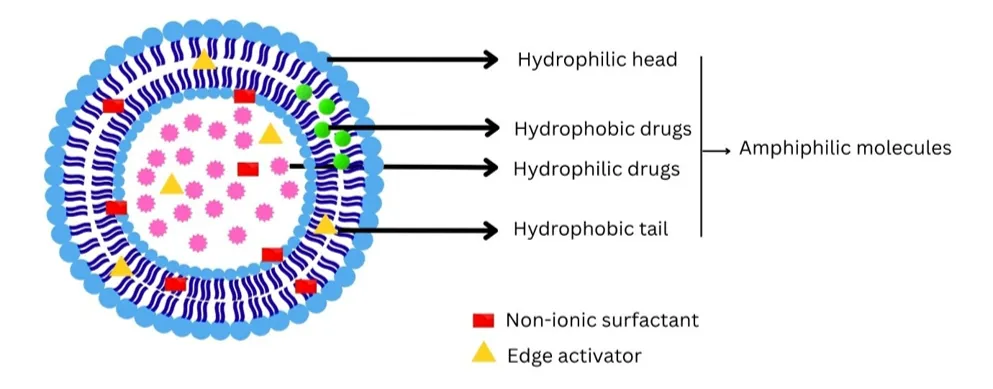
Figure 1. Structure of Spanlastics Vesicle
2.3 Composition of Spanlastics
The composition of spanlastics consists of two major parts:
1. Non-ionic surfactants
2. Edge activators (EAs)
The above-mentioned substances act together to create deformable vesicles suitable for drug transport.
Non-ionic surfactants
Surfactants belonging to the Span series, Span 20, 40, 60, and 80, are the primary non-ionic agents employed for bilayer formation. Being biocompatible, non-toxic and capable of forming stable vesicles, they represent widely used components. Among them, Span 60 is the most commonly used due to its high stability and ability to form well-structured bilayers.
Edge activators (EAs)
Edge activators, primarily Tween 80 and sodium cholate, are included to enhance vesicle flexibility. They destabilise tightly packed surfactant bilayers, making membranes more flexible and deformable. [11,12]
2.4 Mechanism of Spanlastics
The mechanism behind spanlastics mainly relies on edge activators that provide flexibility to the vesicular bilayer by breaking its rigidity. This allows vesicles to move through the narrow intercellular spaces due to the hydration gradient across the skin. Spanlastics first interact with the skin through adsorption on the stratum corneum, and then the deformability of vesicles enables their movement through intercellular pores. According to the literature, vesicles can penetrate the skin by fusion with the skin lipids or movement through intercellular channels.
Drug delivery is accomplished using two mechanisms:
(i) penetration into deeper dermal layers through breaking the skin lipid matrix, thus improving permeability; and
(ii) transport of drug-containing vesicles to deeper skin layers, where drug release occurs. [13,14]
2.5 Advantages
1. Spanlastics exhibit non-immunogenic and biodegradable properties.
2. Drug bioavailability is increased through reduced drug degradation and targeted delivery.
3. Therapeutic effectiveness is improved due to reduced drug loss and preservation of drug activity.
4. Vesicles possess suitable physicochemical properties that enhance drug entrapment and stability.
5. Spanlastics exhibit good compatibility and low toxicity because of the presence of non-ionic surfactants.
6. They exhibit enhanced permeability, especially across biological membranes like the cornea.
7. They are compatible with multiple administration routes, including topical, oral, parenteral, and ocular delivery.
8. They increase the circulation time of drugs by delaying their excretion.
9. They are economically feasible and easy to prepare due to the availability of raw materials.[15]
2.6 Disadvantages
1. Spanlastics require further in vivo and clinical evaluation.
2. Possibility of drug leakage during storage.
3. They may face challenges in large-scale industrial production.
4. May exhibit poor aqueous solubility depending on the drug.[9]
3. METHOD FOR THE PREPARATION OF SPANLASTICS
Spanlastics are formulated using various techniques based on the nature of the drugs and the intended use. The most common methods include forming vesicles using non-ionic surfactants and edge activators to increase vesicle flexibility and drug entrapment.
3.1 Ethanol Injection Method
In this procedure, the drug and non-ionic surfactant (Span 60) were dissolved in ethanol and sonicated briefly. The resulting ethanolic solution was then injected dropwise under continuous stirring (800–1600 rpm) into a pre-warmed aqueous phase containing the edge activator (Tween 80) at 70–80°C. As ethanol diffused quickly into the aqueous phase, vesicles formed spontaneously. The dispersion was further stirred and diluted with distilled water to obtain spanlastic vesicles of appropriate size.[16]
3.2 Thin Film Hydration Method
In this widely used method, the surfactant (Span 60) is dissolved in an organic solvent such as chloroform in a round-bottom flask. Using a rotary evaporator maintained near 55°C, the organic solvent is evaporated under vacuum, leaving behind a thin lipid film on the flask surface. Subsequently, the deposited film is hydrated with an aqueous solution of drug and edge activator. The system is rotated again to facilitate complete detachment and dispersion of the film, followed by stabilisation at low temperature to form spanlastic vesicles.
3.3 Ether Injection Method
Here, the surfactant is first dissolved in diethyl ether, which is gradually introduced into a heated aqueous phase held near 60°C. On contact with water, the ether rapidly volatilises, resulting in vesicle formation, and complete solvent removal yields unilamellar structures.[17]
3.4 Hand Shaking Method
After dissolving the surfactant in an organic solvent, the solvent is evaporated under vacuum to leave a thin film, which is then hydrated using an aqueous drug solution with gentle agitation. During hydration, the amphiphilic molecules swell as they take up water to finally self-assemble into bilayer vesicles encapsulating the drug.[18]
3.5 Sonication Method
Sonication is used to reduce vesicle size and improve uniformity. The vesicular dispersion is exposed to ultrasonication through a probe sonicator. This results in the breakdown of large vesicles into small, uniformly dispersed nanovesicles.[1]
3.6 Microfluidization Technique
This is an advanced technique that uses high pressure to drive two fluid streams comprising drug and surfactant through microchannels. The impact of these two streams creates high shear forces, resulting in the generation of small and uniform vesicles. This technique is highly reproducible and scalable.[18]
3.7 Modified Spraying Method
This technique involves dissolving the surfactant in ethanol to make the organic phase, which is sprayed into a high-temperature aqueous phase containing sucrose. This process increases drug entrapment efficiency, resulting in the formation of nanosized spanlastics.[7]
3.8 Extrusion Method
In this technique, the vesicular dispersion is passed through polycarbonate membranes of defined pore size under controlled conditions. This process helps in achieving uniform vesicle size distribution and improved formulation consistency.[19]
4. CHARACTERISTICS OF SPANLASTICS
4.1 Vesicle Size
Vesicle size in spanlastics is a critical determinant of drug delivery outcomes, including drug permeation and stability. Dynamic light scattering (DLS) is typically used to determine vesicle size, and the vesicle suspension is properly diluted with distilled water to avoid aggregation. Vesicle size is usually reported in terms of the mean diameter of the vesicles, and the smaller vesicles generally exhibit better permeability and drug delivery performance. [20-22]
4.2 Polydispersity Index (PDI)
PDI serves as an indicator of how uniformly sized the vesicle population is. The PDI is determined along with the vesicle size using the dynamic light scattering technique. A low PDI indicates a homogeneous formulation with a narrow size distribution, which represents an ideal characteristic of a drug delivery system. [20,23]
4.3 Zeta Potential
Zeta potential reflects the surface charge of vesicles and is closely linked to their colloidal stability. This can be determined through the measurement of the electrophoretic mobility using a zeta potential analyser. Higher absolute values indicate better stability, and values above ±30 mV generally suggest good stability. [24,25]
4.4 Drug Content
The determination of drug content in spanlastic formulations is done by dissolving 0.2 mL of spanlastic dispersions, which contain 2mg of drug, in 25mL of methanol with constant stirring. The resultant solution is analysed for drug content by UV spectrophotometry at λmax. [26,27]
4.5 Entrapment Efficiency (%EE)
Entrapment efficiency is typically assessed via the ultracentrifugation approach. The suspension is centrifuged at 15,000 rpm for one hour while maintained at 4°C to obtain separation of free drug (supernatant) and drug-entrapped vesicles (sediment). The supernatant is isolated, diluted appropriately, and quantified spectrophotometrically at λmax to establish the free-drug concentration.[28]
%EE can be calculated as follows:
%EE = (Total drug – Free drug in supernatant / Total drug) x 100
4.6 Morphological analysis
The morphological analysis of spanlastic vesicles is done by transmission electron microscopy (TEM). The technique offers information about vesicle structure and dimensions. Spanlastics show spherical morphology with uniform distribution. [29,30]
4.7 In Vitro Drug Release
To characterise the release behaviour of the encapsulated drug, in vitro release studies are performed on spanlastic vesicles, commonly employing either Franz diffusion cell setups or dialysis membrane techniques. In Franz diffusion studies, the formulation is introduced into the donor compartment separated by a semipermeable membrane, while the receptor compartment contains a buffer solution kept at 37 ± 0.5°C with continuous agitation. In the dialysis method, the formulation is enclosed within a dialysis bag and immersed in dissolution medium maintained at physiological temperature. Samples are collected at fixed time intervals and analysed spectrophotometrically to determine cumulative drug release. [31,32]
4.8 Deformability Index
Deformability of spanlastic vesicles is measured through the deformability index (DI), which indicates the ability of vesicles to penetrate biological barriers. The determination of the deformability index is performed through extrusion of vesicle dispersion through a polycarbonate membrane with a specified pore size. It is evaluated based on the ability of vesicles to pass through the membrane. A high deformability index is an indication of the flexibility of vesicles and thus enhances drug delivery. [23,33]
The deformability index is determined using the formula:
DI = J × (rv / rp) ²
Where:
J = volume of vesicle suspension passed through membrane
rv = size of vesicle post membrane passage
rp = membrane pore size
4.9 Stability Studies
The stability of spanlastic formulations is assessed by storage of the formulations at 4°C and 25 ± 2°C. Samples are collected at defined time points (1, 2, and 3 months) and analysed for vesicle size, PDI, zeta potential, and %EE, and physical changes such as colour or odour. [34,35]
4.10 Number of Vesicles Per Cubic Millimetre
Vesicle count per cubic millimetre is quantified with the help of a haemocytometer. Spanlastic dispersion is appropriately diluted with distilled water, and the number of vesicles present within 80 grid squares is enumerated under a microscope.[28]
4.11 Differential Scanning Calorimetry (DSC)
A calibrated DSC instrument is used to thermally analyse both the spanlastic formulation and a physical blend of drug with excipients. The samples are loaded into a standard aluminium pan and heated from 10°C to 300°C at 10°C/min under a steady nitrogen purge (25 mL/min).[36]
4.12 Permeability Studies
In some studies, ex vivo permeability studies are conducted using biological membranes such as excised cornea or skin. These studies help evaluate the permeation capability of spanlastic formulations and their potential for enhanced drug delivery across biological barriers.[37]
5. FACTORS AFFECTING THE PHYSICOCHEMICAL PROPERTIES OF SPANLASTIC VESICLES
There are several parameters that affect the physicochemical characteristics of spanlastic vesicles. These include the size, shape, stability, and drug entrapment capability of the vesicles.
5.1 Membrane Additives
The incorporation of surfactants, drugs, and other additives into the formulation is an important step in increasing vesicle stability. The inclusion of compounds like Tween (surfactant) increases the fluidity of membranes, thereby enhancing vesicle permeation through biological membranes. The additives also influence the shape and permeability of vesicles.
5.2 Drug Properties
The properties of the drugs, including molecular mass, chemical nature, hydrophilicity, lipophilicity, and the HLB value, affect vesicle formation and entrapment efficiency. When drug molecules interact with the surfactant's polar head region, this may lead to enlargement of vesicles due to repulsive forces within the bilayer. [38,39]
5.3 Hydration Temperature
The temperature of hydration influences the size as well as the structure of vesicles. At lower temperatures, vesicles may adopt polyhedral structures, whereas high temperatures favour the formation of spherical vesicles. Vesicles can form aggregates when the temperature is lowered after formation. [38,40]
5.4 Surfactant Type and Concentration
Higher HLB value surfactants have a tendency to form larger vesicles with low entrapment efficiency. A low HLB value will form smaller vesicles with high entrapment efficiency. An optimum HLB value of about 8.6 is considered suitable.
5.5 Surfactant Packing Structure (Critical Packing Parameter, CPP)
Vesicle architecture is governed by how surfactant molecules pack together, quantified through the Critical Packing Parameter (CPP).
CPP = V / (lc × a₀)
Here, V denotes hydrophobic chain volume,
lc denotes critical chain length, and
a₀ denotes the area of the hydrophilic headgroup.
In general, CPP predicts the structure as spherical micelles (CPP ≤ 0.5), bilayer vesicles (0.5-1), and inverted micelles (CPP > 1). [38,39]
5.6 Edge Activators
Edge activators like Tween 80 increase membrane fluidity and flexibility, which allows the vesicles to be deformable, resulting in improved drug permeability. Edge activators with low HLB values promote the formation of small, deformable vesicles, but highly hydrophilic or lipophilic edge activators can destabilise the vesicles.[18]
5.7 Entrapment Efficiency (% EE)
Entrapment efficiency refers to the capability of vesicles to contain the drug. The entrapment efficiency depends on the surfactant structure, drug properties, method of preparation, and presence of edge activators. An optimal balance of membrane elasticity enhances entrapment efficiency.
5.8 Sonication Time
Vesicle size and uniformity are strongly influenced by sonication duration. Increased sonication time reduces vesicle size due to higher energy input, but too much sonication can cause leakage of the drug, which affects the drug entrapment capacity. Thus, optimisation is required.[41]
6. APPLICATIONS OF SPANLASTICS
Nanovesicles have been developed primarily in cosmetic science and have become extensively researched in drug delivery because they can carry drugs that are both water-soluble and water-insoluble. Advantages of Spanlastics include biocompatibility, non-toxicity, stability, and low cost. In addition, they may be utilised as co-delivery systems and can increase the permeability and retention of drugs.
6.1 Ocular Drug Delivery System
Spanlastics act as ocular drug delivery systems because they help in bypassing precorneal and corneal barriers. Spanlastics have the ability to deliver drugs to both the front and back regions of the eye, delivering hydrophilic and lipophilic drugs. [42,43]
6.2 Oral Drug Delivery
Although oral administration remains the most favoured route, it is often limited by poor drug solubility, hepatic first-pass metabolism, and reduced bioavailability. Spanlastics assist in overcoming these challenges by enhancing drug stability and absorption. Pravastatin-loaded spanlastics exhibit increased bioavailability and sustained release of the drug.[44]
6.3 Topical Drug Delivery
Spanlastics are employed for the topical delivery of drugs for treating skin disorders like fungal infections and for cosmetic applications.
6.4 Transdermal Drug Delivery
Spanlastics are employed in transdermal applications since they can circumvent the first-pass effect and increase bioavailability. They provide sustained drug delivery and increase drug effectiveness.[24]
6.5 Intranasal Drug Delivery
Intranasal drug delivery via spanlastics can deliver medications into the brain via systemic circulation, olfactory and trigeminal routes. This approach is useful for delivering drugs across the blood-brain barrier. [26,45]
6.6 Protein and Peptide Delivery
Protein and peptide drugs like insulin and bacitracin are useful in limited applications due to poor bioavailability and instability. Spanlastics increase their stability and prevent their degradation, thereby increasing their efficacy.[46]
6.7 Vaccine Delivery
Spanlastics help in overcoming stability and degradation issues associated with vaccines.[47]
6.8 Gene Delivery
Spanlastics are utilised as carriers for gene delivery to help overcome some of the constraints involved in the process of delivering genetic material like DNA.[48]
6.9 Miscellaneous Applications
Spanlastics have found applications in the administration of drugs including sodium stibogluconate. They have shown enhanced uptake into target organs including the liver, spleen, and bone marrow compared to the standard formulations. [49,50]
FUTURE PROSPECTS
Spanlastics have positioned themselves as a noteworthy vesicular carrier system due to their superior permeation and deformability properties. However, further research on formulation optimisation is required for stability improvement and better scalability of spanlastic formulations. Also, clinical trials need to be conducted to ensure their safety, efficacy, and therapeutic utility. Further research should focus on developing targeted and responsive spanlastic delivery systems. Moreover, their role in transdermal and ocular distribution of poorly soluble drugs and biomolecules deserves much attention. Overall, spanlastics are anticipated to contribute significantly to the advancements of novel drug delivery technologies.
CONCLUSION
Spanlastics are an innovative and effective vesicular drug delivery system, with multiple advantages compared to traditional carriers. Spanlastics have a unique feature of deformability due to non-ionic surfactants and edge activators that help in enhancing permeability and delivery of drugs at the desired locations. Several advantages are associated with these vesicular carriers, including increased bioavailability, controlled drug release, decreased toxicity, and improved patient compliance. Moreover, their biocompatibility, stability, and cost-effectiveness contribute to their application in pharmaceuticals. Although there are certain drawbacks in the form of stability issues, scale-up challenges, and the need for clinical validation, spanlastics are highly promising vesicular drug delivery carriers with great prospects.
CONFLICT OF INTEREST
The author declares no conflicts of interest.
REFERENCES



Harshitha Shri S, Gowthaman R, Gokulkumar M, Spanlastics: An Emerging Generation of Flexible Nanovesicular Drug Delivery Systems, Int. J. of Pharm. Sci., 2026, Vol 4, Issue 9, 471-482. https://doi.org/10.5281/zenodo.22260910
 10.5281/zenodo.22260910