

We use cookies to ensure our website works properly and to personalise your experience. Cookies policy
Department of Pharmaceutical Quality Assurance, Abhinav Education Society's College of pharmacy, Narhe, Pune.
Background: Myopia, or nearsightedness, has reached pandemic proportions globally, particularly among school-age children in urban and East Asian populations. While conventional management strategies such as corrective lenses, orthokeratology, pharmacological agents, and refractive surgery effectively address visual symptoms, none decisively arrests the underlying pathophysiology of progressive axial elongation and scleral biomechanical failure. Stem cell therapy represents an innovative, potentially curative paradigm that could address these root mechanisms.Objectives: This comprehensive narrative review synthesises current evidence on the pathomechanisms of myopia, evaluates conventional and emerging myopia control strategies, and critically appraises the scientific rationale, preclinical data, and translational challenges of stem cell-based therapies for halting myopia progression.Methods: A comprehensive search of PubMed, Medline, Cochrane Library, ClinicalTrials.gov, and Google Scholar was conducted for publications from 1990 to April 2025. Keywords included myopia, nearsightedness, scleral biomechanics, stem cell therapy, mesenchymal stem cells (MSCs), dopaminergic signalling, orthokeratology, atropine, and axial elongation. Studies were selected based on relevance to the subject.Key Findings: Two principal stem cell strategies have emerged as biologically plausible for myopia: (1) mesenchymal stem cell (MSC)-based scleral reinforcement via subscleral injection to restore structural integrity, and (2) dopaminergic stem cell transplantation to reconstitute the retina-sclera signalling loop disrupted in myopic eyes. Preclinical animal models demonstrate promising results; however, human clinical trials are currently absent. Existing conventional therapies—including low-dose atropine (0.01%), orthokeratology, and multifocal lenses—offer incremental benefit with important limitations.Conclusion: Stem cell therapy holds transformative potential as a single-intervention strategy for long-term myopia control. Rigorous preclinical validation, standardised animal models, and carefully designed early-phase clinical trials are required before clinical translation can be responsibly achieved.
Myopia, commonly referred to as nearsightedness, is a refractive error characterised by the convergence of parallel light rays anterior to the retina when the eye is in a state of accommodation at rest. The condition results from excessive axial elongation of the globe, excessive corneal or lenticular curvature, or a combination of these structural anomalies.1,2 Objects at distance appear blurred while close objects are perceived with clarity a distinction that has shaped both the clinical definition and patient experience of the condition for centuries.
Over the past four decades, myopia has undergone an epidemiological transition from a manageable refractive inconvenience to a global public health crisis of considerable magnitude.3 The global prevalence of myopia was estimated at 1.4 billion persons in 2000 and is projected to afflict approximately 4.76 billion individuals nearly half the world's population by 2050.4 This trajectory is particularly alarming in East Asian nations, where myopia among university students now approaches 95.5% in cities such as Shanghai, with approximately 19.5% qualifying as highly myopic (≥ –6.0 dioptres).5 Taiwan has documented that up to 80% of students are myopic on completion of elementary schooling, while Singapore, Hong Kong, South Korea, and Japan exhibit comparably elevated figures.6
In the United States, the prevalence of myopia has nearly doubled over three decades, rising from approximately 25% between 1971–1972 to 41.6% between 1999–2004.7 In India, once considered a region of relatively low myopia prevalence, urbanisation, digital technology proliferation, and intensified academic pressures are driving rapidly escalating rates among school-age children.
The clinical significance of myopia extends far beyond the inconvenience of refractive error. High myopia (≥ –6.0 dioptres) constitutes a major risk factor for vision-threatening complications including pathologic myopia, posterior staphyloma, myopic macular degeneration, choroidal neovascularisation, rhegmatogenous retinal detachment, glaucoma, and early-onset cataracts.8 Each of these complications carries the potential for irreversible visual impairment or blindness, rendering effective myopia control in childhood a matter of critical public health importance.
Current myopia management relies on a spectrum of interventions including corrective optical devices (spectacles, contact lenses), pharmacological agents (atropine), orthokeratology (Ortho-K), and surgical correction.9 While these strategies provide effective refractive correction and can modestly slow axial elongation, none addresses the underlying pathomechanisms scleral biomechanical deterioration and disrupted retino-scleral signalling that drive progressive myopia. The concept of stem cell therapy offers a fundamentally different and potentially disease-modifying approach.
Stem cells, by virtue of their capacity for self-renewal, multilineage differentiation, paracrine signalling, and trophic support, represent a compelling therapeutic tool for conditions characterised by tissue degeneration or aberrant development.10 This review comprehensively examines the pathophysiological basis of myopia, evaluates current management modalities, and critically appraises the emerging evidence base for stem cell-based therapeutic strategies aimed at preventing myopia progression.
2. EPIDEMIOLOGY AND PUBLIC HEALTH BURDEN OF MYOPIA:
2.1 Global Prevalence and Projections
The epidemiological pattern of myopia demonstrates a striking gradient, with prevalence in East Asian urban populations reaching near-universal proportions, compared to substantially lower rates in sub-Saharan Africa and rural South America.5,6 A pivotal landmark study comparing 6- and 7-year-old children of Chinese ethnicity residing in Sydney, Australia (prevalence: 3.3%) versus Singapore (prevalence: 29.1%) compellingly illustrated the dominant role of environment over ethnicity.11 This single comparison encapsulates the essence of the modern myopia pandemic: urbanisation, near-work demands, and reduced outdoor time drive incidence irrespective of genetic substrate.
Studies of Inuit families in North America demonstrated that, following the introduction of mandatory education, myopia prevalence among children rose to approximately 60%, contrasting with virtually no myopia in their uneducated parents.12 Similarly, populations in regions with minimal educational infrastructure including rural communities in Ethiopia and Brazil exhibit correspondingly low myopia rates.12 These natural experiments provide compelling epidemiological evidence of the environmental aetiology of the myopia pandemic.
2.2 Socioeconomic and Educational Determinants
Among the most robustly established risk factors for myopia is educational attainment. A landmark analysis of 421,116 young Singaporean males demonstrated a strong positive correlation between years of schooling and both myopia prevalence and severity.13 This relationship has been replicated across diverse global cohorts and is hypothesised to reflect the prolonged ciliary muscle stress and reduced dopaminergic retinal signalling associated with sustained near work in artificial indoor environments.
Educational pressure, particularly the demands of mastering complex logographic writing systems such as Chinese hanzi or Japanese kanji, may impose additional ocular strain beyond that of alphabetic literacy. This has been proposed as a contributing factor to the disproportionate myopia burden in East Asian societies, though it requires further systematic investigation.3
2.3 Economic and Disability-Adjusted Life Year (DALY) Burden
Myopia and its complications impose a substantial global economic burden. Refractive correction costs, lost productivity, and treatment of sight-threatening sequelae collectively represent billions of dollars annually. Vision loss attributable to high myopia is a leading cause of visual disability worldwide, and is projected to become the leading cause of permanent blindness by 2050 if current trends continue.4 Beyond direct medical costs, myopia-associated psychological morbidity including anxiety related to vitreous floaters, fear of progressive vision loss, and dependence on corrective devices further diminishes quality of life.8
3. PATHOPHYSIOLOGY OF MYOPIA:
3.1 Axial Elongation and Scleral Biomechanics
The cardinal structural event in myopia development is excessive axial elongation of the eyeball. In emmetropic adults, the axial length averages approximately 23–24 mm. Myopic eyes demonstrate progressive elongation, reaching 26 mm or more in high myopia, with each millimetre of axial elongation corresponding to approximately –3 dioptres of refractive error.8 This elongation is mechanistically driven by biomechanical compromise of the sclera the connective tissue coat enveloping the globe.
The myopic sclera is characterised by reduced rigidity, decreased collagen fibril density and diameter, altered proteoglycan composition, and increased matrix metalloproteinase (MMP) activity mediating extracellular matrix (ECM) degradation.14 These changes render the sclera susceptible to creep under normal intraocular pressure, thereby perpetuating axial elongation. Posterior scleral thinning, most pronounced at the posterior pole, predisposes to the formation of posterior staphyloma a hallmark of pathologic myopia associated with degenerative macular changes.
3.2 Retino-Scleral Signalling Cascade
Ocular growth is regulated by a sophisticated emmetropisation mechanism in which visual experience drives biochemical signalling from the retina, through the retinal pigment epithelium (RPE), choroid, and ultimately to the sclera, modulating its growth rate.15 Form-deprivation and imposed defocus are the principal experimental models through which this pathway has been characterised.
Dopamine occupies a central position in the retino-scleral signalling axis. Amacrine cells the principal intraretinal source of dopamine release dopamine in response to light stimulation, particularly bright outdoor illumination.16 Dopamine, acting via D2 receptors, inhibits axial elongation. In form-deprived eyes, retinal dopamine levels are significantly reduced, correlating with accelerated myopia development.17 Local intravitreal injection of dopamine in form-deprived rabbit eyes has been demonstrated to slow scleral thinning and myopia progression.18 Similarly, administration of L-DOPA a dopamine precursor used in Parkinson's disease management inhibits form-deprivation myopia in guinea pigs.19
This dopaminergic mechanism provides the mechanistic rationale for the well-established protective effect of outdoor exposure against myopia, as outdoor environments provide luminance intensities of 10,000–100,000 lux compared to typical indoor illuminance of 300–500 lux, sufficient to robustly stimulate retinal dopamine synthesis.20
3.3 Genetic Architecture of Myopia
Twin studies across diverse ethnicities confirm significant heritability of myopia, with estimates ranging from 50% to 80% in different populations.21 Genome-wide association studies (GWAS) have identified over 30 loci associated with refractive error, with risk score analysis demonstrating a ten-fold increase in myopia risk among individuals carrying the highest genetic burden.22 Notable identified genes include SCO2, which encodes a copper homeostasis protein, though the mechanistic link between this gene and myopia development remains incompletely understood.
The "double-hit hypothesis" has been proposed to integrate genetic and environmental determinants: myopia-susceptibility loci compromise the functional reserve of retinal or scleral cells, which, when additionally challenged by environmental stressors (intensive near work, reduced outdoor exposure), precipitate the phenotypic expression of progressive myopia.3 This model has important therapeutic implications, suggesting that interventions targeting either the genetic predisposition or the environmental second hit may effectively prevent disease expression.
3.4 Role of Muscarinic Receptors and Accommodation
Accommodative stress has been implicated in myopia through both clinical observation and experimental data. Cycloplegia produced by atropine a non-selective muscarinic antagonist prevents the development of experimental myopia in primates and remains among the most efficacious pharmacological interventions for myopia control in children.23 However, the precise mechanism by which atropine exerts its anti-myopic effect remains debated, as it may act through muscarinic receptors on the sclera directly, rather than exclusively through cycloplegia.
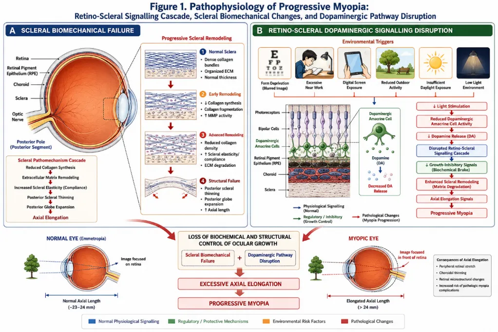
Figure 1. Pathophysiology of Progressive Myopia.
Schematic illustrating the dual pathomechanisms underlying myopia progression: (A) scleral biomechanical failure characterised by reduced collagen density, increased elasticity, and progressive thinning of the posterior sclera; (B) disruption of the retino-scleral dopaminergic signalling cascade, in which reduced retinal dopamine production secondary to form deprivation or insufficient light exposure attenuates the biochemical brake on axial elongation. Adapted and extended from Janowski et al. (2015).
4. CONVENTIONAL MYOPIA MANAGEMENT STRATEGIES:
4.1 Optical Correction
Standard spectacle lenses and soft contact lenses remain the most widely utilised modality for myopia correction globally. They effectively restore distance visual acuity but do not alter the course of axial elongation. Single-vision spectacles may, paradoxically, impose peripheral hyperopic defocus a proposed stimulus for axial elongation and are therefore not recommended as myopia control interventions.24
4.2 Orthokeratology (Ortho-K)
Orthokeratology involves the overnight use of specially designed rigid gas-permeable contact lenses that temporarily reshape the corneal epithelium, providing unaided daytime vision while simultaneously creating a pattern of peripheral myopic defocus hypothesised to retard axial elongation.9 The primary effect on refraction is reversed within 72 hours of lens cessation, confirming its temporary mechanical action.
Multiple controlled trials demonstrate that orthokeratology slows axial elongation by approximately 43–55% compared to single-vision control lenses. A five-year follow-up study demonstrated sustained but diminishing efficacy beyond four years.25 Limitations include the requirement for nightly lens wear, risk of infectious keratitis and corneal abrasions both of which are particularly concerning in young children and qualification criteria excluding approximately 50% of myopic children.3
4.3 Low-Dose Atropine
Low-dose atropine particularly at concentrations of 0.01% has emerged as the pharmacological myopia control strategy with the most favourable efficacy-to-side-effect ratio.9 The Atropine in the Treatment of Myopia (ATOM) studies demonstrated that 0.01% atropine reduced myopia progression by approximately 50–60% over two years while minimising pupillary dilation and accommodation loss.26 However, discontinuation of atropine treatment frequently results in accelerated myopic rebound, and the molecular mechanisms underlying its anti-myopic effect remain incompletely elucidated.3
Higher concentrations of atropine (0.5–1.0%) achieve the greatest efficacy, producing near-complete arrest of myopia progression through cycloplegia, but are associated with unacceptable side effects including significant photophobia, loss of near accommodation, and patient non-compliance necessitating years of continuous treatment.
4.4 Multifocal and Dual-Focus Lenses
Multifocal and bifocal spectacle lenses, as well as dual-focus soft contact lenses, have been investigated as optical myopia control strategies. These lenses introduce simultaneous myopic peripheral defocus theoretically modulating the peripheral retinal stimulus for axial elongation.9 Clinical trials with dual-focus soft contact lenses demonstrate approximately 37–54% reduction in axial elongation compared to single-vision lenses.27 Effects are modest, cumulative, and imperfectly sustained.
4.5 Increased Outdoor Exposure
Prospective studies and meta-analyses consistently demonstrate that 80–90 additional minutes of outdoor time daily reduces the annual incidence of myopia by approximately 50%.28 The mechanism is principally attributed to high outdoor luminance stimulating retinal dopamine release, though ultraviolet light effects on ocular growth and vitamin D-mediated pathways have also been proposed, without consistent confirmatory evidence.3
4.6 Posterior Scleral Reinforcement (PSR) Surgery
Posterior scleral reinforcement surgery involves the surgical implantation of a reinforcing material typically cadaveric fascia lata, donor sclera, or synthetic polymer scaffolds at the posterior aspect of the globe to mechanically limit further axial elongation.29 Case series from China and Eastern Europe describe arrest of myopia progression in a proportion of patients, but the procedure carries significant operative risks including cilioretinal artery occlusion, binocular diplopia from extraocular muscle weakness, and impaired venous outflow.3 Its application is therefore restricted to severe, rapidly progressive myopia where conservative measures have failed.
Table 1. Comparison of Current Myopia Control Strategies
|
Modality |
Efficacy (Axial Length Reduction) |
Duration of Effect |
Key Limitations |
|
Single-vision spectacles |
None |
N/A |
May worsen peripheral defocus |
|
Low-dose atropine 0.01% |
~50–60% |
Years (rebound on cessation) |
Long-term use; rebound effect |
|
Orthokeratology |
~43–55% |
4–5 years |
Infectious risk; compliance; 50% ineligible |
|
Dual-focus contact lenses |
~37–54% |
While worn |
Modest effect; contact lens risks |
|
Outdoor exposure |
~50% incidence reduction |
Ongoing |
Behaviour change; not curative |
|
PSR Surgery |
Variable; arrest in some |
Potentially permanent |
Operative risks; limited access |
|
Stem Cell Therapy (proposed) |
Potentially high (TBD) |
Potentially life-long |
Investigational; no human trials yet |
5. STEM CELL BIOLOGY: FOUNDATIONS FOR OCULAR APPLICATIONS:
5.1 Classification and Properties of Stem Cells
Stem cells are undifferentiated or partially differentiated cells characterised by two fundamental biological properties: the capacity for self-renewal through mitotic division, and the ability to differentiate into one or more specialised cell types.30 Stem cells are broadly classified by their developmental potency as totipotent (capable of forming all cell lineages including extraembryonic tissues), pluripotent (embryonic stem cells [ESCs] and induced pluripotent stem cells [iPSCs]), multipotent (including MSCs, haematopoietic stem cells, and neural stem cells), oligopotent, and unipotent.
Mesenchymal stem cells (MSCs), derivable from bone marrow, adipose tissue, umbilical cord blood, and the sclera itself, are of particular relevance to myopia therapy.31 MSCs exhibit multipotent differentiation capacity into mesodermal lineages including osteoblasts, chondrocytes, and fibroblasts the latter being the principal cell type responsible for scleral collagen synthesis. Beyond direct cell replacement, MSCs exert potent paracrine, anti-inflammatory, and trophic effects via secretion of growth factors, cytokines, and extracellular vesicles.
5.2 Stem Cells in Ocular Disease: Established Applications
The eye has emerged as a particularly attractive organ for stem cell therapy trials, owing to its immunologically privileged status, accessibility, precise anatomical boundaries, and availability of sensitive functional outcome measures.32 The subretinal transplantation of human embryonic stem cell-derived retinal pigment epithelium (hESC-RPE) in patients with age-related macular degeneration and Stargardt's disease has demonstrated preliminary evidence of safety, graft survival, and functional improvement in small, early-phase studies.33
Stem cell therapy for corneal limbal stem cell deficiency is among the most clinically advanced applications, with autologous limbal epithelial stem cell transplantation achieving successful restoration of corneal epithelial integrity in a substantial proportion of treated eyes.34 For retinitis pigmentosa, photoreceptor replacement strategies utilising ESC- and iPSC-derived photoreceptor precursors have demonstrated partial functional recovery in murine models, paving the way for early human trials. These precedents provide proof-of-concept for the application of stem cell strategies to additional ocular conditions, including myopia.
5.3 Scleral Stem Cells: An Endogenous Source
A particularly significant discovery for myopia therapy is the identification of endogenous multipotent stem and progenitor cells within the sclera itself.35 Tsai and colleagues demonstrated the presence of scleral multipotent stem/progenitor cells in murine scleral tissue capable of differentiating into fibroblasts, chondrocytes, and adipocytes in vitro. This finding raises the compelling possibility that endogenous scleral stem cells could be pharmacologically stimulated to proliferate and differentiate into collagen-producing fibroblasts, reinforcing scleral biomechanics from within an approach that would circumvent the requirement for exogenous cell transplantation entirely.
6. STEM CELL-BASED STRATEGIES FOR MYOPIA: SCIENTIFIC RATIONALE AND EVIDENCE:
6.1 Concept and Overview
Janowski and colleagues (2015), in a seminal conceptual paper published in Stem Cells, were among the first to formally propose and articulate the scientific rationale for stem cell therapy as a disease-modifying intervention for progressive myopia.3 Their framework identified two convergent pathomechanisms amenable to stem cell intervention: (1) structural scleral weakness, addressable by MSC-based scleral reinforcement; and (2) disrupted retino-scleral dopaminergic signalling, addressable by transplantation of dopamine-producing stem cells.
The authors proposed that a single, minimally invasive subscleral injection of appropriately prepared stem cells in childhood could, in principle, provide lifelong protection against myopia progression analogous in conceptual terms to childhood vaccination against infectious disease.3 This vision of a "one-time intervention" with enduring biological effect represents a profound departure from existing myopia control paradigms requiring years of compliance with pharmacological or optical treatments.
6.2 MSC-Based Scleral Reinforcement
6.2.1 Biological Rationale
As discussed, the myopic sclera is characterised by weakness, thinning, and reduced collagen content features shared with connective tissue disorders amenable to MSC therapy. MSCs derived from bone marrow, adipose tissue, or umbilical cord possess robust capacity to differentiate into type I collagen-producing fibroblasts under appropriate culture conditions.14 Transplanted MSCs within the subscleral space would be expected to: (a) physically contribute to scleral structural reinforcement; (b) differentiate into fibroblasts that synthesise and deposit extracellular matrix, increasing collagen fibril density; and (c) provide paracrine signals that modulate local inflammatory and remodelling processes, creating a more favourable scleral biomechanical environment.
6.2.2 Delivery Route: Subscleral Injection
The proposed delivery route for MSC-based scleral reinforcement is subscleral injection deposition of the cell suspension into the anatomical space between the sclera and the choroid, adjacent to the posterior pole.3 This approach leverages recently developed micro-needle-based delivery systems that permit minimally invasive access to the suprachoroidal/subscleral space, as demonstrated in animal models for the treatment of acute posterior uveitis with triamcinolone acetonide.36 The subscleral space has the critical advantage of positioning transplanted cells within the retina-sclera signalling loop enabling dynamic, physiologically integrated therapeutic responses in contrast to retrobulbar injection, which provides only external mechanical support without participation in the biochemical signalling environment.
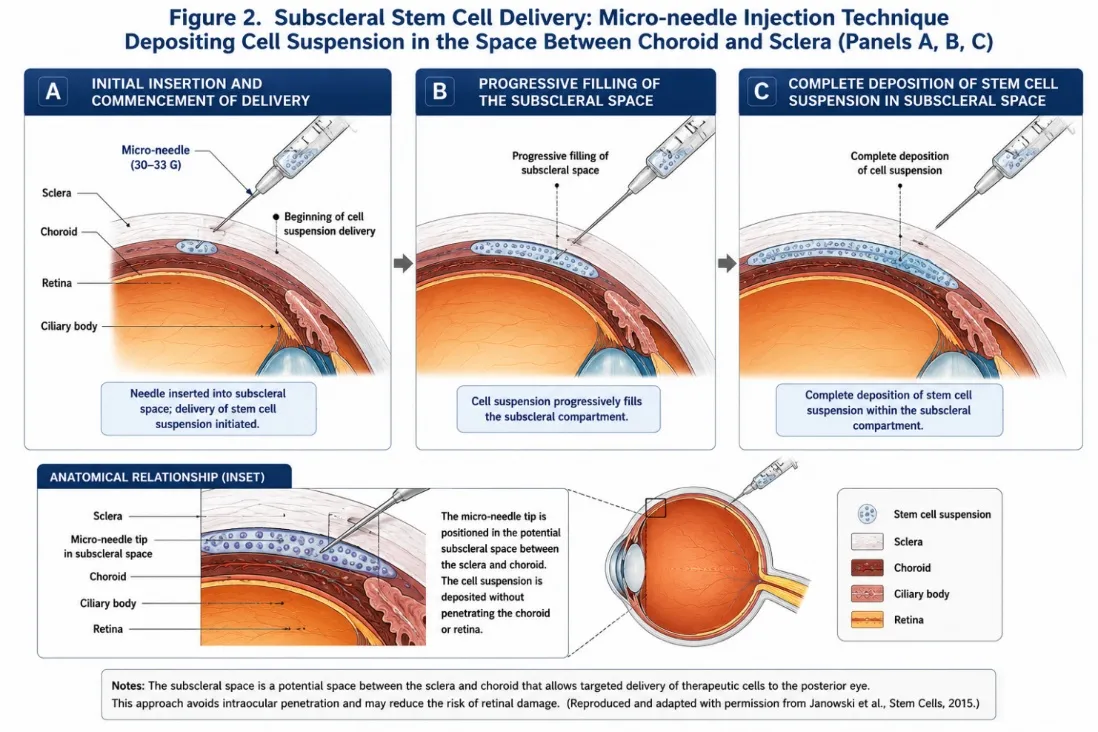
Figure 2. Subscleral Stem Cell Delivery System.
Sequential depiction of subscleral stem cell injection technique. (A) Initial insertion of the micro-needle with commencement of cell suspension delivery; (B) progressive filling of the subscleral space with the cell payload; (C) complete deposition of stem cell suspension within the subscleral compartment. Inset demonstrates the anatomical relationship of the needle tip to the sclera, choroid, ciliary body, and retina. Reproduced and adapted with permission from Janowski et al., Stem Cells, 2015.
6.2.3 Autologous versus Allogeneic MSCs
Autologous MSC transplantation wherein cells are derived from the patient's own bone marrow or adipose tissue, cultured, and reinjected offers the theoretical advantage of immunological compatibility, avoiding the requirement for immunosuppression.3 However, autologous MSCs derived from genetically predisposed individuals would carry the same myopia-susceptibility loci potentially contributing to their cellular dysfunction. The alternative allogeneic MSC transplantation from healthy donors would circumvent this limitation but necessitates immunosuppressive regimens with their attendant risks, given increasing evidence that MSCs are not universally immunoprivileged.37 A third approach ex vivo genetic correction of autologous cells prior to transplantation represents a theoretically elegant but technically demanding solution that may become feasible as gene editing technologies mature.
6.3 Dopaminergic Stem Cell Transplantation
6.3.1 Dopamine as a Therapeutic Target
The substantial preclinical evidence base for dopamine's role in myopia control, reviewed above, strongly motivates investigation of dopaminergic stem cell strategies. The eye naturally requires dopamine from retinal amacrine cells to regulate postnatal ocular growth.16 In myopic eyes, this endogenous dopamine production is insufficient. Restoring dopamine signalling through transplanted dopaminergic cells could therefore constitute a biologically coherent approach to limiting axial elongation.
6.3.2 Sources of Dopaminergic Cells
Several sources of dopamine-producing cells are available for therapeutic application:3
(i) Fetal mesencephalic dopaminergic neurons: The first dopaminergic cells transplanted clinically, in Parkinson's disease patients, with demonstrated efficacy and a quarter-century of evidence base. Ethical and practical limitations restrict their widespread use.
(ii) Embryonic stem cell-derived dopaminergic neurons: Floor-plate-based differentiation protocols reliably generate functional midbrain dopaminergic neurons from human ESCs, representing a scalable allogeneic source.
(iii) Induced pluripotent stem cell-derived dopaminergic neurons: Patient-specific iPSCs can be reprogrammed from somatic cells and differentiated into dopaminergic neurons, offering the prospect of personalised, autologous dopaminergic cell therapy.
(iv) Genetically engineered MSCs: Lentiviral transduction of MSCs with tyrosine hydroxylase (TH) the rate-limiting enzyme in dopamine biosynthesis has been demonstrated to be effective in experimental Parkinson's disease models. This approach combines the structural scleral support of MSCs with dopaminergic signalling, theoretically addressing both pathomechanisms of myopia simultaneously.
6.3.3 Optogenetic Enhancement
A particularly innovative refinement of the dopaminergic stem cell approach employs optogenetics the engineering of cells to express light-activated ion channels or enzymes to create dopamine-producing cells whose activity is gated by light stimulation.3,38 This would reconstitute the physiological coupling between outdoor light exposure and retinal dopamine release, effectively amplifying the protective effect of sunlight on ocular growth regulation while preventing constitutive dopamine toxicity in the dark.
7. PRECLINICAL EVIDENCE AND ANIMAL MODELS
7.1 Animal Models of Myopia
Two principal experimental paradigms are used to induce myopia in animal models:3
Form-Deprivation Myopia (FDM): Induced by occluding the eyelids or fitting diffuser lenses that degrade retinal image quality without imposing a specific defocus signal. FDM robustly produces axial elongation across multiple species including mice, rats, guinea pigs, rabbits, chicks, and primates.
Lens-Induced Myopia (LIM): Produced by fitting negative-power lenses that impose hyperopic defocus on the retina, stimulating compensatory axial elongation. LIM more closely models the environmental near-work conditions associated with human myopia.
The porcine model has been proposed as particularly suitable for preclinical stem cell therapy studies, given the comparable size of porcine and human eyes, and the availability of transgenic swine with targeted knockout of myopia-related genes through NIH National Swine Resource and Research Center resources.3
7.2 Evidence from Dopaminergic Interventions
The preclinical evidence base for dopaminergic modulation of myopia is substantial. Retinal dopamine levels in form-deprived eyes are reduced relative to controls, and this reduction temporally precedes the development of myopia.17 The dopamine agonist apomorphine, applied locally or systemically, produces a dose-dependent anti-myopic effect in primate and chick models, acting through D2 receptors in the retina or retinal pigment epithelium.39 Direct intravitreal injection of dopamine in lid-suture myopic rabbits significantly slowed myopia progression.18
L-DOPA administration in guinea pig models of form-deprivation myopia inhibited myopia development, paralleling observations from retrospective analyses of children receiving L-DOPA for amblyopia treatment, who exhibited attenuated myopia progression.19 GABA antagonists have been shown to have an additive effect with dopaminergic agonists in preventing form-deprivation myopia, suggesting potential for combination strategies.40
7.3 Subscleral Delivery: Proof-of-Concept Studies
The feasibility of subscleral drug and biomaterial delivery has been demonstrated in multiple preclinical models. Gilger and colleagues demonstrated that triamcinolone acetonide delivered to the suprachoroidal space via micro-needle injection effectively treated acute posterior uveitis in a porcine model without causing haemorrhage or retinal detachment.36 Biomaterial scaffolds delivered to the subscleral space via similar routes have demonstrated stable retention over observation periods relevant to a therapeutic timeline.3 These data establish proof-of-concept for the delivery route proposed for MSC transplantation, though stem cell-specific pharmacokinetics, survival, and integration dynamics require dedicated investigation.
7.4 Gaps in the Preclinical Evidence Base
Despite the compelling biological rationale, several critical gaps in the preclinical evidence base must be acknowledged. No published study has yet specifically evaluated MSC transplantation into the subscleral space in a myopia model, examined the fate and integration of transplanted dopaminergic cells in the myopic eye, or directly compared the efficacy of stem cell strategies to standard-of-care myopia control interventions in a head-to-head preclinical trial.3 These studies represent the necessary next steps in the translational pathway and should be prioritised in the research agenda.
8. TRANSLATIONAL CHALLENGES AND SAFETY CONSIDERATIONS:
8.1 Cell Sourcing, Expansion, and Quality Control
The clinical translation of stem cell therapies for myopia requires resolution of several practical challenges beginning at the level of cell manufacturing. Autologous MSC derivation from bone marrow aspiration or adipose tissue carries procedural morbidity, and the resultant cells exhibit significant inter-patient variability in expansion capacity, differentiation potential, and paracrine activity.41 Allogeneic off-the-shelf cell products would offer greater scalability and consistency, but necessitate immunological characterisation and potentially immunosuppressive management.
Standardised good manufacturing practice (GMP)-compliant protocols for cell expansion, quality control testing (sterility, identity, potency, karyotypic stability), and cryopreservation are prerequisites for any clinical application. The stem cell therapy landscape for other ocular conditions has navigated these challenges with variable success, and the lessons learned inform the myopia field.
8.2 Immune Rejection and Immunosuppression
The immunoprivileged status of the eye conferred by the blood-retinal barrier, local immunosuppressive cytokine milieu, and anterior chamber-associated immune deviation (ACAID) has been invoked as a potential advantage for intraocular stem cell transplantation. However, the subscleral space may not share the same degree of immunoprivilege as the intravitreal or subretinal compartments.37 MSCs have historically been considered immunoevasive, but this assumption has been increasingly challenged by evidence of T-cell-mediated rejection in multiple clinical contexts, necessitating careful immunological assessment in future preclinical studies.
8.3 Tumorigenicity and Genotoxicity
The pluripotency that confers therapeutic flexibility on ESC- and iPSC-derived cells simultaneously introduces the risk of teratoma formation should undifferentiated cells persist in the transplant product. Rigorous differentiation protocols, negative selection strategies, and inclusion of inducible safety switches (e.g., conditional transgene silencing systems for off-target dopamine production) are essential safety considerations.3 The use of more restricted progenitor or terminally differentiated cells reduces but does not eliminate oncological concerns associated with ex vivo expansion. Long-term post-transplantation follow-up in preclinical models for evidence of neoplastic transformation is mandatory.
8.4 Long-term Efficacy and Durability
A fundamental question for stem cell myopia therapy is the expected durability of effect. Myopia typically progresses through adolescence, with a period of relative stabilisation in the late teenage years. A stem cell intervention delivered in early childhood (5–8 years of age) would ideally provide protection through this entire vulnerable window a period of a decade or more.3 MSC survival in ocular tissues beyond 12 months has not been systematically characterised. The possibility of waning effect necessitating re-treatment must be considered and is particularly relevant given that repeat subscleral injections in the context of an already thin myopic sclera carry cumulative procedural risks.
8.5 Regulatory and Ethical Framework
Stem cell therapies for myopia present unique regulatory and ethical challenges. Myopia, unlike conditions such as age-related macular degeneration or retinitis pigmentosa, is not inherently blinding in its mild or moderate forms. The ethical calculus for exposing children to an investigational cellular intervention with attendant risks in a condition managed by safer established alternatives must be carefully navigated, with the benefit-risk threshold appropriately calibrated to severity.3
Regulatory pathways for advanced therapy medicinal products (ATMPs) in the European Union and analogous cell and gene therapy frameworks in the United States, India (CDSCO), and Japan (PMDA) impose rigorous requirements for preclinical safety data packages before Investigational New Drug (IND) or equivalent applications can be submitted. The International Society for Stem Cell Research (ISSCR) guidelines provide an ethical framework for the responsible progression of stem cell therapies from bench to bedside.
9. FUTURE DIRECTIONS AND RESEARCH AGENDA:
9.1 Priority Preclinical Studies
The immediate research priorities for stem cell myopia therapy are clear: (1) in vitro characterisation of MSC differentiation into scleral fibroblasts and collagen-producing phenotypes under myopia-relevant culture conditions; (2) in vivo assessment of MSC subscleral injection safety and tolerability in non-myopic large animal models with human-scale eyes (porcine); (3) efficacy evaluation of subscleral MSC transplantation in form-deprivation and lens-induced myopia porcine models, with axial length, scleral biomechanics, and collagen content as outcome measures; and (4) parallel evaluation of dopaminergic cell transplantation in equivalent models, with retinal dopamine levels and downstream signalling as mechanistic endpoints.
9.2 Combination Strategies
The two proposed stem cell strategies for myopia scleral reinforcement and dopaminergic restoration are biologically complementary, addressing structural and signalling deficits respectively. Their combination in a single co-transplantation regimen, potentially using TH-transduced MSCs that simultaneously provide structural support and dopamine production, may offer superior and more durable efficacy than either strategy alone.3 Anti-myopic growth factors including bFGF, which promotes survival of dopaminergic cells, and strategies targeting TGF-β signalling a proposed susceptibility pathway for severe myopia represent additional combinatorial opportunities.
9.3 Gene Therapy as a Complementary Modality
Gene therapy delivery of corrective genetic material to restore normal function of myopia-susceptibility gene products represents a complementary but distinct therapeutic approach.3 The genetic heterogeneity of myopia, with over 30 identified susceptibility loci and likely many more yet to be discovered, renders a universal gene therapy approach challenging. Personalised exome-based strategies to correct patient-specific mutations, delivered via the same subscleral route proposed for cell therapy, are theoretically feasible but technically demanding. Advances in CRISPR-Cas9 base editing and epigenome editing may enable more targeted interventions as the molecular genetics of myopia are further characterised.
9.4 Artificial Intelligence and Predictive Modelling
Artificial intelligence (AI) and machine learning algorithms, applied to large-scale longitudinal datasets of paediatric refractive error, axial length, environmental exposure data, and genetic profiles, have the potential to transform myopia risk stratification and personalised treatment planning. AI-based prediction of myopia onset and progression trajectory could identify children most likely to benefit from aggressive intervention including experimental stem cell strategies thereby optimising the benefit-risk balance at an individual patient level.
9.5 Biomarkers of Treatment Response
Identification and validation of biomarkers capable of predicting, monitoring, and confirming therapeutic response to stem cell interventions will be essential for clinical trial design and regulatory evaluation. Candidate biomarkers include: retinal dopamine levels (assessable by novel non-invasive imaging correlates), scleral biomechanical parameters (by ultrasound elastography or MRI-based methods), choroidal thickness (by optical coherence tomography), and systemic MSC engraftment markers. A validated biomarker framework will also support regulatory approval by providing objective surrogate endpoint data.
10. PROPOSED CLINICAL TRANSLATION ROADMAP
A responsible translational roadmap for stem cell therapy in myopia encompasses the following sequential phases:
Phase 0 In vitro characterisation (Years 1–2): Optimise MSC and dopaminergic cell culture, differentiation, and quality criteria; assess gene-engineered cell lines for dopamine production and safety.
Phase 1 Preclinical Small animal proof-of-concept (Years 2–4): Assess subscleral MSC and dopaminergic cell delivery in rodent and guinea pig myopia models for short-term safety, biodistribution, and preliminary efficacy.
Phase 2 Preclinical Large animal efficacy (Years 4–7): Pivotal porcine model studies with human-scale eyes, assessing axial length, scleral biomechanics, retinal function, and safety over 18–24 months.
Regulatory Submission IND/CTA (Year 7–8): Compilation of complete preclinical data package; engagement with regulatory agencies (FDA, EMA, CDSCO); submission of IND/CTA application for Phase I first-in-human trials.
Phase I Human Trial Safety and tolerability (Years 8–11): Open-label dose-escalation study in adults with high progressive myopia (≥ –6D), assessing safety, procedure tolerability, pharmacokinetics, and preliminary activity.
Phase II Human Trial Efficacy (Years 11–15): Randomised controlled trial in children aged 8–12 years with rapidly progressing moderate-to-high myopia, comparing stem cell therapy to best standard-of-care comparator, with axial length as the primary endpoint.
DISCUSSION
The myopia pandemic represents one of the defining public health challenges of the 21st century, with global projections indicating that nearly 5 billion individuals will be affected by 2050. The personal, societal, and economic costs of unchecked myopia progression particularly the downstream burden of blinding complications in adulthood demand urgent investment in genuinely disease-modifying therapies.
Current myopia control strategies, while representing meaningful clinical advances, are constrained by several fundamental limitations. They require sustained compliance over years to decades, offer only partial efficacy in arresting axial elongation, and are associated with rebound progression on cessation. None addresses the root structural and signalling pathology driving myopia.9 Posterior scleral reinforcement surgery provides the most direct structural intervention but is invasive, technically demanding, and associated with significant morbidity.29
Stem cell therapy, as conceptually articulated by Janowski et al.3 and extended by subsequent evidence reviewed in this paper, offers the prospect of a biologically integrated, potentially curative approach. The convergence of MSC biology with scleral pathophysiology, and of dopaminergic stem cell science with the retino-scleral signalling literature, provides a coherent and compelling scientific basis for further investigation. The subscleral delivery route offers both anatomical accessibility and physiological integration, and the minimally invasive micro-needle technology required for delivery is already in clinical development for other indications.
The comparison with childhood vaccination deserves further elaboration. A vaccine administered once in childhood confers protection that persists across the vulnerable developmental window and into adulthood. A stem cell intervention offering equivalent biological durability reinforcing the sclera and restoring dopaminergic signalling for the critical 5–18 year developmental period would transform the management of myopia from a condition requiring lifelong management to one that can be effectively addressed with a single early intervention.3
Nevertheless, the significant gaps in the current evidence base demand intellectual honesty. No preclinical study has yet directly tested MSC or dopaminergic stem cell transplantation in a myopia model. No human clinical data exist. The biological questions of cell survival, integration, durability, and immune compatibility in the specific context of the subscleral space in myopic eyes remain entirely unanswered. These gaps do not undermine the scientific rationale but identify the work that must be done before clinical translation can be responsibly pursued.
The parallel advances in iPSC technology, gene editing, biomaterial scaffolding, optogenetics, and AI-driven patient stratification provide an enabling ecosystem within which the necessary preclinical programme can be efficiently designed and executed. The convergence of these technologies in the next decade may substantially accelerate the translational timeline.
CONCLUSION
Myopia has attained pandemic status globally, with its trajectory unchecked by current management strategies that address symptoms without resolving the underlying pathophysiology. Stem cell therapy particularly MSC-based scleral reinforcement and dopaminergic cell transplantation offers a scientifically grounded, potentially transformative approach to arresting myopia progression at its biological roots.
The scientific rationale is robust, integrating decades of scleral biomechanics research with the established dopaminergic myopia literature and the growing body of evidence supporting MSC applications in connective tissue disorders.3 The technological infrastructure for subscleral cell delivery, cell manufacturing, and safety monitoring is increasingly available.
However, the translational pathway is long, and the field is at an early conceptual stage. Systematic preclinical validation in appropriate animal models, safety and biodistribution studies, regulatory engagement, and thoughtful first-in-human trial design are all required before stem cell therapy can be responsibly offered to myopic patients. The paediatric target population demands the highest evidentiary and ethical standards.
The vision of a childhood stem cell intervention that prevents the lifetime burden of progressive myopia and its blinding complications is compelling and scientifically plausible. The research community, regulatory agencies, and funding bodies should recognise this as a priority area warranting coordinated, well-resourced investigation. The next decade may determine whether this vision becomes clinical reality.
REFERENCES

Pratima Sagar, Sharvari Chavan, Stem Cell Therapy in Myopia: Current Evidence, Emerging Strategies, and Future Directions: A Systematic Narrative Review, Int. J. of Pharm. Sci., 2026, Vol 4, Issue 7, 4115-4131, https://doi.org/10.5281/zenodo.21470885
 10.5281/zenodo.21470885