

We use cookies to ensure our website works properly and to personalise your experience. Cookies policy
1Department of Chemistry, MMH College Ghaziabad {CCSU Meerut}, UP, India.
2Department of Chemistry, KMGGPG College Badalpur, {CCSU Meerut}, UP, India.
3Assistant Professor, Department of Chemistry, KMGGPG College Badalpur, {CCSU Meerut}, UP, India.
Tetradentate Schiff base ligand (L) from 2,5-hexanedione and 2-aminophenol has been synthesised. The ligand (L) was characterized by FT-IR, 1H NMR and mass spectrometry. the computational methods such as Density Functional Theory (DFT), molecular docking, and ADME studies have been employed to provide the theoretical assessment of the quantum calculations and biological activity. frontier molecular orbitals, energy gap, bond length and bond angle of optimised geometry of Schiff base (L) were all examined using molecular modeling techniques. The efficacy of Schiff base ligand inhibition against the estrogen receptor (3ERT) and CDK2 (3QTR) was predicted using a molecular docking analysis. Additionally, an in-silico ADME analysis was conducted to assess the pharmacokinetic properties of the Schiff base ligand.
Hugo Schiff, who initially reported the reversible acid-catalyzed condensation reaction involving primary amines and carbonyl compounds, is recognized with synthesizing the Schiff base [1]. The development of coordination chemistry has been greatly aided by the interesting family of heterocyclic molecules known as Schiff base ligands, which include azomethine bond (C=N) [2]. In coordination chemistry, Schiff base ligands with tetra dentate (ONNO) donor sites have become one of the most extensively studied ligands due to their adaptability and broad range of complexing with various metal ions, which further enhanced their biological activity [3]. One of the most important public health concerns is cancer, which is also the second most common cause of death worldwide, behind cardiac arrest. Approximately 10 million fatalities and 19.3 million new instances of cancer were reported globally, according to current data [4]. Promising compounds for cancer treatment are transition metal complexes having anticancer potential. Therefore, several research groups are actively focused on developing novel chemotherapeutic Schiff bases and related metal complexes [5]. In addition to this, they show Antimicrobial[6], anti-inflammatory[7], anti-tuberculosis[8], anti-cancer[9], and antioxidant activities[10] Schiff bases have been used as ligand synthesis intermediates to produce metal complexes with a variety of distinct structures [11].
In this research article, we have been synthesised Schiff base ligand derived from 2,5-hexanedione and 2-aminophenol and characterized different spectroscopic methods. The Density Functional Theory (DFT), molecular docking, and ADME of 2-aminophenol derived Schiff base ligand have been studied.
2 MATERIALS AND METHODS
2.1. Chemicals and instruments
The chemicals and solvents are of AR grade and were purchased from TCI and CDH and used as received with no further purification. The IR spectra recorded using NICOLET iS50 FT-IR with diamond ATR was used in the range 4000-400 cm-1. 1H-NMR spectra (Bruker Avance Neo) were collected at 400 MHz using DMSO-d₆ as solvents. HRMS analysis using Waters Xevo G2-XS QTOF instrument.
3 Experimental sections
3.1. Preparation of ligand L
Hot solution of hexane-2,5-dione (1mmol) in 20 mL ethyl alcohol was mixed with a hot solution of 2-aminophenol (2 mmol) in 20 mL ethyl alcohol. Small amount of CH3COOH was added in reaction mixture. The Reaction mixture refluxed for 4hr (figure 1) and then filtered hot reaction mixture. Cool to at room temperature and leave to for slow evaporation for 4-5 days. Brown colour power obtained and washed with cold ethyl alcohol, after that filtered and dried at room temperature. Mp:120°C
| Figure 1: Scheme of Synthesis of Schiff Base (L) |
|---|
RESULTS AND DISCUSSION
4.1. Characterization of Ligands L (FT-IR, HNMR, HRMS)
The proposed structure of the synthesized bis-Schiff base ligand (L) was systematically evaluated using FTIR, 1H-NMR, and Mass spectral analyses. The zoomed-in HRMS spectrum supports the symmetric 1:2 bis-Schiff base ligand by showing a distinct molecular ion peak at m/z = 297.1128 (figure 3) corresponding directly to the protonated molecular ion [M+H]+ (Theoretical: 297.1603). A broad band at 3390 cm-1 corresponds to phenolic -OH groups. The strong, sharp absorption band at 1589 cm-1 is assigned to the conjugated azomethine (vC=N) stretching vibration, shifted to a lower wavenumber due to extended resonance with the aromatic rings show in figure 2. the intense peak at 746 cm-1 validates the ortho-disubstituted nature of the terminal aromatic rings. 1H-NMR spectrum in DMSO-d6. The two phenolic -OH protons appear downfield at delta 10.30 and 9.96 ppm due to geometric isomerism or hydrogen bonding. The eight aromatic protons (Ar-H) are resolved as multiplets at delta 7.18–6.85 ppm (4H) and 6.64–6.36 ppm (4H) (figure 4). In the aliphatic region, the terminal methyl groups (-C=N-CH)3) appear as two distinct singlets at delta 2.33 and 2.17 ppm (6H total), while the central methylene backbone (CH2-CH2) integrates for four protons as a multiplet between delta 1.90 and 1.62 ppm.
Analytical Data for Schiff base (L):
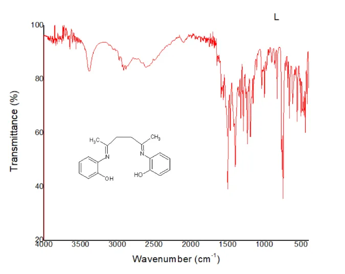
Figure 2: IR Spectrum of Schiff Base (L)
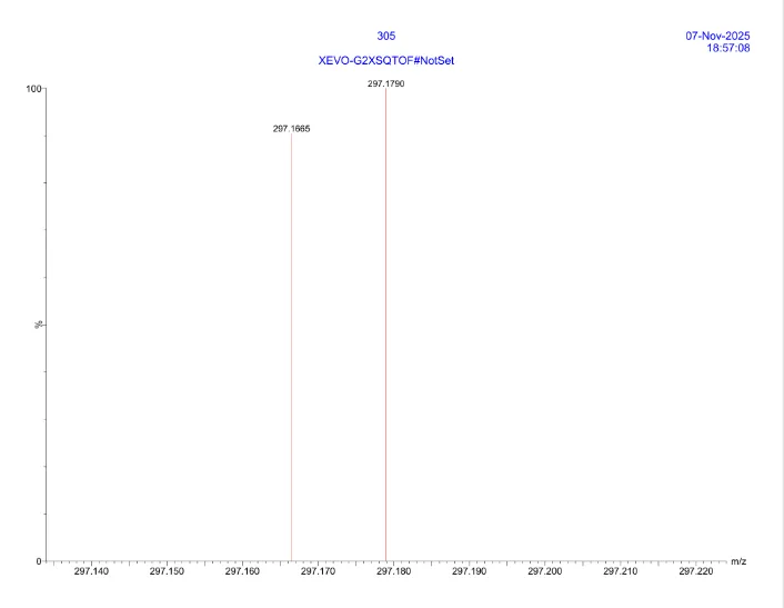
Figure 3: Mass Spectrum of L
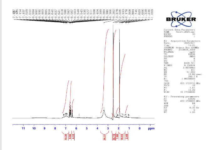
Figure 4: 1H-NMR Spectrum of L.
5. Computational Studies
5.1 Molecular Modelling
Density functional theory (DFT) is an effective method for computing the electronic structure of atoms, molecules, and solids in chemistry and physics. Calculations of electronic structure are crucial for interpreting chemical structure and reactivity. Using DFT, the structure of the prepared Schiff base ligand (L) was optimized at B3LYP level in the ground state. The RIJCOSX approximation was used to speed up the calculations.[12] The conformations of the Schiff base were clarified through studies for energy minimization using the Avogadro (1.2.0) with ORCA software (4.2.1). The basis set def2-SVP was used for calculation.
5.1.1 Geometry optimization and bonding analysis
Geometric optimization was used to obtain broad structural information of the ligands. The optimized ligand structure [The ball-stick models] is represented in [Figure 5]. The characteristic azomethine bond length C=N is observed at 1.276 Å (for C5-N1) and 128.1 Å (for C3-N2) . Table 1 provides a summary of the bond lengths and bond angles of the optimized molecular structures.
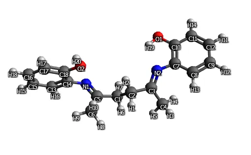
Figure 5: Geometry Optimised Structure of Schiff Base (L) [Colour Code: C-grey, N-blue, O-red,]
Table 1: Optimised Geometry of the Schiff Base (L) (Bond Lengths in Angstroms and Bond Angles in Degrees)
| Atoms | Bond length | Atoms | Bond angle |
|---|---|---|---|
| C1-C2 | 1.545 | C1-C5-N1 | 118.4 |
| C1-C5 | 1.509 | C1-C5-C6 | 117.2 |
| C5-N1 | 1.276 | C2-C3-N2 | 116.9 |
| C2-C3 | 1.510 | C1-C2-C3 | 114.7 |
| C3-N2 | 1.281 | N2-C7-C10 | 113.8 |
| C5-C6 | 1.512 | C7-C10-O1 | 119.0 |
| C3-C4 | 1.511 | C2-C1-C5 | 109.3 |
| N2-C7 | 1.398 | N2-C3-C4 | 126.4 |
| C10-O1 | 1.346 | N1-C5-C6 | 124.3 |
| N1-C14 | 1.401 | C14-C16-O2 | 117.1 |
5.1.2 Frontier molecular orbital analysis:
The highest occupied molecular orbitals (HOMO), that indicate the ability to donate an electron, and the lowest unoccupied molecular orbitals (LUMO), which indicate the ability to accept an electron, are referred to as "Frontier molecular orbitals" (FMO) [13]. A significant quantity of intramolecular charge transfer via a conjugated pathway through the electron donor component to the corresponding electron acceptor component is indicated by a small energy gap (ΔE) [14]. Figure 6 shows frontier molecular orbitals of Schiff base (L) calculated from DFT approach.
| LUMU of Ligand = -0.8442 eV Band Gap (∆E ) = 4.6762eV HOMO of Ligand = -5.5204 eV |
|---|
Figure 6: The Frontier Molecular Orbitals of Schiff Base (L) Calculated from DFT Approach.
5.1.3 Thermochemistry
DFT is used to calculate the thermodynamic characteristics of the synthesized Schiff base (L), which is illustrated in Table 2.
Table 2: Thermodynamic Data Calculated from DFT
| Electronic energy -956.70424660 Eh Zero-point energy 0.34570413 Eh Total thermal energy -956.33769249 Eh Total Enthalpy -956.33674828 Eh Final entropy term 0.06949853 Eh Final Gibbs free energy -956.40624681 Eh |
|---|
5.2 Molecular docking study
three-dimensional structures of target protein were taken from Protein Data Bank. The AutoDock tools program [15] was used for molecular docking. PyMOL and Discovery Studio software 2025 [16] were used for the visualization purpose to visualize different interactions between the target protein and Schiff base ligand.[17]
The free energy of binding for the Schiff base ligand against the MCF-7 the estrogen receptor (PDB ID: 3ERT) and Cyclin-dependent kinase 2 (PDB ID: 3QTR) were found to be −7.4 and −8.3 respectively, given in table 3. The interaction is stronger when the binding energy is more negative [18]. 3D and 2D interaction between the Schiff base (L)and 3ERT and 3QTR shows in figure 7 and 8 respectively, obtained from Discovery Studio Visualizer 2025. The ligand showed hydrophobic interaction of MET343, LEU525, LEU384, MET388, LEU525, ALA350, LEU387, LEU391 with 3ERT. The ligand also shows hydrophobic interaction with ALA144, LEU134, ILE10, LEU134, ALA31 and PHE82 of 3QTR.
Table 3: Docking Parameters of Schiff Base Ligand (L) with Cancer Protein 3ERT and 3QTR
| Target | Binding energy (kcal.mol-1) |
Interactions | ||||
|---|---|---|---|---|---|---|
| H-Bond | Hydrophobic | |||||
| Alkyl | Pi-sigma | Pi-Alkyl | Pi-Pi Stacked | |||
| 3ERT | -7.4 | LEU346 | MET343 LEU525 LEU384 MET388 |
LEU525 LEU525 |
ALA350 ALA350 LEU387 LEU391 |
|
| 3QTR | -8.3 | ILE10 | ALA144 LEU134 |
ILE10 LEU134 |
ALA31 | PHE82 |
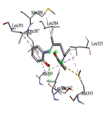
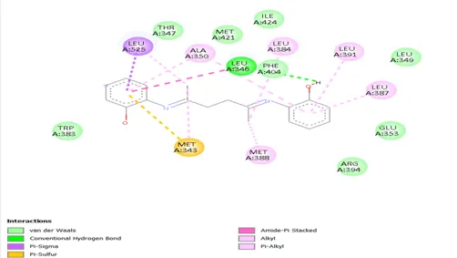
Figure 7: 3D and 2D Interaction of Schiff Base Ligand (L) with 3ERT Receptor
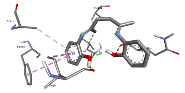
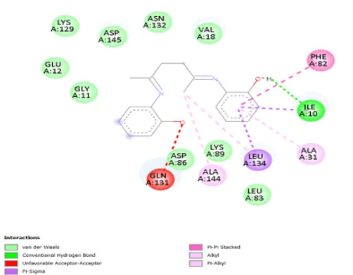
Figure 8: 3D and 2D Interaction of Schiff Base Ligand (L) with 3QTR Receptor
6. In-silico drug-likeness and ADME prediction
Schiff base (L) ADME profiles were predicted using the online tool SwissADME [13]. The Schiff base (L) has high gastrointestinal (GI) absorption as SwissAdme predictions parameters shows in table 4. There are number of H-bond acceptors is 4 and Number of H-bond Donar is 2. and follows Lipinski rule (0 violation). The L is expected to cross the blood-brain barrier. The Schiff base has a predicted bioavailability score of 0.55. Figure 9 shows bioavailability radar reflects.
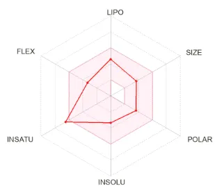
Figure 9: Bioavailability Radar Reflects
Table 4: The Predicted ADME Properties of Schiff Base (L) by SwissAdme
| Compounds | Molecular weight | No. of H-bond acceptors |
No. of H-bond donor | TPSA | BBB permeant |
GI absorption | Bioavailability Score | Lipinski |
|---|---|---|---|---|---|---|---|---|
| Schiff base (L) | 296.36 g/mol | 4 | 2 | 65.18 Ų | Yes | high | 0.55 | Yes; 0 violation |
CONCLUSION
Schiff base ligand (L) has been synthesised and characterized by FT-IR, 1H NMR and mass spectrometry. Density Functional Theory (DFT), molecular docking, and ADME studies have been employed to provide the theoretical calculations and in-silico biological activity. Frontier molecular orbitals, energy gap (∆E = 4.6762eV), bond length and bond angle of optimised geometry of Schiff base (L) were calculated. The characteristic azomethine (C=N) bond length in optimized structure of Schiff base is observed at 1.276 Å (for C5-N1) and 128.1 Å (for C3-N2). The interaction of Schiff base (L) against the estrogen receptor (3ERT) and CDK2 (3QTR) was predicted using a molecular docking analysis. The binding energy for the Schiff base ligand against the MCF-7 the estrogen receptor (3ERT) and Cyclin-dependent kinase 2 (3QTR) were found to be −7.4 and −8.3 respectively. Additionally, an in-silico ADME analysis was conducted.The Schiff base has predicted bioavailability score of 0.55 and biological experiments are required to validate the obtained results.
ACKNOWLEDGEMENT
The authors are thankful and grateful to the Principal of MMH College Ghaziabad, Uttar Pradesh (UP), India and Principal of KMGGPG College Badalpur, UP, India for their support and assistance. Seema Nagar gratefully acknowledges the Council of Scientific and Industrial Research, New Delhi, India for a Senior Research Fellowship (SRF) to her [CSIR file no.– 08/0306 (16413)/2023-EMR-I]. Authors acknowledge the USIC-DU for FT-IR, 1HNMR and IIT Jammu for HRMS characterization.
Competing interests
The authors declare no competing interests
REFERENCES

Seema Nagar, Smriti Raizada, Neha Tripathee, Synthesis, Characterization, DFT, in Silico ADME, and Molecular Docking Studies of a Schiff Base Ligand Against Cancer Targets, Int. J. of Pharm. Sci., 2026, Vol 4, Issue 11, 146-157. https://doi.org/
 10.5281/zenodo.23876969