
We use cookies to ensure our website works properly and to personalise your experience. Cookies policy
A and E College of Pharmacy, Samastipur, Bihar
To develop diclofenac sodium gel using high molecular weight hydroxypropyl methylcellulose (HPMC) and Carbopol 934P for topical and systemic delivery. Diclofenac sodium gel was prepared with HPMC K100M and Carbopol 934P as gelling agents. The formulations were examined for pH, spreadability, consistency, viscosity, homogeneity, drug content and stability. In vitro drug release was evaluated using Franz diffusion cell. Carrageenan induced rat paw oedema model was used for the evaluation of the anti-inflammatory activity of the gels. A commercial diclofenac sodium gel product was used as the reference drug. Formulations containing glycerin as permeation enhancer gave drug release patterns comparable to that of the reference product. The drug content of F2, F5 and F9 was 99.81, 99.75 and 99.96 %, respectively. Accelerated stability results showed no significant variation in the appearance and drug release after storage for 3 months. Diclofenac sodium gel containing HPMC K100M and Carbopol 934P exhibited pronounced anti-inflammatory activity and could be further developed for topical and systemic delivery.
Drug delivery through the skin has been a promising concept for a long time because the skin is easy to access, has a large surface area with vast exposure to the circulatory and lymphatic networks and the route is non-invasive[1]. Transdermal delivery is of great importance for drugs that may cause systemic side effects, e.g., non-steroidal anti-inflammatory drugs (NSAIDs) [2].
Diclofenac sodium is an effective NSAID often used in the treatment of acute and chronic arthritic conditions. It is prescribed for long-term treatment of rheumatoid arthritis, osteoarthritis and ankylosing spondylitis [3].
There is great interest to develop non-oral dosage forms of diclofenac sodium to minimize its gastric side effects and to provide relatively consistent drug levels at the application site for prolonged periods [4].
Topical delivery of diclofenac sodium using various formulations has been described in the literature. However, effective permeation of the drug through skin is difficult to achieve due to its intrinsically poor permeability, though this is relatively good compared to other commonly used NSAIDs [5]. Skin permeation enhancers can improve drug skin penetration [6].
HPMC K100M and Carbopol 934P have been used as hydrophilic polymers in the formulation of gels for transdermal drug delivery. One of their most important characteristics is high swellability, which has a significant effect on the release kinetics of the incorporated drug [7]. Cellulose ethers, especially HPMC K100M, are frequently used as the basis for sustained release hydrophilic matrix tablets [8].
The overall drug release mechanism of HPMC K100M-based pharmaceutical devices strongly depend on the design (composition and geometry) of the particular delivery system. On imbibing water, HPMC K100M swells, resulting in dramatic changes in the viscous nature of the polymer, drug concentration and increased dimension of the dosage system. Upon contact with water, the incorporated drug dissolves and diffuses out of the device. Depending on the chain length and degree of substitution of the HPMC type used, the polymer itself dissolves more or less rapidly.
Diffusion, water uptake and erosion are the most important rate-controlling mechanisms of commercially available controlled release products [9].
A series of grades based on the molecular fraction of these polymers are used at concentrations between 1 and 5 % in topical gel formulation. Carrageenan-induced paw oedema in mice has been widely used for the evaluation of antiinflammatory activity of drugs [10].
The aim of this study was to develop suitable topical formulations of diclofenac sodium using HPMC K100M and Carbopol 934P as gelling agents and glycerin as permeation enhancers.
An "in vitro diffusion study" is a laboratory experiment where the movement of molecules (diffusion) through a membrane, like a biological tissue or a synthetic barrier, is observed outside of a living organism, allowing researchers to analyze how a substance might pass through a specific barrier under controlled conditions, typically using a diffusion cell apparatus to measure the rate of diffusion across the membrane.
2.1 Key points about in vitro diffusion studies:
2.1.1 "In vitro" means "in glass":
This signifies that the experiment is conducted outside a living organism, usually in a controlled laboratory setting.
2.1.2 Diffusion process:
Molecules naturally move from an area of high concentration to an area of low concentration, which is the principle behind diffusion.
2.1.3 Diffusion cell:
A specialized device used to study diffusion, typically consisting of a donor compartment where the substance is placed, a membrane separating the compartments, and a receptor compartment where the diffused substance is collected.
2.2 Applications of in vitro diffusion studies:
Important factors in an in vitro diffusion study:
The choice of membrane (e.g., animal skin, synthetic membrane) significantly impacts the results.
Concentration of the substance being tested and the vehicle used to dissolve it.
The medium used to collect the diffused substance, often a buffer solution.
Maintaining a consistent temperature throughout the experiment.
If you've dealt with osteoarthritis pain in the past, you may be familiar with diclofenac. Diclofenac is a non-steroidal anti-inflammatory drug (NSAID) and it falls in the same class as drugs such as ibuprofen, naproxen and aspirin. Diclofenac eases pain and reduces inflammation like other NSAID medications. It works by blocking the production of certain enzymes in our body called COX1 and COX2. These enzymes are responsible for the production of chemicals called prostaglandins which cause pain and inflammation. So, when you block the production of COX 1 and 2, you reduce the production of prostaglandins, resulting in less pain and inflammation.
3.1 What is over-the-counter diclofenac gel used for?
Diclofenac sodium is the active ingredient in Voltaren Arthritis Pain, an over-the-counter arthritis pain relief gel. OTC diclofenac gel is used to relieve pain from arthritis in the knees, ankles, feet, elbows, wrists, and hands.2
Over-the-counter diclofenac sodium topical gel can be good option for people who don't want to take oral NSAIDs or they can't swallow pills.
3.2 How should I use diclofenac gel?
Over-the-counter topical diclofenac comes as a 1% gel to apply on up to two joint areas, four times daily.2 These areas can include the foot, ankle, knee, hand, wrist or elbow. It's recommended to apply your OTC diclofenac gel around the same time every day. Some basic tips for application for Voltaren Arthritis Pain diclofenac gel:
Diclofenac sodium gel may cause a severe allergic reaction, especially in people allergic to aspirin. Symptoms include:3
Make sure to keep your diclofenac sodium topical gel in the container it came in and out of reach of children or pets. It's important to keep it out of sight and high enough that children can't reach it or see it.
3.3 Who should use diclofenac gel?
Before using topical diclofenac gel, you should read the product label to determine if it is right for you. People with certain underlying medical conditions are advised to talk to their doctor before use so it is important to understand if this includes you. It may not be right for you if you already take oral NSAIDs, you have sensitive skin, or you have several affected joints.4
There are also several drugs’ interactions you should consider if you're considering using OTC diclofenac gel. Ask your doctor or pharmacist before using diclofenac gel if you are taking any other pain reliever or are planning on taking any other pain reliever, especially prescription or nonprescription NSAIDs (aspirin, ibuprofen, naproxen, or others).
Now that you know more about diclofenac gel, the active ingredient in Voltaren, make sure to visit our arthritis resource center to learn more about managing your arthritis symptoms.
Content contained in this article is not meant to be used as a direct indication of Voltaren product use or results.
Fig.1: Voltaren Gel (Diclofenac Sodium Topical Gel)
Fig.2: Diclofenac Sodium Topical Gel
Fig.3: Diclofenac Sodium Topical Gel
4. All the key aspects of in vitro evaluation of Diclofenac Sodium Gel:
4.1. Formulation Preparation [11]
4.2. Drug Release Studies
4.3. Permeation Studies
4.4. Viscosity and Texture Analysis
4.5. Stability Testing
4.6. Release Mechanism and Model Fitting
4.7. Drug Content and Uniformity
4.8. Microbial Testing
4.9. Irritation Testing
5.1. MATERIALS AND METHODS
5.1.1. Materials[12]
Diclofenac sodium, was procured from Research fine lab Hyderabad. Mumbai. Methyl paraben, propyl paraben, isopropyl alcohol, menthol were obtained from Samar chemicals, Nagpur. Triethanolamine from Merck chemical Mumbai, Propylene glycol Carbopol 940P from Research Fine Lab, Mumbai. Hpmc k4m from Merck Chemical Mumbai and marketed gel from Intas Pharmaceuticles Ltd, Ahmedabad
5.1.2. Methods:
Formulation of Topical gel containing Diclofenac Sodium was prepared by direct compression technique using varying concentrations of different grades of polymers with carbopol 940 and Hpmc k4m. All the ingredients were accurately weighed and passed. Then, most of the ingredients were buy from summer chemicles, Nagpur.
5.1.3. Extraction of Ginger oleoresin: [13]
Ginger oleoresin is extracted from dried crushed ginger in soxhlet extractor using ethyl ether as a solvent and linseed oil are two key ingredients added to propose formulation for antiinfllamatiory effect.
5.1.4. Two different procedures are applied to formulate gel
1) formulation of hpmc gel Diclofenac was dissolved in propylene glycol. Menthol was dissolved in Isopropyl alcohol. The whole amount of HPMC was sprinkled on drug solution with slow stirring then methyl paraben and propyl paraben were added. The mixture of drug solution and polymer was kept aside for six hours to seven hours, for adequate swelling of polymer. The oil phase consisting of linseed oil, ginger oleoresin and methyl salicylate was added slowly in above aqueous gel with continuous stirring with overhead stirrer. The gel was packed in aluminium collapsible tube. Ur
2) formulation of carbopol gel Carbopol was added in given quantity of water and kept it for 8 hours for adequate swelling of polymer. Triethanolamine was added for neutralize the above Carbopol base for the proper pH adjustment. Diclofenac was then added in propylene glycol at 65 0C.After that methyl and propyl paraben was added in the solution of propylene glycol, cooled to room temperature and the resultant was added in carbopol base with the help of mechanical stirrer by rotating at 600 rpm.
5.1.5. Solubility analysis
The solubility of diclofenac sodium was determined using various solvents of different polarities including methanol, 95 % ethanol, water, glacial acetic acid, ether, chloroform and phosphate buffer (pH 6.8). The vials containing the solvent and excess diclofenac sodium were kept in a shaker for 24 h. It was filtered through Whatman filter paper. The filtrate was analyzed by UV spectrophotometry to determine the amount of diclofenac sodium.
5.1.6. Fourier transform-infrared spectroscopy (FTIR) study
FT-IR study was used to check compatibility and interaction between the drug and the polymers. The spectra of pure diclofenac sodium and the physical mixture of diclofenac sodium and polymer, embedded in KBr discs,.were recorded in the range of 4000 cm-1 and 400 cm-1 using IR spectroscopy (Shimadzu, IR Affinity- 1).
5.1.7. Preparation of diclofenac sodium gel [14]
For formulations F1, F2 and F3, 1 g of diclofenac sodium was dissolved in 15 ml of glycerin with the aid of mild heat (solution A). Weighed quantity of HPMC K100M was added to 75 ml of distilled water and stirred until dissolved (solution B). Solutions A and B were mixed thoroughly and the final weight made up to 100 g. For formulations F4, F5 and F6, 1 g of diclofenac sodium was dissolved in 15 ml of glycerin with the aid of mild heat (solution A). Weighed quantity of Carbapol 934P was added to 75 ml of distilled water, stirred until dissolved and then neutralized with 10 % NaOH (solution B). Solutions A and B were mixed thoroughly and the final weight made up to 100 g. For formulations F7, F8 and F9, 1 g of diclofenac sodium was dissolved in 15 ml of glycerin with the aid of mild heat, and methyl paraben and propyl paraben added (solution A). Weighed quantity of sodium alginate was added to 75 ml of distilled water and stirred until dissolved (solution B). Solutions A and B were mixed thoroughly and the final weight made up to 100 g. The composition of the formulations is outlined in Table 1.
The three different hydrophilic polymers were used for study and in each formulation code the combination of two polymers were used by keeping one of the polymers constant. The different nine formulation of diclofenac sodium gel (F1-F9) with their code are listed in table 1. The gel were kept in plastic well closed container and stored at room temperature until the time of analysis.
5.1.8. In vitro evaluation of diclofenac gels [15]
The diclofenac sodium gels were subjected to evaluation for the following parameters - pH, spreadability, consistency, viscosity, homogeneisity [14], drug content, in vitro drug release studies, in vivo anti-inflammatory activity by carrageenan induced paw oedema and accelerated stability studies.
Table 1: Composition of diclofenac sodium gel
|
Formulation Code |
Drug (g) |
HPMC K100M (g) |
Carbopol 934P (g) |
10% NaOH (ml) |
Glycerin (ml) |
Propyl Paraben (g) |
Distilled water to (g) |
|
F1 |
1 |
2.0 |
0.25 |
q.s. |
15 |
- |
100 |
|
F2 |
1 |
2.0 |
0.50 |
q.s. |
15 |
- |
100 |
|
F3 |
1 |
2.0 |
0.75 |
q.s. |
15 |
- |
100 |
|
F4 |
1 |
- |
0.50 |
q.s. |
15 |
0.1 |
100 |
|
F5 |
1 |
- |
0.50 |
q.s. |
15 |
0.1 |
100 |
|
F6 |
1 |
- |
0.50 |
q.s. |
15 |
0.1 |
100 |
|
F7 |
1 |
1 |
- |
q.s. |
15 |
0.1 |
100 |
|
F8 |
1 |
1.5 |
- |
q.s. |
15 |
0.1 |
100 |
|
F9 |
1 |
2.0 |
- |
q.s. |
15 |
0.1 |
100 |
5.1.9. Determination of pH of gel formulations
The pH of the gel formulations was measured with a pH meter (Eutech, Cyberscan) using 1 % aqueous solutions of the gels at room temperature.
This parameter was determined with a wooden block and glass slide apparatus.The gel (approx. 20 g) was added to the pan and the time for the upper slide (movable) to separate completely from the fixed was noted. Spreadability was calculated formulas in Eq 1.
S = W×L/T ………… (1)
where S = spreadability, W = weight tide to upper slide, L = length of glass slide, and T = time taken to separate the slide completely from each other
5.1.10. Consistency [16]
Measurement of consistency of the gels was carried out by dropping a cone attached to a holding rod from a fix distance of 10 cm in such way that it falls in the centre of a glass cup filled with the gel. The penetration by the cone was measured from the surface of the gel to the tip of the cone inside the gel. The distance traveled by the cone after 10 s was noted.
5.1.11. Viscosity
The viscosity of the formulations was determined using a Brookfield digital viscometer (model DVII, USA) equipped with spindle S27.. The gel sample (5 g) was placed in the sample holder of the viscometer and allowed to settle for 5 min and the viscosity measured a rotating speed of 50 rpm at room temperature (25 - 27 oC).
5.1.12. Spreadability
Spreadability was performed with extensiometer apparatus. The apparatus consists of two square glass plates, 11 cm on each side. On the outside part of the inferior plate a coordinate paper is attached, on which five concentric circles with perpendicular diameter in millimeters are drawn. The spreadability was determined as follows: 1 g gel was placed between the plates and the upper plate was increasingly loaded with weights at equal time intervals . Based on the results of 3 measurements the mean calculated surfaces were plotted in the form of extensiometric curves. On Ycoordinate the ointment surfaces, in cm2 were marked and on Xcoordinate the loadings value, in grams (g) were marked. [17]
5.1.13. pH
The pH was measured in water solutions of each gel, using a digital pH meter, which was calibrated before each use with standard buffer solutions at pH 4.6 and 8.6. The solutions are prepared by dissolving 2.5 g of each gel in 25 g water [18]
5.1.14. Homogeneity
All the gels formed were tested for homogeneity by visual inspection after the gels have been allowed to set in a container. They were tested for their appearance and presence of any aggregates. [19]
5.1.15. Drug content
A quantity (100 mg) of the gel was dissolved in 100 ml of phosphate buffer of pH 6.8. The volumetric flask containing gel solution was shaken for 2 h on a mechanical shaker to allow the drug to dissolve completely. The solution was filtered and drug content determined spectrophotometrically at 276 nm using phosphate buffer (pH 6.8) as blank.
5.1.16. In-vitro evaluation of diclofenac sodium release
Pretreated skin of albino mice was used in the Franz diffusion cell experiment. The receptor compartment contained 100 ml of phosphate buffer pH 6.8. One gram of the test formulation or reference was applied to the skin over an area of 1.131 cm2 and placed across the donor compartment. The donor cell was exposed to ambient temperature and covered with parafilm to prevent evaporation. The temperature of the diffusion medium was maintained at 37 ± 1 oC while the buffer solution was stirred continuously with a Teflon-coated magnetic bar at 500 rpm. Samples (1 ml each) were withdrawn from the release medium at 30, 60, 90 and 120 min and replaced with an equal volume of fresh buffer solution to maintain sink conditions. The samples were analyzed spectrophotometrically at 276 nm against their respective blank. [20]
5.1.17. Accelerated stability studies
All the selected formulations were subjected to accelerated stability test over a period of three months as per ICH guidelines at a temperature of 40 ± 2 oC/ and 75 % relative humidity (RH). All the formulations were analyzed for changes in appearance, pH and drug content as described above.
5.1.18. Statistical analysis
The results were expressed as mean ± standard deviation. The data were analyzed by one way analysis of variance (ANOVA) followed by Bonferroni’s multiple comparison. A level of significance of p < 0.05 was set to determine any significance. [21]
6.1. Characterization of Formulations
The prepared formulations shared a smooth and homogeneous appearance. The HEC diclofenac sodium gels were transparent while Vurdon gel was white viscous, opalescent. All preparations were easily spreadable, with acceptable bioadhesion and fair mechanical properties. At table 2 are shown the values of pH, viscosity and drug content for each gel. The pH values ranged from 7.33 ± 0.016 to 8.35 ± 0.136, which are considered acceptable to avoid the risk of irritation after skin application. Viscosity is an important physical property of topical formulations, which affects the rate of drug release; in general, an increase of the viscosity vehicles would cause a more rigid structure with a consequent decrease of the rate of drug release.
6.2. Spreadability
Mean results of three measurements expressed in the form of spreadability curve are shown at figure 4.
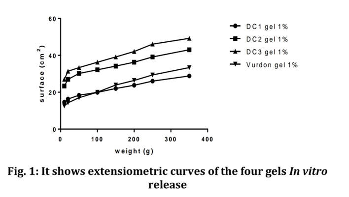
Fig. 4: It shows curves of the four gels In vitro release
The three DC formulations were found to express good spreadabilty compared with that of commercial gel. Considering the stability studies and physiochemical parameters, batch DC1 and DC3 were selected for in vitro permeability release studies as well as compared with the marketed gel. The results are shown at the figures 5 and 6.
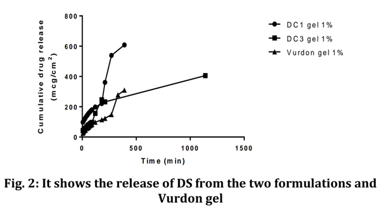
Fig. 5. It shows release of DS from the two formulations and Vurdon Gel
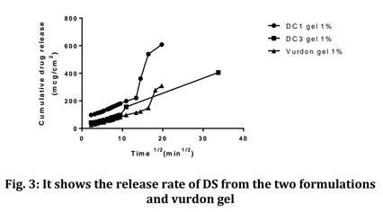
Fig. 6. It shows release rate of DS from the two formulations and Vurdon Gel
When the amounts of drug released per unit area (µg/cm2) were plotted against the square root of time, a linear relationship was obtained for each gel, showing that the release of drug from the gels could be well described by the Higuchi model, where the ratecontrolling step is the process of diffusion through the gel matrix. It is possible to calculate the steady state flux (J) from the slope of the linear portion (5-300 min) of the graph of the release rate of drug.
6.3. Drug-excipient compatibility
There were no significant changes in the peak pattern of the IR spectra of pure diclofenac sodium and of the combined drug/polymer mixture (fig 4), which implies that there was no interaction between the drug and the polymers. [22]
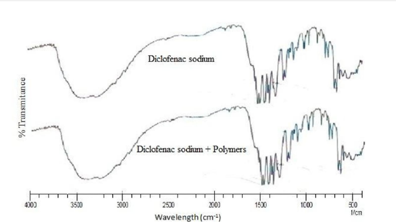
Fig 7: FT-IR spectra of pure diclofenac sodium and diclofenac sodium/polymer mixtures
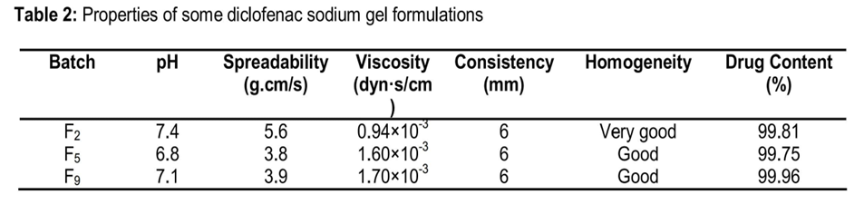
6.4. Physical properties of gel [23]
Precipitation occurred in some of the gel batches (F1, F3, F4, F6, F7 and F9) of polymer-based gel containing diclofenac sodium which was probably due to incompatibility. Hence, these batches were discarded and remaining batches (F2, F5 and F8) were used in further studies.
The results for pH, spreadability, viscosity, consistency, homogeneity and drug content are shown in Table 2. Spreadability data indicate that the gel is easily spreadable by a small amount of shear. Consistency reflects the capacity of the gel to get ejected in uniform and desired quantity when the tube is squeezed. Consistency in terms of distance travelled by the cone was 6 mm. The gel formulations were homogeneous in texture and fell within a pH range of 6.8 to 7.4 which is within the normal skin pH in healthy people. The results show that there was no significant difference between the viscosities of the gel formulations and that of the reference.
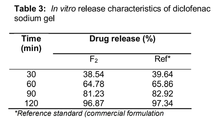
6.5. In vitro diclofenac sodium release
Diclofenac sodium release from the F2 formulation was comparable with that of the reference standard (commercial formulation), as Table 3 shows. The pH of F2 and the commercial gel was was 6.8.
6.6. In vivo anti-inflammatory activity of diclofenac gel
The results of the anti-inflammatory test (Fig 2) indicate there was no significant difference between the anti-inflammatory activities of the test formulations and the reference [24]
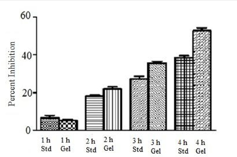
Fig 8: Inhibition of rat paw edema by reference standard (Std) and test (Gel) formulations
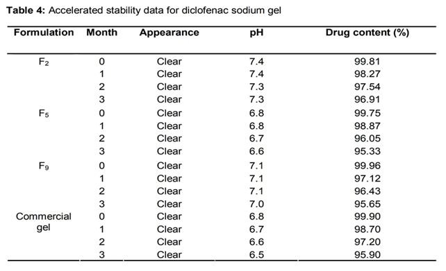
6.7.Stability of the gels
The results of the accelerated stability studies are shown in Table 4. The appearance of the gels remained clear and no significant variation in pH was observed after subjecting the formulations to stability stress for 3 months. Drug content was 96.91, 95.33 and 95.65 % formulations F2, F5 and F9, respectively, after the 3-month period.
The aim of this study was to develop suitable topical gel formulations of diclofenac sodium gel using HPMC K100M and Carbopol 934P as a gelling agents and glycerin as permeation enhancer. The apparent viscosity of the test formulations was comparable to that of the reference standard. Due to the viscous and hydrophilic nature of HPMC K100M and Carbopol 934P, the complex might have expanded more in water than the individual polymers would in water and hence the increase in solution viscosity. The use of glycerin as permeation enhancers significantly increased drug release rate. It is generally agreed that in vitro drug release data for topical formulation cannot be used to accurately predict permeation across the skin, due to the barrier properties of stratum corneum which can be altered by the presence of various permeation enhancers in the formulation. The difference between the anti-inflammatory activities of the reference standard and test gels after 1 and 2 h was not significant, but after 3 and 4 h, the test gel exhibited significantly higher activity than that of standard formulation [25]
Table No. 5: Formulation batches from F1 toF3 Gel with Hydroxy propyl Methyl Cellulose
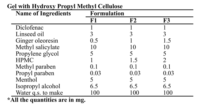
Table No. 6 Formulation batches from F4 to F7 Gel with Carbopol
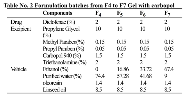
Table No. 7 Organoleptic Characterization of formulation Batches gels
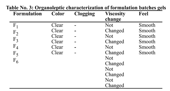
Table No. 8: Skin Irritation and Apperance test
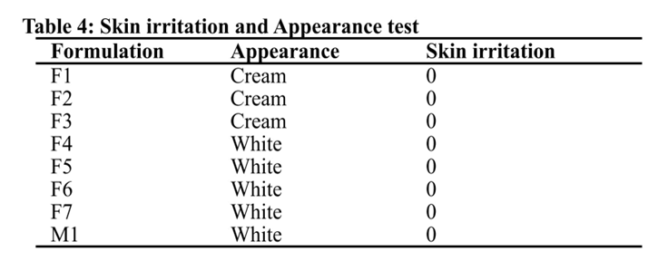
Table No. 9 Drug Content of formulation Batches gels
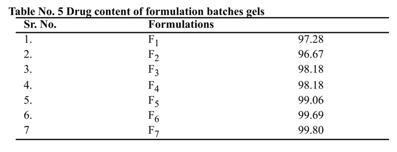
Table No. 10 pH of gels
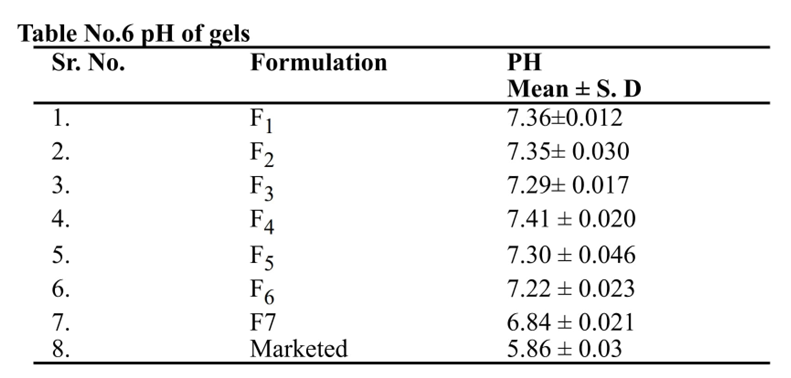
Table No. 11 Viscosity of various formulations at fixed time
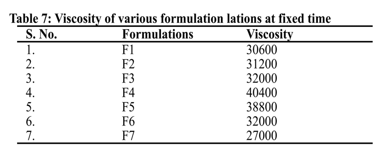
Table No. 12 Viscosity observation of various formulations
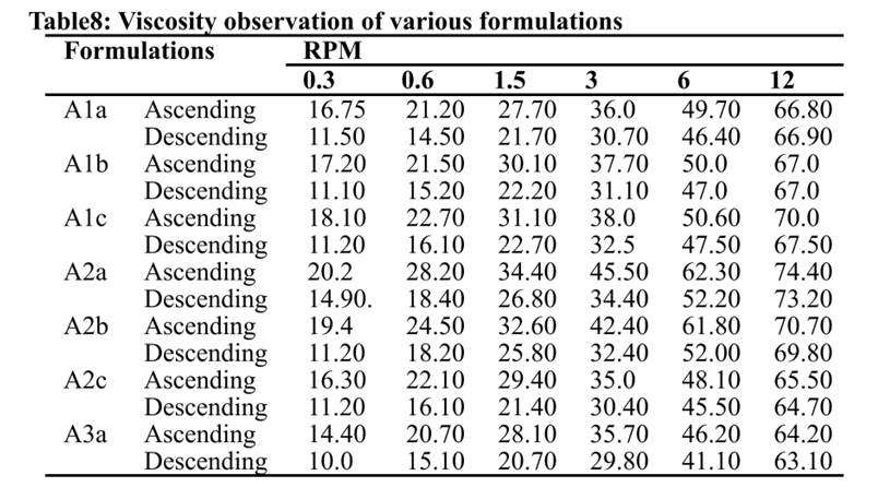
Table No. 13 Spreadability of Various formulations
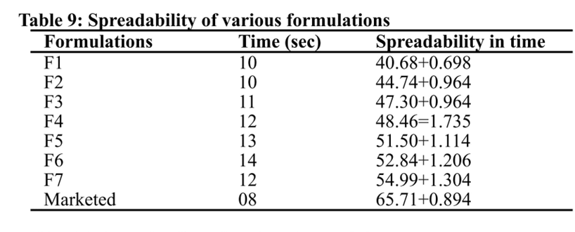
Table No. 14 Extrudability of various formulations
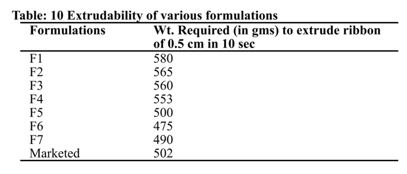
Table 15: Cumulative % Drug Release Profile of Diclofenac Gels.
|
Time (Hr) |
Formulation |
|||||||
|
F1 |
F2 |
F3 |
F4 |
F5 |
F6 |
F7 |
Marketed |
|
|
1 |
14.36 |
18.5 |
8.78 |
18.37 |
18.52 |
15.72 |
31.40 |
20.83 |
|
2 |
17.13 |
25.19 |
20.78 |
22.74 |
24.96 |
18.60 |
36.40 |
25.80 |
|
3 |
21.69 |
31.47 |
29.07 |
31.43 |
31.78 |
25.33 |
44.27 |
36.41 |
|
4 |
28.01 |
37.05 |
37.66 |
36.18 |
38.59 |
30.43 |
51.37 |
43.92 |
|
5 |
32.99 |
43.86 |
43.47 |
40.68 |
42.03 |
36.71 |
59.09 |
49.58 |
|
6 |
41.07 |
49.57 |
52.32 |
46.65 |
50.98 |
42.59 |
67.67 |
57.71 |
|
7 |
47.04 |
56.86 |
60.41 |
59.09 |
57.87 |
50.35 |
71.34 |
65.58 |
|
8 |
52.00 |
62.81 |
67.06 |
63.12 |
60.71 |
58.70 |
73.69 |
75.01 |
CONCLUSION
The release penetration enhancer and other materials are cheap, readily available, safe, having wide regulatory acceptance and easy to handle for economic point of view. It may beneficial to adopt such simple technology for the commercial production of Diclofenac gel. The future scope of this study is that formulation should be subjected for long-term stability and in-vivo performance study and anti-inflammatory activity.
REFERENCES

Nand Lal Thakur, Sana Nusrat Praween, In-Vitro Evaluation of Diclofenac Sodium Gel, Int. J. of Pharm. Sci., 2025, Vol 3, Issue 9, 3667-3684. https://doi.org/10.5281/zenodo.17235413
 10.5281/zenodo.17235413