Abasaheb Kakade College of B. Pharmacy Bodhegaon Tal. Shevgaon Dist. Ahilyanagar.
For the past few decades, there has been a considerable research interest in the area of drug delivery using particulate delivery systems as carriers for small and large molecules.Particulate systems like nanoparticles have been used as a physical approach to alter and improve the pharmacokinetic and pharmacodynamic properties of various types of drug molecules. They have been used in vivo to protect the drug entity in the systemic circulation, restrict access of the drug to the chosen sites and to deliver the drug at a controlled and sustained rate to the site of action. Various polymers have been used in the formulation of nanoparticles for drug delivery research to increase therapeutic benefit, while minimizing sideeffects. Here, we review various aspects of nanoparticle formulation, characterization, effect of their characteristics and their applications in delivery of drug molecules and therapeutic.....1 genes.Nanocarriers like polymeric nanoparticles, mesoporous nanoparticles, nanomaterials, carbon nanotubes, dendrimers, liposomes, metal nanoparticles, nanomedicine, and engineered nanomaterials are used as carriage systems for targeted delivery at specific sites of affected areas in the body. Nanomedicine has rapidly grown to treat certain diseases like brain cancer, lung cancer, breast cancer, cardiovascular diseases, and many others. These nanomedicines can improve drug bioavailability and drug absorption time, reduce release time, eliminate drug aggregation, and enhance drug solubility in the blood. Nanomedicine has introduced a new era for drug carriage by refining the therapeutic directories of the energetic pharmaceutical elements engineered within nanoparticles. In this context, the vital information on engineered nanoparticles was reviewed and conferred towards the role in drug carriage systems to treat many ailments. All these nanocarriers were tested in vitro and in vivo. In the coming years, nanomedicines can improve humanhealth more effectively by adding more advanced techniques into the drug deliverysystem....2Nanomaterials are at the leading edge of the rapidly developing field of nanotechnology. Their unique size-dependent properties make these materials superior and indispensable in many areas of human activity.5
In 1959, Feynman was the first physicist to introduce the notion of nanotechnology in the lecture entitled “There’s Plenty of oom at the Bottom”. This concept initiated remarkable developments in the area of nanotechnology [15]. Nanotechnology is the study of extremely tiny things and is basically the hub of all science disciplines including physics, chemistry, biology, engineering, information technology, electronics, and material science [16]. The structures measured with nanotechnology range from 1–100 nm at the nanoscale level [17]. Nanoparticles have different material characteristics because of submicroscopic size and also provide practical implementations in a wide range of fields include study of extremely tiny things and is basically the hub of all science disciplines including physics, chemistry, biology, engineering, information technology, electronics, and material science [16]. The structures measured with nanotechnology range from 1–100 nm at the nanoscale level [17]. Nanoparticles can be defined as objects ranging in size from 1 to 100 nm that due to their size may differ from the bulk material. Presently, different metallic nanomaterials are being produced using copper, zinc, titanium, magnesium, gold, alginate and silver.....5 Nanoparticles are being used for diverse purposes, from medical treatments, using in various branches of industry production such as solar and oxide fuel batteries for energy storage, to wide incorporation into diverse materials of everyday use such as cosmetics or clothes.Nanoparticles and based formulations have occupied several important application domains that have direct relevance to our routine. The range of nanoparticle applications extends from drugs and drug delivery to the development of efficient solar cells....4 Given the nature of their applications, it is crucial to characterize these nanoparticlesfor several physical, chemical, and biological properties....5
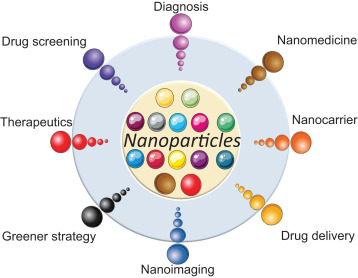
Fig No 01: Nanoparticles
1.1 Microparticles and Nanoparticles
In particulate drug delivery, the distinction is often made between micro- and nanoparticles. (In some cases, the importance of this distinction may be related to the recent rise of nano-specific institutions and funding mechanisms.) For the purposes of this article, the terms ‘‘microparticle’’ and ‘‘nanoparticle’’ refer to particles where the dimensions of the particle are measured in micrometers and nanometers respectively. Some authors reserve the term ‘‘nanoparticles’’ for specific size cut-offs, with some justification: the larger the particles are, the more likely they are to behave as microparticles, and to require the same production processes. The difference in size between micro- and nanoparticles has numerous effects. It is important to bear in mind that some of the distinctions between particles of different sizes may not hold across classes of particles......11
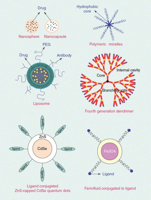
Fig No 02: Schematic of Different Nanotechnology -Based Drug dDelivery Systems
Nanotechnology has gained huge attention over time. The fundamental component of nanotechnology is the nanoparticles. Nanoparticles are particles between 1 and 100 nanometres in size and are made up of carbon, metal, metal oxides or organic matter [1]. The nanoparticles exhibit a unique physical, chemical and biological properties at nanoscale compared to their respective particles at higher scales. This phenomena is due to a relatively larger surface area to the volume, increased reactivity or stability in a chemical process, enhanced mechanical strength, etc. [2]. These properties of nanoparticles has led to its use various applications. The sscan be either a zero dimensional where the length, breadth and height is fixed at a single point for example nano dots, one dimensional where it can possess only one parameter for example graphene, two dimensional where it has length and breadth for example carbon nanotubes or three dimensional where it has all the parameters such as length, breadth and height for example gold nanoparticles. The nanoparticles are of different shape, size and structure. It be spherical, cylindrical, tubular, conical, hollow core, spiral, flat, etc. or irregular and differ from 1 nm to 100 nm in size. The surface can be a uniform or irregular with surface variations. Some nanoparticles are crystalline or amorphous with single or multi crystal solids either loose or agglomerated [4]. Numerous synthesis methods are either being developed or improved to enhance the properties and reduce the production costs. Some methods are modified to achieve process specific nanoparticles to increase their optical, mechanical, physical and chemical properties [3]. A vast development in the instrumentation has led to an improved nanoparticle characterisation and subsequent application.
2.Nanoparticles Types :
1. Silver:
These are proved to be most effective because of their good antimicrobial efficacy against bacteria, viruses and other eukaryotic microorganisms.5,6Among all the nanomaterials they are most widely used as antimicrobial agents, for sunscreen lotions, water treatment and in textile industries etc.7,8 By using the plants i.e. Capsicum annuum, Azedarach indica 10 and Carica papaya the successful biosynthesis of silver nanoparticles have been reported.
2. Gold:
For identification of protein interactions in immunochemical studies gold nanoparticles (AuNPs) are used. In DNA fingerprinting they are used as lab tracer to detect existence of DNA in a sample. Aminoglycoside antibiotics i.e. streptomycin, gentamycin and neomycin are also detected by using these nanoparticles. Detection of cancer stem cells, diagnosis of cancer and identification of different classes bacteria done by using Gold nano rods.12
3. Alloy:
From the bulk samples, structural properties of alloy nanoparticles are different.14 Silver flakes are most commonly used due to their highest electrical conductivity among other metal fillers, their oxides also have relatively greater conductivity.15 By both metals and over ordinary metallic NPs bimetallic alloy nanoparticles properties are more advantages.
4. Magnetic:
Magnetic nanoparticles are known to be biocompatible i.e. maghemite and magnetite. For magnetic resonance imaging (MRI), guided drug delivery, targeted cancer treatment (magnetic hyperthermia), gene therapy, stem cell sorting and manipulation and for DNA analysis they have been actively considered......13
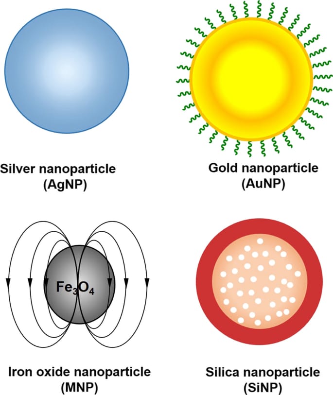
Fig No 3: Types of Nanoparticles
3. Nanoparticles Preparation Methods:
For the preparation of nanoparticles, the selection of the appropriate method is based on the drug to be loaded and physicochemical properties of the polymer. The primary preparation methods of nanoparticles includes:
3.1 Emulsion-Solvent Evaporation Method
The nanoparticles are mostly prepared by using this method. Two steps are mainly involved in this method. In an aqueous phase, emulsification of the polymer solution required in the first step. While in the second step, evaporation of polymer solution occurs and nanospheres are formed by inducing the polymer precipitation. Collection of nanoparticles is done by ultracentrifugation and to remove free drug or residue, washed with distilled water and for storage these are lyophilized..................13
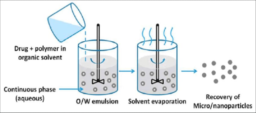
Fig No 04: Emulasion Solvent Evaporation Method
3.2 Double Emulsion and Evaporation Method
Poor entrapment of hydrophilic drugs is the main drawback of this method. Therefore to encapsulate hydrophilic drug the double emulsion technique is engaged, in which aqueous drug solutions is added to organic polymer solution with vigorous stirring to form w/o emulsions. With continuous stirring to form mixed emulsion (w/o/w), this w/o emulsion is added into another aqueous phase. Then by the evaporation solvent is removed and by centrifugation at high speed nano particles can be isolated. Before lyophilisation the prepared nanoparticles must be washed.......13
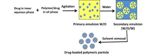
Fig No 05: Double Emulsion and Evaporation
Method
3.3 Salting Out Method
By using salting-out from aqueous solution the water-miscible solvent is separated using this method.29 Initially in a solvent, polymer and drug are dissolved which is consequently containing the salting out agent (electrolytes, such as calcium chloride and magnesium chloride or sucrose as non- electrolytes) and polyvinylpyrrolidone(PVP) or hydroxyethyl cellulose as a colloidal stabilizer into an aqueous gel are emulsified. This oil in water emulsion is diluted with water or with an aqueous phase to increase the diffusion of solvent, which indicates the formation of nanospheres........13,14
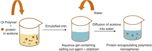
Fig No 06: Salting Out Method
3.4 Emulsions Diffusion Method
To prepare nanoparticles, emulsions diffusion method is another method which is commonly used. The encapsulating polymer is dissolved in a solvent which is partially miscible with water such as propylene carbonate, benzyl alcohol and the initial thermodynamic equilibrium of both liquids saturated with water should be ensured. Subsequently, The polymer-water saturated solvent phase is emulsified in an aqueous solution containing stabilizer, leading to solvent diffusion to the external phase and according to the oil-topolymer ratio nanospheres or nanocapsules are formed. Finally, according to boiling point the solvent is removed by evaporation or filtration.......13
3.5 Solvent Displacement/Precipitation method
Solvent displacement includes from an organic solution, the precipitation of a preformed polymer and in the aqueous medium the diffusion of the organic solvent in the presence or absence of surfactant. In a semi-polar water miscible solvent such as acetone or ethanol, polymers, drug and lipophilic surfactant are dissolved. Then solution is poured or injected using the magnetic stirring, into stabilizer containing aqueous solution. By the rapid solvent diffusion nano particles are formed. Then under reduced pressure solvent is removed from the
Suspension ........13
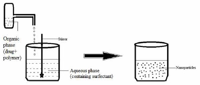
Fig No 07: Solvent Displacement/Precipitation method
3.6 Spontaneous emulsification or solvent diffusion method
This is a modified version of solvent evaporation method. In this method, the water miscible solvent along with a small amount of the water immiscible organic solvent is used as an oil phase. Due to the spontaneous diffusion of solvents an interfacial turbulence is created between the two phases leading to the formation of small particles. As the concentration of water miscible solvent increases, a decrease in the size of particle can be achieved. Both solvent evaporation and solvent diffusion methods can be used for hydrophobic or hydrophilic drugs. .....16
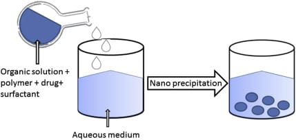
Fig no 08: Spontaneous emulsification or solvent diffusion method
3.7 Coacervation or ionic gelation method
The nanoparticles preparation is carried by using biodegradable hydrophilic polymers such as chitosan, gelatin and sodium alginate. Developing a method for preparing hydrophilic chitosan nanoparticles by ionic gelation. In this method, positively charged amino-group of chitosan interacts with negative charged tripolyphosphate to form coacervates with a size in the range of nanometer. .........16
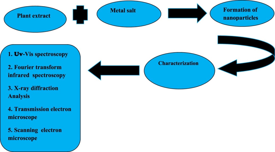
Fig No 09: General Method Of Nanoparticles Using Plant
4. Nanotechnology in Medicine:
1. Nanoparticles for diagnostic and screening purposes.......15
One of the first applications of nanotechnology will likely be to improve the use of fluorescent markers for diagnostic and screening purposes. While fluorescent markers are routinely used in basic research and clinical diagnostic applications,there are several inherent disadvantages with current techniques, including the requirement for colour-matched lasers, the fading of fluorescence even after a single use and the lack of discriminatory capacity of multiple dyes due to the tendency of the different dyes to bleed together.......15
To make very early detection of cancer tumors easier, researchers are developing a nanoparticle intended. The nanoparticles release "biomarkers" molecules when the glue to cancer tumors is detected and the biomarkers are known as peptides. The idea is that even at initial stages of cancer, each nanoparticle carries several peptides which results in a high concentration of these biomarkers will, enabling early detection of the disease. The color of the nanorod shifts when proteins accumulate on the nanorods. The test is designed to be done fast and inexpensively for early detection of a problem.......13
2. Drug Delivery: In medicine one application of nanoparticle is presently being developed which involves deliver the drugs, heat, light or other elements to specific types of cells in form of nanoparticles (such as cancer cells). Nanoparticles are engineered so that they are drawn to disease cells, which will allow direct treatment of those cells. This technique will not only decrease damage to healthy cells in the body but will enable earlier detection of disease............13 Anti-Microbial Techniques Staph infections can be fought by nanoparticle cream which contains nitric oxide gas, known to kill bacteria. Studies on mice have shown that using the nanoparticle cream to release nitric oxide gas at the site of staph abscesses significantly reduced the infection. If an infection is started by the harmful bacteria releasing the antibiotics, coating with nano capsules containing antibiotics, burn dressing will open. Quicker treatment of an infection is done which reduces the number of times a dressing has to be changed.......13
3. Development of artificial receptors
An early goal of nanomedicine is to provide insight into how biological molecular receptors function and then to build artificial binding sites capable of specifically recognising proteins.......15
4 . DNA sequencing using nanopore
This technique was also used to sequence a complete codon in an individual DNA strand tethered to a nanopore. In principle, nanopore detection and characterisation of single molecules represents a new method for directly reading information encoded in linear polymers. If single-nucleotide resolution can be achieved, it is possible that nucleic acid sequences could be determined at rates exceeding 1000 bases per second......15
5. Gene therapy
Gene delivery using a polymeric vehicle has also been explored recently. Polymeric nanospheres are capable of translo-cation into the cytoplasm of a cell, but transport to the nucleus has not been established
5. Synthesis of Nanoparticles:
The nanoparticles are synthesised by various methods that are categorised into bottom-up or top-down method. A simplified representation of the process is presented in Figure.
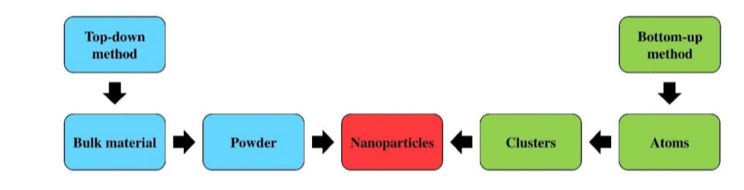
Fig No 10: Synthesis process
Bottom-up method
Bottom-up or constructive method is the build-up of material from atom to clusters to nanoparticles. Sol-gel, spinning, chemical vapour deposition (CVD), pyrolysis and biosynthesis are the most commonly used bottom-up methods for nanoparticle production.
Top-down method
Top-down or destructive method is the reduction of a bulk material to nanometric scale particles. Mechanical milling, nanolithography, laser ablation, sputtering and thermal decomposition are some of the most widely used nanoparticle synthesis methods.
Table No 1: Synthesis of Nanoparticles
|
Category |
Method |
Nanoparticles |
|
Bottom up |
Sol gel Spinning Chemicalvapour disposition Pyrolysis Biosynthesis |
Carbon,metal and metal oxide based Organic polymers Carbon and metal based Carbon and metal oxide based Organic polymers and metal based |
|
Top- Down |
Mechanical milling Nanolithography Laser ablation Sputtering Thermal Decomposition |
Metal, oxide and polymer based Metal based Carbon based , and metal oxide based Metal based Carbon and metal oxide based |
6.Approaches:
Fig No 11: Approaches of NPs Synthesis
Approaches of Nanoparticle (NP) Synthesis
Nanoparticles can be synthesized by three major approaches:
1. Top-Down / Physical Approaches
These methods start with bulk materials and break them down into nanoparticles.
(Think: big → small)
Examples:
Mechanical Milling – Grinding bulk material into nanosized particles.
Electrospinning – Producing nanofibers using electric force.
Laser Ablation – High-power laser breaks material into nanoparticles.
Sputtering – Ejecting atoms from a surface to form nanoparticles.
Electron Explosion – Using electron beams to fragment material.
Sonication – Ultrasonic waves break materials into nanoparticles.
Pulsed Wire Discharge – High-voltage pulse vaporizes wire, forming nanoparticles.
Arc Discharge – Electric arc causes vaporization and nanoparticle formation.
Lithography – Patterning materials on a nanoscale.
2. Bottom-Up Approaches
These methods build nanoparticles from atoms or molecules.
(Think: small → big)
They are further divided into:

Fig No 12: Methods Of Nanoparticle Preparation
A. Chemical Methods
These rely on chemical reactions to form nanoparticles.
Examples:
Chemical Vapor Deposition (CVD) – Gas-phase chemicals react to form nanoparticles.
Sol–gel Process – Formation of nanoparticle gels from solutions.
Co-precipitation – Precipitating nanoparticles from chemical solutions.
Molecular Condensation – Molecules assemble to form nanoparticles.
Hydrothermal – Using high temperature & pressure in water to grow nanoparticles.
B. Green/Biological Methods
These eco-friendly methods use natural sources such as plants or microorganisms.
Types:
1. Plants: Plant extracts act as reducing/stabilizing agents.
2. Microorganism: Algae Bacteria, Fungi
Actinomycetes
(These organisms naturally produce nanoparticles by biological processes.)
3. Biomimetic Methods
These mimic biological systems using: Proteins, Cells, Pollens, Enzymes
These biological components help in reducing metal ions and stabilizing nanoparticles.
7. Characterisation of Nanoparticle:
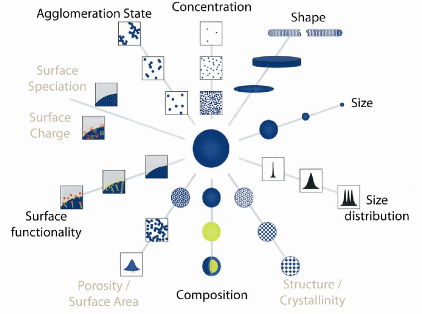
Fig No 13: Characterisation Of Nanoparticles
1. Size
The particle is one of the most basic and important measurement for nanoparticle characterisation. It determines the size and distribution of the particle and whether it falls under nano or micro scale. The particle size and distribution is most commonly measured using electron microscopy. The images of Scanning Electron Microscope (SEM) and Transmission Electron Microscope (TEM) are used for the measurement of particles and clusters whereas laser diffraction methods are used for measuring bulk samples in solid phase [22].
2. Surface area
The surface area is also a significant factor in nanoparticle characterisation. The surface area to volume ratio of a nanoparticle has a huge influence on its performance and properties. The surface area is most commonly measured using BET analysis. A simple titration is sufficient for the surface area analysis of particles in liquid phase, but it is a labour intensive process. Hence nuclear magnetic resonance spectroscopy (NMR) is used. A modified SMPS and differential mobility analyser (DMA) is used for the measurement of surface are of nanoparticles in gaseous phase.
3. Composition
The chemical or elemental composition determines the purity and performance of the nanoparticle. Presence of higher secondary or undesired elements in the nanoparticle may reduce its efficiency and also lead to secondary reaction and contamination in the process. The composition measurement is usually carried out by X-ray photoelectron spectroscopy (XPS) [23]. Some techniques involve chemical digestion of the particles followed by wet chemical analysis such as mass spectrometry, atomic emission spectroscopy and ion chromatography. The particles in gaseous phase are collected either by filtration or electrostatically and spectrometric or wet chemical techniques are used for the analysis [24].
4. Surface morphology
The nanoparticles possess various shapes and surface structures that plays a key role in exploiting its properties. Some of the shapes include spherical, flat, cylindrical, tubular, conical and irregular shapes with surface like crystalline or amorphous with uniform or irregularities on the surface. The surface is generally determined by electron microscopy imaging techniques like SEM and TEM [25]. The particles in liquid phase are deposited on a surface and analysed whereas the particles in gaseous phase are capture electrostatically or by filtration for imaging using electron microscopy.
5. Surface charge
The surface charge or the charge of a nanoparticle determines its interactions with the target. Generally a zeta potentiometer is used for the measurement of surface charges and its dispersion stability in a solution [22]. A Differential Mobility Analyser (DMA) is used for the charge determination of nanoparticles in gaseous phase.
6. Crystallography
Crystallography is the study of atoms and molecules arrangement in crystal solids. The crystallography of nanoparticles are carried out by a powder X-ray, electron or neutron diffraction to determine the structural arrangement [26].
7. Concentration
The concentration of nanoparticles in gaseous phase is measured to determine the volume of air or gas required for the process. The concentration, size and distribution of nanoparticles in a unit volume ofair or gas marks the performance or its efficiency. The concentration measurements are usually made through a Condensation Particle Counter (CPC).
8. Characterisation Techniques:
1. X-Ray Diffraction Method (XRD):
X-ray diffraction (XRD) is a non-destructive type of analytical technique which provides valuable insight about the lattice structure of a crystalline substance like unit cell dimensions, bond angles, chemical composition and crystallographic structure of natural and manufactured materials [1]. XRD is based on the principle of constructive interference of x-rays and the sample concerned which should be crystalline. The x-rays which are generated by a CRT are filtered, collimated and then directed towards the sample. The interaction that follows produces constructive interference based on Bragg’s law which relates wavelength of the incident radiations to the diffraction angle and lattice spacing
2. Scanning Electron Microscopy (SEM):
A scanning electron microscope (SEM) is a type of electron microscope that images a sample by scanning it with a high-energy beam of electrons in a raster scan pattern [1], [2]. The electrons interact with the atoms that make up the sample producing signals that contain information about the sample's surface topography, composition, and other properties such as electrical conductivity. SEM can produce very high-resolution images of a sample surface, revealing details about less than 1 to 5 nm in size. Due to the very narrow electron beam, SEM micrographs have a large depth of field yielding a characteristic three-dimensional appearance useful for understanding the surface structure of a sample. Under vacuum, electrons generated by a source are accelerated in a field gradient. The beam passes through Electromagnetic Lenses, focusing onto the specimen. As result of this bombardment different types of electrons are emitted from the specimen. A detector catches the secondary electrons and an image of the sample surface is constructed by comparing the intensity of these secondary electrons to the scanning primary electron beam. Finally the image is displayed on a monitor.
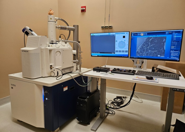
Fig No 15: SEM
3. Ultraviolet- Visible Microscopy: Ultraviolet-visible spectroscopy or ultraviolet-visible spectrophotometer (UV- Vis) involves the spectroscopy of photons in the UV-Visible region. It uses light in the visible and adjacent near ultraviolet (UV) and near infrared (NIR) ranges. In this region of the electromagnetic spectrum, molecules undergo electronic transitions. UV-Vis Spectrophotometers are mainly used to measure transmission or absorption in liquids and transparent or opaque solids. It does so by sending a beam of light through the sample and then monitoring the remaining light in a detector. In the case of a UV-Vis spectrophotometer the light is in the wavelength of 800 - 200nm, probing electronic transitions in the sample. It is hard to reach a lower wavelength than 200nm as oxygen starts to absorb light below that wavelength. When the light passes through the sample some of the molecules in the sample will absorb lights at various wavelengths of this spectrum, depending on their chemical bonds and structure. As a rule, energetically favored electron promotion will be from the highest occupied molecular orbital (HOMO) to the lowest unoccupied molecular orbital (LUMO), and the resulting species is called an excited state. When sample molecules are exposed to light having an energy that matches a possible electronic transition within the molecule, some of the light energy will be absorbed as the electron is promoted to a higher energy orbital. A spectrophotometer records the wavelengths at which absorption occurs, together with the degree of absorption at each wavelength. The resulting spectrum is presented as a graph of absorbance versus wavelength.
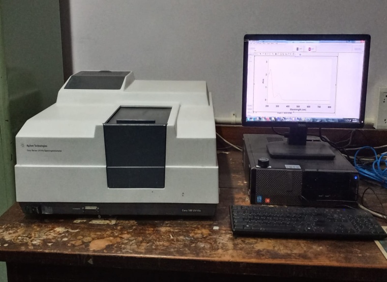
Fig No 16: ULTRAVIOLET-VISIBLE SPECTROSCOPY
9. Evaluation of Nanoparticles:
Evaluating nanoparticles is essential to understand their size, shape, surface properties, stability, composition, and biological behavior. These methods are usually grouped into physical, chemical, surface, functional, and biological characterization techniques....18
1. Zeta potential
The Zeta potential of a nanoparticle is commonly used to characterized the surface charge property of nanoparticles. It reflects the electrical potential of particles and is influenced by the composition of the particle and the medium in which it is dispersed. Nanoparticles with a zeta potential above (±) 30 mV have been shown to be stable in suspension, as the surface charge prevents aggregation of the particles. [16]
2.Particle Shape
SEM characterizes the nanosuspension before going for evaluation; the nanosuspension is lyophilized to form solid particles. The solid particles are coated with platinum alloy using a sputter coater. [17]
3. Particle size
Particle size and size distribution are the most important characteristics of nanoparticle systems. They determine the in vivo distribution, biological fate, and toxicity and targeting ability of nanoparticle system. [18]
4. Drug Entrapment Efficiency
The nanoparticles were separated from the aqueous medium by ultracentrifugation at 10,000 rpm for 30 min at 50C. Then the resulting supernatant solution was decanted and dispersed into phosphate buffer saline pH 7.4. Thus the procedure was repeated twice to remove the unentrapped drug molecules completely. The amount of drug entrapped in the nanoparticles was determined as the difference between the total amount of drug used to prepare the nanoparticles and the amount of drug present in the aqueous medium. Drug Entrapment efficiency (%) = Amount of released from the lysed nanoparticle X 100 Amount of drug initially taken to prepare the Nanoparticles .[19]
5. PR spectroscopy as an orthogonal method
Another orthogonal method for NP characterization, but restricted to metallic NPs such as gold and silver NPs, is UV/Vis spectroscopy. While the aforementioned instrumental methodologies, i.e. DLS, AsFlFFF, and nES-GEMMA are less common, UV/Vis spectroscopy is available in more or less every chemical laboratory. Its utility for GNP characterization is based on the sensitivity of the SPR band to minute molecular alterations on the surface. It provides not only quantitative information about GNP concentrations but also qualitative information about size, shape, and surface modifications of nanoparticles.....[18]
10.Properties of Nanoparticles:
Important physicochemical properties of nanoparticles:
physicochemical properties of NPs As discussed earlier, various physicochemical properties suchas large surface area, mechanically strong, optically active and chemically reactive make NPs unique and suitable applicants for various applications. Some of their important properties are discuss in the following section......14
Particle shape, size, and distribution
• Particle roughness and topography
• Surface area and surface chemistries
• Stability, dispersion, swelling, agglomeration, and aggregation
• Purity
• Reactivity and hydrophobicity......4
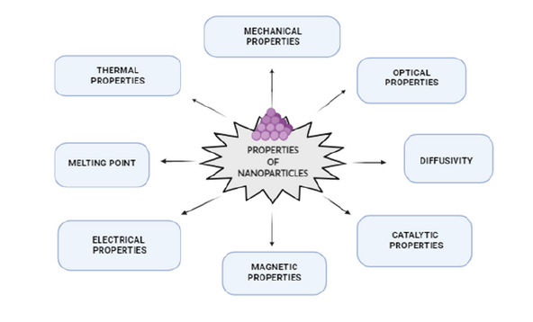
Fig No 17: Properties of Nanoparticles
11.Advantages:
1. After parenteral administration to achieveboth passive and active drug targetingparticle size and surfacecharacteristics ofnanoparticles can be easily manipulated.
2. To achieve high drug therapeutic efficacyand less side effects, during thetransportation they control and sustainrelease of the drug and at the siteof localization, altering distribution of thedrug and subsequent clearance of the drug.
3. By attaching targeting ligands to surfaceof particles or use of magnetic guidancesite-specific targeting can be achieved.
4. Including oral, intra-ocular ,parenteral andnasal, the system can be used for variousroutes of administration.
5. Within the body, drug delivery to tinyareas can be achieved better bynanoparticles.
6. Engineering enables researchers toexercise precisely on this scale andpreviously control over the biomaterials and physical features of polymers........13Nanoparticles can aid in efficient drug delivery by improving aqueous solubility of poorly soluble drugs and increase bioavailability for organized release of drug molecules, and accurate drug targeting.
7. For targeted drug delivery, the surface properties of nanoparticles can be altered for proteins, small molecules, peptides, and nucleic acids loaded nanoparticles are not recognized by immune system and targeted to particular tissue types efficiently.1
12.Applications of Nanoparticulate Delivery Systems:
1.Tumor targeting using nanoparticulate delivery systems .........1
2. Long circulating nanoparticles: Studies show nanoparticles containing a coat of PEG not only have a prolonged half-life in the blood compartment but also be able to selectively extravasate in pathological sites such as tumors or inflamed regions with a leaky vasculature.......1,2
3. Nanoparticles for oral delivery of peptides and proteins...1
4. Tissue engineering
5. Commercial exploration:
Most major and established pharmaceutical companies have internal research programs on drug delivery that are on formulations or dispersions containing components down to nano sizes. Colloidal silver is widely used in antimicrobial formulations and dressings. The high reactivity of titania nanoparticles, either on their own or then illuminated with UV light, is also used for bactericidal purposes in filters. Enhanced catalytic properties of surfaces of nano-ceramics or those of noble metals like platinum are used to destruct dangerous toxins and other hazardous organic materials........5
6. Future directions
As it stands now, the majority of commercial nanoparticle applications in medicine are geared towards drug delivery.In biosciences, nanoparticles are replacing organic dyes in the applications that require high photo-stability as well as high multiplexing capabilities. There are some developments in directing and remotely controlling the functions of nano-probes, for example driving magnetic nanoparticles to the tumour and then making them either to release the drug load or just heating them in order to destroy the surrounding tissue. The major trend in further development of nanomaterials is to make them multifunctional and controllable by external signals or by local environment thus essentially turning them into nano-devices.......5
Table No 2: Applications of Nanoparticulate Delivery Systems
|
Applied Field |
Application |
|
Nanomedicine |
Nano durgs, medical devices, tissue engineering |
|
Chemicals and cosmetics |
Nanoscale chemicals,and compounds,paints,coatings,etc |
|
Materials |
Nanoparticals,carbon nanotubes,biopolymers.points,coatings |
|
Food sciences |
Processing,neutraceutical foods,nanocapsules |
|
Environment and energy |
Water and air purification filters,fuel cells,photovoltaic |
|
Milling and energy |
Biosensors,weapons,sensory enhancements |
|
Scientific tools |
Semiconductors chips,memory storage,photonica,optoelectronics |
13.Challenges and Limitations:
Nanoparticles have already been effectively utilized as drug delivery systems, providing notable advantages in targeted drug delivery and the possibility of combining diagnosis with treatment, making them essential tools in nanomedicine. Nevertheless, numerous technical obstacles remain in developing various methods, such as virus-like systems for delivering drugs inside cells, designing biomimetic polymers, managing sensitive drugs, and implementing features like active targeting, bioresponsive triggered release, and intelligent delivery systems that interact with the body. Additional areas include nanochips for controlled nanoparticle release and carriers made from advanced polymers for delivering therapeutic peptides and proteins. Drug delivery techniques have been created to administer medications and control their dosage and release timing. Most major internal research initiatives concentrate on drug delivery formulations and dispersions that incorporate nanoscale components.
Scalability: Methods developed at the laboratory level need to be adapted for large-scale industrial manufacturing.
14.Future Perspectives:
1.Clinical Validation: Comprehensive in vivo and clinical studies are essential to assess long-term safety, photostability, skin absorption, and potential allergic reactions. The findings from these investigations will aid in obtaining regulatory approval and gaining public trust in herbal-based nanocosmetics. Combination with Other Herbal Extracts: Merging Tulsi with other medicinal plants like Aloe vera, Neem, or Green Tea might produce synergistic benefits, leading to multifunctional sunscreens with anti-aging, moisturizing, and antimicrobial qualities.
2.Eco-Friendly and Scalable Production: Developing energy-efficient, solvent-free, and large-scale green synthesis methods is vital for commercial viability. Fine-tuning factors such as pH, extract concentration, and temperature will help achieve consistent particle size and reproducibility on an industrial scale.
3.Smart Delivery and Multifunctional Applications: Future studies could investigate embedding Tulsi–ZnO nanoparticles into smart delivery systems for targeted release of antioxidants or therapeutic agents. These systems could be used not only in sunscreens but also in wound healing, dermatological treatments, and skincare products aimed at pollution protection.
4.Environmental and Toxicological Assessments: Long-term studies on ecological impact are necessary to ensure that nanoparticles remain non-toxic to aquatic and terrestrial life after disposal, supporting sustainability throughout their lifecycle.
5.Regulatory and Commercial Progress: Standardizing the phytochemical content in Tulsi extracts, maintaining quality control, and adhering to international cosmetic safety regulations will be key for product certification and successful entry into the global market.
CONCLUSION
Nanocarriers as drug delivery systems are designed to improve the pharmacological and therapeutic properties of conventional drugs. The incorporation of drug molecules into nanocarrier can protect a drug against degradation as well as offers possibilities of targeting and controlled release. Due to small dimensions, nanocarriers are able to cross the blood-brain-barrier (BBB) and operate on cellular level......7They can reduce the toxicity and other adverse side effects in normal tissues by accumulating drugs in target sites......7,8 Due to their incredible properties, nanoparticles have become significant in many fields in recent years such as energy, healthcare, environment, agriculture etc. Nanoparticle technologies have great potentials,being able to convert poorly soluble, poorly absorbed and labile biologically active substance into promising deliverable substances. A real therapeutic breakthrough can be achieved solely by carrying out painstaking studies in the field of nano-therapy. Using nanosystems in therapies of diseases may contribute to achieving an effective cancer treatment. Moreover, immobilization of homing devices, such as folic acid, epidermal growth factor or antibodies, to the surface of nanoparticles, improves selectivity of drug carriers. The key applications of nanoparticles in medicine are diagnosis and target therapy, however, their wider use is still the future.7
REFERENCES


Mirza Saniya Sameer Baig, Mundhe Rutuja T, Mhaske Sachin, Zine Sapna, Hemant Gangurde, Review on Nanoparticles: Their Types, Preparation Methods, Applications And Evaluation, Int. J. of Pharm. Sci., 2026, Vol 4, Issue 2, 2069-2088. https://doi.org/10.5281/zenodo.18628655
 10.5281/zenodo.18628655
