

We use cookies to ensure our website works properly and to personalise your experience. Cookies policy
Department of Pharmacology, Indira School of Pharmacy, Pune.
Vitiligo is a long-term acquired depigmentary condition that causes white patches to form on the skin and mucosal surfaces due to the selective loss of melanocytes. It affects 0.1–2% of the world's population and has a major social and psychological impact on those who are impacted. Genetic susceptibility, immunological processes, oxidative stress, neurological factors, and metabolic abnormalities are all part of the complex and multifaceted etiology of vitiligo. The most well acknowledged pathogenic mechanism among them is autoimmune-mediated melanocyte destruction. Tumor necrosis factor-alpha (TNF-?), interleukin-17 (IL-17), interferon-gamma (IFN-?), and chemokines like CXCL9 and CXCL10 are among the biomarkers linked to vitiligo activity that have been identified by recent developments in immunology and molecular biology. The two main kinds of vitiligo are segmental and non-segmental, and each has a unique clinical appearance, course, and response to treatment. Dermoscopy and a differential assessment of other hypopigmented illnesses complement the clinical diagnosis. Using topical corticosteroids, calcineurin inhibitors, phototherapy, surgical grafting methods, depigmentation therapy, and newly developed targeted immunotherapies like Janus kinase (JAK) inhibitors, current treatment approaches seek to stop the disease's development and encourage repigmentation. Ayurvedic and conventional methods have also demonstrated beneficial effects in the treatment of illness. The epidemiology, pathophysiology, categorization, biomarkers, and accessible therapeutic methods for vitiligo are all thoroughly covered in this review, which also highlights new developments and potential directions for diagnosis and therapy.
Vitiligo is a chronic autoimmune disease characterized by depigmentation of the skin, leading to the development of white patches on various parts of the body. In this disease, melanocytes responsible for skin pigmentation are destroyed because of humoral and cell-mediated immunity1. Extensive research has been conducted worldwide to understand better this disease's pathogenesis and development. We need to understand the processes involved to develop better prophylactic and therapeutic measures against it. Vitiligo is now widely accepted to be an autoimmune disease with a multifactorial aetiology because of significant advancements made in identifying the factors involved over the years2. Skin loses colour because of a disease called vitiligo(vit-il-EYE-go). As early as the Aushooryan Dynasty, circa 2200 B.C. vitiligo was put into writing and referred to as Kilăsa. The Egyptian Ebers Papyrus also describes vitiligo dating back to 1550 B.C3.
The two primary forms of the condition that were officially recognized by an international agreement in 2011 are nonsegmental vitiligo (NSV) and segmental vitiligo (SV). The term "vitiligo" is used to describe all types of NSV, including acrofacial, mucosal, generalized, universal, mixed, and other uncommon varieties. A significant decision made by the consensus was to clearly separate SV from other forms of vitiligo, as this distinction has important effects on how the condition is expected to progress4. However, recent clinical and research findings indicate that vitiligo is caused by the body's immune system attacking melanocytes, the cells responsible for skin colour, not only in the skin but also in mucous membranes, the eyes, hair follicles, and the ears. This can be especially distressing for individuals with darker skin tones, as the contrast between normal and affected skin is more noticeable. Vitiligo is found all around the world, with a prevalence rate ranging from 0.1% to 2%, and it affects both men and women equally5. The primary method for diagnosing vitiligo is based on clinical observations, but it is important to rule out other conditions that cause lighter or depigmented skin, such as post-inflammatory hypopigmentation, nevus depigmentosus, nevus anaemics, and idiopathic guttate hypomelanosis. A key consideration in diagnosis is steroid-induced depigmentation, which can occur after accidental use of steroids in the skin and may appear similar to vitiligo. Dermoscopy, a non-invasive technique, provides valuable insights into the condition's stage and can help guide treatment strategies6.
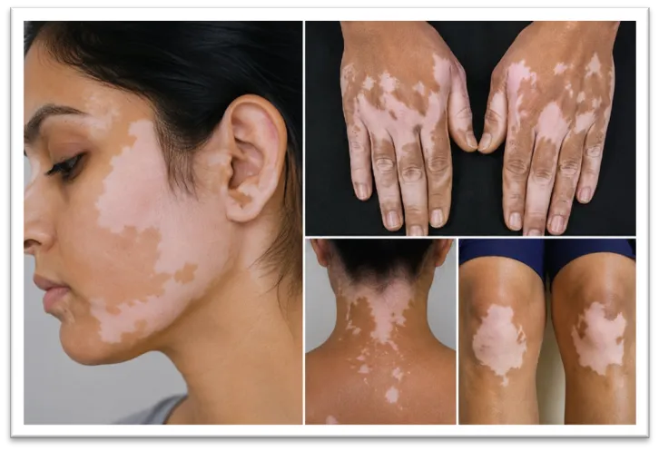
Fig 1: Vitiligo disease
During the process of wound healing, reduced expression of the adhesion molecule E-cadherin is thought to promote the movement of melanocytes. This phenomenon has been observed in areas of skin that regain pigmentation after punch grafting procedures. Additionally, increased levels of heparinase following grafting may lead to a decrease in heparan sulfate at the dermo-epidermal junction (DEJ), which can enhance factors involved in melanocyte activity and pigmentation7.
Epidemiology
Vitiligo affects 0.5 to 2 percent of people all over the world. It seems to affect males and females. Both adult men and women and boys and girls get vitiligo. Non-segmental vitiligo can start at any age. It usually happens in young people between 10 and 30 years old8. Then 150 million people around the world have vitiligo. Every country has people with vitiligo. The number of people with the condition is different in each country. Vitiligo is found over the world but it is more common in some places. Africa, the Middle East and some parts of South Asia like India have people with vitiligo. In some areas of India up to 8.8 percent of the population has vitiligo9. It is hard to know how many people have vitiligo. The biggest study on vitiligo looked at 47,033 people living on the island of Bornholm, in Denmark. Found that 0.38 percent of them had vitiligo. Worldwide between 0.1 and 2 percent of people have vitiligo. Vitiligo can start at any age. In 70 to 80 percent of cases it starts before the person is 30 years old10.
Etiology and Pathogenesis
The exact cause of vitiligo is not known. It often occurs with autoimmune diseases. There are ideas about how it develops and the cause is likely due, to many factors. Vitiligo has penetrance, genetic heterogeneity and multiple susceptibility loci. Studies of families and twins have shown that inheritance is complex and involves both genetic factors. The age at which vitiligo starts may also be influenced by factors. The genes that contribute to vitiligo may include those involved in making melanin controlling autoantibodies and responding to stress. Vitiligo’s cause is still not well understood and involves a mix of environmental factors. The condition often runs in families11.
The most accepted and understood explanation is autoimmune mediation. This theory suggests that melanocytes are destroyed by autoimmune effector mechanisms such as memory cytotoxic T cells or circulating autoantibodies directed against melanocyte surface antigens when their response becomes misdirected. Vitiligo has been associated with autoimmune disorders; Graves' disease and Hashimoto's thyroiditis (thyroid disease) occur in patients with vitiligo more frequently than would be expected by chance. Other autoimmune endocrinopathies associated with vitiligo include Addison's disease and diabetes mellitus. There are others that need further study such as autoimmune polyglandular syndrome, alopecia areata, systemic lupus erythematosus, inflammatory bowel disease, rheumatoid arthritis, psoriasis and pernicious anaemia12.
According to the neural hypothesis, neurochemicals released by nerve endings can harm melanocytes or decrease melanin synthesis. Additionally, it suggests that the catalase gene is involved in the pathophysiology of vitiligo. Nearly all living organisms have the peroxisome enzyme catalase. It shields cells against very reactive oxygen radicals by promoting the breakdown of hydrogen peroxide into water and oxygen. Patients with vitiligo have reduced catalase enzyme activity in both lesional and non-lesional skin13.
The reason we get vitiligo is because of something called hydrogen peroxide. This is when our body makes much of it and it hurts our melanocytes. Melanocytes are the things that give our skin colour. When we make melanin, which's the stuff that makes our skin colour sometimes bad things are made too. These bad things can hurt our melanocytes if we do not have good things to protect them. Some people think that vitiligo happens because our melanocytes are not growing right. This could be because they are not getting what they need to grow or because they are just not working right. It could also be because of the way we are born our genetics. Most people think that vitiligo is when our body attacks itself which is called an illness. We are not really sure how all of this works together. There are different kinds of vitiligo and we cannot really explain why they all happen. We do know that things, around us like the air we breathe and the food we eat can make a difference in whether or not we get vitiligo14.
According to the oxidative stress theory, intra-epidermal buildup of reactive oxygen species the most well-known of which is hydrogen peroxide (H2O2), whose concentration can reach up to one millimole is the primary cause of vitiligo. Melanocytes undergo apoptosis and die as a result of H2O2 changing the mitochondria at this concentration. Patients with vitiligo frequently have altered redox status indicators. Malondialdehyde (MDA), selenium, vitamins C and E, glutathione peroxidase (GPx), superoxide dismutase (SOD), and catalase (CAT) are significant indications of significance. MDA is a result of lipid peroxidation and a sign of oxidative stress. Selenium, an essential antioxidant found in erythrocytes, is necessary for GPx activity. Superoxide radicals are converted into oxygen (O2) and water (H2O) by CAT, while SOD neutralizes them and lessens their toxicity. Patients with vitiligo had significantly greater amounts of SOD, decreased erythrocyte GPx activity, low levels of the enzyme CAT, and low levels of vitamins C and E in their blood and epidermis15.
Symptoms
The depigmentation of skin patches is the most significant recognized symptom of vitiligo. The patches are little at first, but they will eventually get bigger. The cheeks, hands, and wrists are where the skin lesions are most frequently seen. Patients with this illness frequently experience depression as well16.
Classification
Currently, segmental vitiligo and non-segmental vitiligo are the two main subtypes of vitiligo.

Segmental vitiligo is an acquired chronic pigmentation condition characterized by unilaterally dispersed white patches that may or may not resemble a dermatome. It is less prevalent, begins earlier than non-segmental vitiligo, and quickly depletes the follicular melanocyte reserve, causing bleaching of the hair17. refers to the existence of white patches that are restricted to one side of the skin. The patches in a given region frequently proliferate quickly, but they are then caught and may stay that way for a considerable period of time18.
This kind of vitiligo is unilaterally spread and may resemble a dermatomal segment. This subtype is commonly prevalent in younger age groups (15–30%), with an average age of 18. Additionally, a combination of treatments, including excimer laser and topical tacrolimus, with or without systemic corticosteroids, was found to be beneficial19. On the other hand, lesions appear to be more responsive if treated early, and medical intervention may help prevent future disease development. For those with active disease, topical medicine (corticosteroids, calcineurin inhibitors, or JAK inhibitors) and targeted UVB treatment are therefore recommended20.
With 80–90% of cases, non-segmental vitiligo (NSV) is the most common kind of vitiligo. Bilateral, frequently symmetrical white patches that enlarge with time are the hallmark of this long-term acquired pigmentation disease, which typically indicates a substantial loss of functioning melanocytes in the epidermis and some in the hair follicles. Examples of NSV include focal, mucosal (when affecting many mucosal areas), acrofacial, generalized, universal, mixed, and more. A variety of depigmented patches affecting the buccal, vaginal, and oral mucosa are referred to as mucosal vitiligo. Patches that are primarily restricted to the face and distal extremities are indicative of acrofacial vitiligo. The fingers and the perioral and periorbital regions of the face are most impacted. One type of vitiligo that is widely distributed in many locations is called generalized vitiligo. When more than 80% of the body's surface is depigmented, the disease is known as global vitiligo21.
A single macule or patch without a segmental distribution that persists for two years is called focal vitiligo, which may be a precursor to generalized vitiligo. refers to discoloration on different parts of the body. This kind is typically distinguished by the skin damage's continuous spread across a wide region. It may be differentiated from localized vitiligo using the same pattern that is commonly seen on patients' skin22.
Treatment for Vitiligo
Vitiligo is a chronic condition that lasts a lifetime. The unsightly contrast between the affected and normally pigmented skin and the photosensitivity of the untreated depigmented skin, which can result in burns and skin cancer, are two disadvantages of vitiligo. The ideal treatment should stop the disease from progressing and produce an aesthetically acceptable repigmentation.
Topical steroids (TCs) are still the primary treatment for vitiligo. TCs are regarded mainly for their accessibility, cost, and efficacy, despite their numerous side effects, including as telangiectasia and atrophy. They seldom achieve more than 50–75% repigmentation and are time-consuming, needing several daily administrations. They may not detect any improvement for up to a year23.
Strong to moderately potent topical corticosteroids are used. But vitiligo requires long-term use of these drugs, sometimes much longer than the usual "safe" prescription periods for inflammatory dermatoses. The result is peri-lesional hypopigmentation, atrophy, hypertrichosis, and other severe side effects that restrict treatment. It is clear that the dosage formulations now in use do not offer site-specific drug delivery24.
Phototherapy and topical and oral immunomodulators such corticosteroids and calcineurin inhibitors are common therapies for vitiligo. Topical calcineurin inhibitors (TCI) and moderate to high potency corticosteroids (TCS), which both reduce the cellular immune response, are the first-line treatments for vitiligo. In a randomized controlled trial, the topical steroid combination (betamethasone) with a narrow-band UVB (NB-UVB) and topical calcipotriol therapy substantially improved repigmentation at six months compared to NB-UVB alone and NB-UVB with topical calcipotriol25.
Topical immunomodulators are novel medicinal chemicals that can increase or decrease the skin's inflammatory and immunological responses through immunologic pathways. Tacrolimus and pimecrolimus are two inhibitory topical immunomodulators used to treat vitiligo. The primary mechanism of action of these drugs in the treatment of vitiligo is calcineurin inhibition, which causes the transcription of several proinflammatory cytokines essential to the pathophysiology of the early immune response to be disrupted and antigen-specific T-cell reactivity to be downregulated26.
Topical vitamin D3 analogs have been used to cure vitiligo because they enhance melanocyte growth, which in turn causes and promotes melanogenesis. They have immunomodulatory properties as well. In actuality, however, they are ineffectual as a stand-alone treatment for vitiligo and, at most, supportive of other therapies like as phototherapy and TCS. The suggested dosage for topical calcipotriol is 100g weekly or 50μg/go twice daily for four weeks (ointment) and eight weeks (cream)27. Vitiligo has been treated with calcipotriol either by alone or in combination with phototherapy. This possible approach prevents cutaneous T cell infiltration, which is a part of the pathogenesis of vitiligo. It also functions effectively in conjunction with TCs, especially in difficult-to-treat areas like the eyelids. However, there is ongoing debate over the actual effects of vitamin D mimics on vitiligo28.
UVB radiation, which has a wavelength of 280–320 nm, is more significant than UVA radiation, which has a wavelength of 320–400 nm. Narrow-band UVB is being used in conjunction with topical corticosteroids (TCS) or calcineurin inhibitors (TCI) to treat a variety of vitiligo subtypes. Vitiligo can be treated with PUVA and UVB (narrow-band UVB (NB-UVB), excimer laser, or lamp) phototherapy. Although PUVA was the first successful phototherapy program, it has some drawbacks, including the inability to use it on Cosmetics 2023, 10, 84 9 of 14 children or pregnant women, as well as phototoxic adverse effects as headache, nausea, dizziness, and skin cancer risk. Compared to PUVA, NB-UVB has shown higher treatment efficacy and fewer, milder side effects29.
Surgery is only an option for treating segmental or stable vitiligo. Skin grafting and micropigmentation are the most common surgical procedures. Before performing the definitive graft on hypopigmented patches that have been stable for at least two years, it is strongly advised to perform a mini-grafting test to evaluate the patient's positive response and the unfavorable occurrence of Koebner's phenomenon at the donor site after two to three months of follow-up. The Koebner phenomenon at the donor site, keloids, hyperpigmentation, "cobblestoning," scarring, and infections are among the side effects of vitiligo surgery. Compared to the control, suction blister, and combination split-thickness suction grafts, split-thickness suction grafting appears to be superior. Hyaluronic acid is increasingly being used in grafting procedures due of its improved biocompatibility. In double-blind research, compared to individuals who got a placebo, 77% of patients who received a hyaluronic acid-enriched cellular transplant achieved repigmentation rates above 70% in the vitiligous areas after a year30.
The removal of pigmented skin is part of the depigmentation therapy for extensive, global vitiligo. There are no long-term side effects, and patients with active vitiligo react better to Q-switched laser therapy than those with stable vitiligo. Applying 20% monobenzyl ether of hydroquinone (MBEH) topically was also effective31.
Pitta Dosha imbalances, which lead to toxins (ama) condensing in deep layers of the skin, are the primary cause of vitiligo, also called as Switra in Ayurveda. Restoring the skin's hue, purifying the blood, and regulating the body's vitality are the basic treatments for this sickness. Poor digestion, which causes the body to accumulate toxins, is one of the primary causes of the sickness. According to Jiva Ayurveda, the body's primary purpose is to restore digestion. In Ayurveda, vitiligo is frequently treated in four phases. The initial stage of purifying therapy (Shodhana Karma) involves the use of Psoralea Corylifolia and Eur phorbianerifolia herbal decoctions. The second stage is oil massage, where the oil is selected according to the patient's condition (roga) and examination (rogiPariksa). The third stage is exposing the lesions to sunlight, depending on the patient's tolerance (Sooryapadasanthapam). Ficushispida (Malayu), Pterocarpusmarsupium (asana), Calllicarpamacrophylla (priyangu), Peusedanumgraveolens (satapuspa), Coleus vettiveroides (ambhasa), an alkaline extract of Buteamonosperma (palasaksara), and an alcoholic jaggery preparation known as phanitha in Ayurveda are all included in the fourth and final step32.
Biomarkers in Vitiligo
Melanocyte depletion in vitiligo is primarily caused by cytotoxic T lymphocytes (CTLs), with activation of the Fas–FasL pathway being a key molecular mechanism. Notably, T helper 1 (Th1) cells, natural killer (NK) cells, and CTLs release interferon-gamma (IFN-γ), a pro-inflammatory cytokine that is crucial to vitiligo pathogenesis and CTL-mediated melanocyte death in depigmented areas. It should come as no surprise that patients with active vitiligo have higher serum concentrations of IFN-γ, which are correlated with the activity and development of the disease33.
Two cell surface receptors, IFNGR1 and IFNGR2, linked to JAK1 and JAK2, respectively, are bound by IFN-γ. This process results in the phosphorylation of STAT proteins, which then enter the nucleus, trigger gene transcription, and increase a number of elements involved in immune cell recruitment and activation. Thus, JAK inhibition promotes melanocyte survival and permits skin repigmentation by reducing IFN-γ-mediated immune activation and preventing immune-mediated melanocyte death34. JAK inhibitors have recently shown clinical effectiveness in vitiligo therapy. Clinical investigations have shown improvements, like as repigmentation and decreased autoimmunity, particularly in early or localized vitiligo. It's interesting to note that psoriasis and vitiligo are two immune-mediated skin disorders that have been treated with JAK inhibitor treatment. But in vitiligo, direct interference with IFN-γ-mediated melanocyte death produced by CTL also occurs, but in psoriasis, blocking the JAK/STAT axis only stops cytokine-mediated activation of Th17 cells35.
In conclusion, IFN-γ plays a crucial role in the pathophysiology of vitiligo by either directly or indirectly inducing melanocyte death and depletion through immune-mediated pathways. Its key significance in the development of vitiligo is highlighted by its participation in both systemic immune dysregulation and local melanocyte dysfunction, making it a viable immunological marker and a crucial target for innovative treatment approaches36.
Immune cells including T cells, monocytes, and dendritic cells are directed to areas of inflammation or damage by chemokines, which are important signaling proteins. Chemokines, particularly those secreted by melanocytes, are essential to the pathophysiology of vitiligo. Studies have revealed that vitiligo patients' skin has higher levels of chemokines linked to a Th1-dominant immune response, especially during the disease's active stages. Important mediators that draw immune cells to the skin and intensify the autoimmune assault on melanocytes include CXCL10 and CXCL9. However, a recent meta-analysis also revealed increased levels of CXCL8, CXCL12, CXCL16, and CCL5 (CC motif chemokine ligand 5), highlighting the complexity of the immunological landscape in vitiligo37.
CCL5 interacts to several receptors, but it has the most affinity for immune cells' CCR5 (C-C chemokine receptor type 5). Nevertheless, lesional CTLs are not the only cells that activate the CCL5CCR5 axis: In vitiligo, CCR5 expression is elevated on regulatory T cells (Tregs), indicating that (dysfunctional) Tregs may be involved in skin depigmentation. Therefore, focusing on the CCL5-CCR5 axis may offer novel vitiligo treatment strategies. Melanocytes and other cell types release CXCL8 (IL-8), a proinflammatory cytokine that attracts neutrophils through CXCR1 and CXCR2. Notably, early migration and cytotoxicity are the main functions of CXCR1+ CD8+ T cells in response to CXCL8. The vitiligo-specific inflammatory response depends on CXCL8's recruitment of neutrophils, which may also help trigger an IL-17-driven Th17 response38.
As the study of chemokines in vitiligo advances, further research on chemokine signaling pathways will be necessary to create efficient therapies that restore pigmentation and restore immunological function in this long-term illness.
Numerous studies have shown a relationship between IL-17 levels and melanocyte activity, indicating that it may play a role in the pathophysiology of vitiligo. According to preclinical research, depigmentation in mouse models has been linked to IL-17 release. Tyrosinase levels, melanin content, and melanocyte count all decrease in response to IL-17 exposure. Anti-IL-17 may have a therapeutic significance in the context of vitiligo, since Bhardwaj et al. showed that inhibiting the IL-17A receptor might increase melanin synthesis and boost melanocyte survival in vitro39.
Acharya et al. did a meta-analysis that included eleven case-control studies that evaluated blood IL-17 concentrations in vitiligo patients in comparison to healthy individuals. The results verified that those with vitiligo had significantly higher levels of circulating IL-17. Additionally, three of these investigations looked at IL-17 levels in lesional skin; consistent with the results above, lesional tissue had greater levels of IL-17 than healthy skin. These results support the idea that vitiligo is associated with an elevated IL-17 pathway, which may contribute to the disease's development. The discovery of lower levels of both IFN-γ and IL-17 in hyperpigmentary diseases lends more credence to this theory. A pilot trial by Speeckaert et al. that examined the effects of Secukinumab, another IL-17A inhibitor, in individuals with active non-segmental vitiligo provides additional data. The study was prematurely terminated since the majority of the patients acquired new lesions. These results suggest that vitiligo may be paradoxically made worse by IL-17 suppression40.
When considered collectively, these clinical data reveal a paradox: whereas IL-17 is increased in vitiligo and causes inflammation, its suppression does not seem to provide therapeutic advantages and may instead exacerbate the condition. As a result, focusing only on IL-17 may be inadequate or even detrimental.
Immune cells, especially activated macrophages and T lymphocytes, are the main producers of TNF-α. It is essential for controlling the immune system, regulating inflammatory reactions, and triggering apoptosis. Numerous autoimmune illnesses have been linked to TNF-α, and new research has shown that it is also involved with vitiligo41.
TNF-α regulation in vitiligo is significantly influenced by genetic variables. Numerous research has looked at the connection between vitiligo susceptibility and certain genetic variants in the TNF-α gene. The TNF-α gene is found on chromosome 6p21.3. The TNF-α -308G/A single nucleotide polymorphism (SNP) is one of the most researched polymorphisms. Such a replacement of guanine (G) to adenine (A) in the TNF-α gene's promoter region may affect transcriptional activity and, in turn, production. For example, it has been discovered that people with vitiligo are more likely to have certain TNF gene variations, indicating a genetic basis for elevated TNF-α levels. Furthermore, in genetically sensitive people, environmental factors including UV radiation, oxidative stress, and infections may cause the production of TNF-α, which can lead to the development or aggravation of vitiligo42.
TNF-α suppression, according to other research, is more likely to cause cutaneous hyperpigmentation than hypo- or depigmentation. Indeed, elevated TNF-α levels appear to be linked to decreased tyrosinase activity, which makes anti-TNF biologic medications a viable therapy option for vitiligo. According to some research, etanercept may be the best medication to use while anti-TNF-α treatment is being considered for people with de novo vitiligo in order to reduce the chance of paradoxical vitiligo responses and prevent the condition from progressing43.
REFERENCES


Yash Wagh, Saee Salvi, Shivanjali Shinde A Comprehensive Review on Vitiligo: Pathogenesis, Classification, Biomarkers, and Therapeutic Approaches, Int. J. of Pharm. Sci., 2026, Vol 4, Issue 5, 7359-7370, https://doi.org/10.5281/zenodo.20411108
 10.5281/zenodo.20411108