View Article

Abstract

Clavulanic acid is an important ?-lactamase inhibitor commonly used with antibiotics to improve their effectiveness against bacterial infections. However, the drug is highly unstable and easily degrades when exposed to heat, light, moisture, and different pH conditions. Because of this instability, there is a need for a simple and rapid method to monitor its stability during pharmaceutical analysis. The present study focuses on the development of a paper-based microfluidic analytical device (µPAD) for the stability-indicating detection of Clavulanic acid.The µPAD was fabricated using filter paper and designed to allow easy movement of small sample volumes through microchannels. A simple colorimetric/UV-based detection method was used to identify the presence of intact and degraded Clavulanic acid. Stability studies were performed under acidic, alkaline, oxidative, thermal, and photolytic conditions. The developed device showed clear changes in color intensity and absorbance at 230 nm, indicating drug degradation under stress conditions. The method demonstrated rapid analysis, low sample consumption, good sensitivity, and reproducible results.Overall, the developed µPAD proved to be a simple, economical, portable, and user-friendly platform for the stability analysis of Clavulanic acid. The study suggests that paper-based microfluidic devices can serve as promising alternatives to conventional analytical techniques for routine pharmaceutical quality control and point-of-care applications.

Keywords

Clavulanic acid, µPAD , Paper-based microfluidics, Stability analysis, Colorimetric detection, UV spectroscopy, Drug degradation, Stability-indicating method

Introduction

× Popup Image

Clavulanic acid is a β-lactamase inhibitor commonly administered in combination with antibiotics such as Amoxicillin to overcome bacterial resistance.[1,2] However, clavulanic acid is chemically unstable and undergoes rapid degradation when exposed to environmental factors such as temperature, pH, and light.[1,2,3] This instability necessitates the development of reliable stability-indicating analytical methods to ensure drug efficacy and safety.[1,4]

Traditional analytical techniques such as High Performance Liquid Chromatography and UV-Visible Spectroscopy are widely used for detection and stability assessment.[3,4] These methods provide high sensitivity and specificity but are limited by their dependence on sophisticated instrumentation, high operational cost, and requirement for skilled personnel, making them less suitable for rapid and on-site analysis.[5,6]

In recent years, Microfluidics has revolutionized analytical science by enabling miniaturized and portable diagnostic systems.[5,7] Among these, paper-based microfluidic analytical devices (µPADs) have emerged as a promising platform due to their low cost, ease of fabrication, portability, and ability to perform rapid analysis using capillary-driven flow without external equipment.[6,8] These features make µPADs highly suitable for point-of-care and field-based applications. [6,9]

Recent studies highlight that µPADs provide an effective alternative to conventional analytical methods by offering rapid, cost-effective, and portable detection systems. These devices have been successfully applied in medical diagnostics, environmental monitoring, and pharmaceutical analysis, and are considered highly promising for decentralized testing.

Despite these advancements, the application of µPADs for stability-indicating detection of pharmaceutical compounds, particularly clavulanic acid, remains limited.[6,10] Therefore, the development of a paper-based microfluidic device for detecting the degradation of clavulanic acid represents a novel and practical approach.[10,11] Such a system can provide a simple, visual, and rapid method for monitoring drug stability, especially in resource-limited laboratory settings.[6,9]

Theory of UV Spectroscopy

Ultraviolet (UV) spectroscopy is an analytical technique used to measure the absorption of ultraviolet light by a substance.[4,7] It is mainly used for the qualitative and quantitative analysis of compounds containing chromophores such as double bonds, aromatic rings, and conjugated systems.

The UV region lies between 200–400 nm of the electromagnetic spectrum.

Far UV region: 10–200 nm

Near UV region: 200–400 nm

Most pharmaceutical and chemical analyses are performed in the 200–400 nm range.

Principle of UV Spectroscopy

UV spectroscopy is based on the principle that molecules absorb ultraviolet radiation and undergo electronic transitions from lower energy levels to higher energy levels.

When UV light passes through a sample:

  • Certain wavelengths are absorbed by the molecule.
  • Electrons get excited from the ground state to an excited state.
  • The amount of absorbed light is measured.
  • The absorbed wavelength depends on the structure of the molecule.

Main components of a UV spectrophotometer:

  1. Light Source
  2. Deuterium lamp (UV region)
  3. Monochromator
  4. Separates light into individual wavelengths.
  5. Sample Holder (Cuvette)
  6. Usually quartz cuvette is used for UV analysis.
  7. Detector
  8. Detects transmitted light.
  9. Recorder/Display
  10. Displays absorbance or spectrum.

Working of UV Spectrophotometer

UV light is produced from the source.

Monochromator selects a specific wavelength.

Light passes through the sample solution.

Sample absorbs some radiation.

Remaining light reaches detector.

Absorbance is measured and spectrum is obtained.[4]

Clavulanic Acid
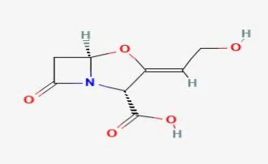


Fig. No 1

  1. General Information

Name: Clavulanic Acid

IUPAC Name: (2R,5R,Z)-3-(2-hydroxyethylidene)-7-oxo-4-oxa-1-azabicyclo[3.2.0]heptane-2-carboxylic acid

Molecular Formula: C₈H₉NO₅

Molecular Weight: 199.16 g/mol

Class: β-lactam compound (β-lactamase inhibitor)

  1. Chemical Structure & Features

Contains a β-lactam ring, essential for its activity

Lacks strong antibacterial activity alone

Structurally similar to penicillins, but functions mainly as an enzyme inhibitor

Possesses an oxazolidine ring fused to β-lactam ring

  1. Physicochemical Properties

Appearance: White to off-white crystalline powder

Solubility: Freely soluble in water, slightly soluble in alcohol

pKa: ~2.7

Melting Point: ~174–177°C (decomposes)

  1. Stability Profile

Degraded by:

Heat

Acidic & alkaline pH

Oxidation

Forms degradation products affecting assay accuracy.[1,2]

Drug Profile

Name: Clavulanic Acid
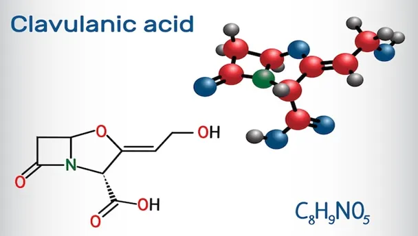


Fig. No 2

IUPAC Name: (2R,5R,Z)-3-(2-hydroxyethylidene)-7-oxo-4-oxa-1-azabicyclo[3.2.0]heptane-2-carboxylic acid

Generic Name: Clavulanic Acid

Category: Beta-lactamase inhibitor

Molecular Formula: C₈H₉NO₅

Molecular Weight: 199.16 g/mol

Nature: Beta-lactam compound

Pharmacological Class

Beta-lactamase inhibitor

Antibacterial adjuvant

Mechanism of Action

Inhibits beta-lactamase enzymes

Protects penicillin antibiotics

Enhances antibacterial activity

Common Combination :Amoxicillin-Clavulanate

Storage

Store in cool, dry place.

Protect from light & moisture.

Materials And Equipment

  1. Chemicals & Reagents

Clavulanic acid reference standard:


Fig. No 3

Used as a benchmark to calibrate and validate the analytical method.

Potassium clavulanate (commercial formulation):

Serves as the test sample for real-world analysis and comparison.

Sodium hydroxide (NaOH):
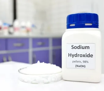


Fig. No 4

Induces alkaline degradation to study stability behavior.

Hydrochloric acid (HCl):
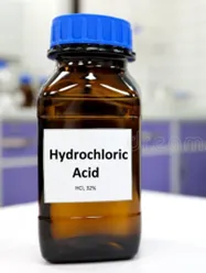


Fig. No 5

Causes acid degradation for stability-indicating analysis.

Hydrogen peroxide (H₂O₂):
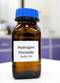


Fig. No 6

Produces oxidative degradation to evaluate drug susceptibility to oxidation.

Distilled/deionized water:
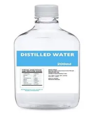


Fig. No 7

Acts as a solvent for preparing all solutions and reagents.

Hydroxylamine hydrochloride:

Reacts with β-lactam ring to form detectable derivatives for analysis.

Ferric chloride (FeCl₃):

Produces a colored complex for colorimetric detection of degraded products.

  1. Materials for µPAD Fabrication

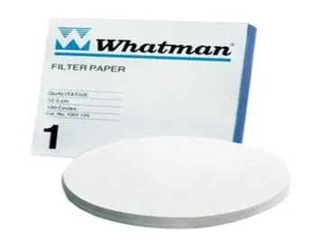
Whatman filter paper (Grade 1):


Fig. No 8

Serves as the substrate for fluid flow and reaction zones in µPAD.

White Soft Paraffin Wax:

Creates hydrophobic barriers to define microfluidic channels.

Hot plate or oven:

Melts wax to penetrate paper and form stable channel boundaries.

Cutter/laser cutter/scissors:

Used to shape and size the paper device accurately.

  1. Instruments & Equipment

Micropipettes:

Deliver precise volumes of samples and reagents onto the µPAD.

UV-visible spectrophotometer:
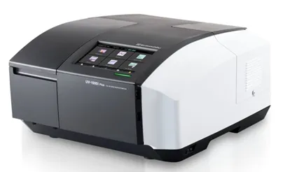


Fig. No 9

Validates colorimetric results by measuring absorbance quantitatively.

Smartphone camera:

Captures images of color changes for portable and low-cost analysis.

Incubator:

Maintains controlled temperature conditions for stability and degradation studies.

Experimental Work

  1. Preparation of Standard Solution

An accurately weighed quantity of clavulanic acid was dissolved in distilled water to prepare a stock solution (100 µg/mL).

Further serial dilutions (10–50 µg/mL) were prepared for UV spectroscopic analysis and comparison.
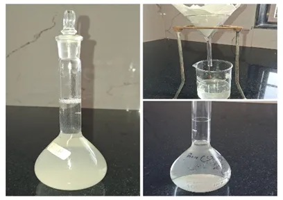
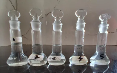


Fig. No 10                                                                   Fig. No 11


  1. Forced Degradation Studies

Forced degradation studies were carried out to evaluate the instability of clavulanic acid under different stress conditions:

Thermal degradation: Sample solution was heated at 60°C for 1–2 hours.

Acidic degradation: Drug solution was treated with 0.1 N HCl and kept for a specific time.

Alkaline degradation: Drug solution was treated with 0.1 N NaOH.

Photolytic degradation: Samples were exposed to UV light.

After treatment, samples were neutralized (if required) and diluted appropriately.
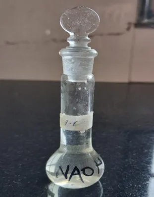


Fig. No 12

  1. UV Spectrophotometric Analysis

The absorbance of both standard and degraded samples was measured using a UV spectrophotometer at the λmax of clavulanic acid.

Decrease in absorbance indicated degradation of the drug.

These results were used as a reference standard for comparison with the paper-based method.

  1. Fabrication of Paper-Based Microfluidic Device (µPAD)

The µPAD was fabricated using Whatman filter paper and wax printing technique:

Hydrophobic barriers were created using wax to form microfluidic channels.

The paper was heated to allow wax penetration, forming defined test zones.

The device was allowed to cool and stored under dry conditions until use.
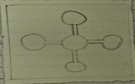


Fig. No 13

  1. Colorimetric Detection Method

A suitable color-forming reagent was selected based on its ability to react with clavulanic acid or its degradation products.

A fixed volume of reagent was added to the test zone of the µPAD.

The sample (standard or degraded) was then applied.

The reaction was allowed to proceed at room temperature.
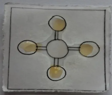


Fig. No 14

  1. Observation and Interpretation

Color development on the paper device was observed visually:

Stable drug: Produced a specific characteristic color

Degraded drug: Produced a different or reduced color intensity

The results were compared with UV spectrophotometric data to validate the method.

  1. Comparison and Validation

The µPAD results were correlated with UV analysis to evaluate:

  • Accuracy
  • Reliability
  • Sensitivity of detection

This confirmed the ability of the paper-based device to act as a stability-indicating method.


RESULT

The present study successfully developed a simple, economical, and rapid paper-based microfluidic analytical device (µPAD) for the stability analysis of Clavulanic acid. The developed µPAD demonstrated effective detection of both intact and degraded drug samples through colorimetric analysis and UV spectroscopic correlation.

Initially, the standard solution of Clavulanic acid was prepared using distilled water and analyzed by UV spectroscopy. During spectral scanning, the maximum absorbance (λmax) of Clavulanic acid was observed at approximately 277 nm. Different concentrations ranging from 10–50 µg/mL showed a gradual increase in absorbance with increasing concentration, indicating good linearity and sensitivity of the analytical method.

The absorbance values obtained were as follows:
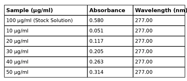


Fig. No 15


The stock solution of 100 µg/mL showed an absorbance of 0.58 at 277 nm, confirming proper detection of the drug and suitability of the UV method for further stability studies.
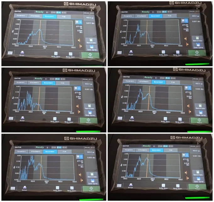


Fig. No 16


Forced degradation studies were then carried out under various stress conditions such as thermal degradation, acidic degradation using HCl, alkaline degradation using NaOH, and photolytic degradation under UV/light exposure. A significant decrease in absorbance was observed in degraded samples compared to the standard solution, confirming degradation of Clavulanic acid. Among the stress conditions, acidic and alkaline degradation showed greater changes due to the instability of the beta-lactam ring present in Clavulanic acid.

The degraded sample of Clavulanic acid showed a noticeable decrease in absorbance at 230 nm, confirming that the drug underwent degradation when exposed to stress conditions like acidic, alkaline, thermal exposure.
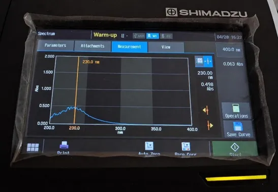


Fig. No 17


The µPAD was successfully fabricated using the wax-printing method on filter paper, which formed effective hydrophobic barriers and microfluidic channels for sample movement. Upon addition of the colorimetric reagent and drug sample onto the detection zone, the intact drug produced a distinct color intensity, whereas degraded samples showed reduced or altered color formation.

The color changes observed on the µPAD correlated well with the absorbance values obtained by UV spectroscopy, indicating that the paper-based device can effectively differentiate between stable and degraded drug samples.


Fig. No 18


The developed µPAD method offers several advantages, including low cost, ease of fabrication, rapid analysis, portability, and minimal reagent consumption. The device also requires simple instrumentation, making it suitable for preliminary stability testing and pharmaceutical quality control applications.

Overall, the study demonstrated that the developed µPAD is a promising and reliable platform for rapid stability analysis of Clavulanic acid. The method has potential for future application in low-resource laboratories, point-of-care testing, and routine pharmaceutical analysis.

DISCUSSION

The present study successfully developed a paper-based microfluidic device (µPAD) for the stability-indicating detection of Clavulanic acid. The device proved to be a simple, low-cost, rapid, and portable alternative to conventional analytical methods like HPLC and UV spectroscopy. Since Clavulanic acid is highly unstable and easily degraded by heat, moisture, light, and pH changes, the developed µPAD effectively helped in monitoring its stability through visible color changes.

The study showed that the device worked efficiently using capillary action without requiring electricity or complex instruments. Advantages such as low reagent consumption, quick analysis, eco-friendliness, and easy handling make the system suitable for pharmaceutical quality control and resource-limited settings. Although some limitations like humidity effects and lower precision compared to advanced instruments were observed, the research demonstrated strong potential for future development of portable and user-friendly pharmaceutical analytical devices.

CONCLUSION

The present research successfully developed a simple and innovative paper-based microfluidic analytical device (µPAD) for the stability analysis of Clavulanic acid. The developed method was found to be rapid, cost-effective, easy to perform, and suitable for identifying both stable and degraded drug samples.

UV spectroscopic studies confirmed the absorbance characteristics of Clavulanic acid, while forced degradation studies showed that the drug is highly sensitive to conditions such as acid, alkali, heat, and light exposure. Degraded samples showed a noticeable decrease in absorbance along with changes in color intensity, confirming drug degradation.

The fabricated µPAD successfully produced visible colorimetric responses and showed good agreement with the UV spectroscopy results. The method required only a small amount of sample and reagents, making it convenient and economical for routine analysis.

Overall, the developed µPAD can be considered a promising platform for rapid stability testing of Clavulanic acid. Because of its portability, simplicity, and low-cost nature, the method may be useful in pharmaceutical quality control laboratories, preliminary screening studies, and resource-limited settings.

REFERENCES

  1. Santos VC, Pereira JF, Haga RB, Rangel-Yagui CO, Teixeira JA, Converti A, Pessoa Jr A. Stability of clavulanic acid under variable pH, ionic strength and temperature conditions. A new kinetic approach. Biochemical Engineering Journal. 2009 Jul 15;45(2):89-93.
  2. Gómez-Ríos D, Ramírez-Malule H, Neubauer P, Junne S, Ríos-Estepa R. Data of clavulanic acid and clavulanate-imidazole stability at low temperatures. Data in brief. 2019 Apr 1;23:103775.
  3. Bostijn N, Hellings M, Van Der Veen M, Vervaet C, De Beer T. In-line UV spectroscopy for the quantification of low-dose active ingredients during the manufacturing of pharmaceutical semi-solid and liquid formulations. Analytica chimica acta. 2018 Jul 12;1013:54-62.
  4. Soum V, Park S, Brilian AI, Kwon OS, Shin K. Programmable paper-based microfluidic devices for biomarker detections. Micromachines. 2019 Aug 2;10(8):516.
  5. Bi H, Fernandes AC, Cardoso S, Freitas P. Interference-blind microfluidic sensor for ascorbic acid determination by UV/vis spectroscopy. Sensors and Actuators B: Chemical. 2016 Mar 1;224:668-75.
  6. Park TS, Cho S, Nahapetian TG, Yoon JY. Smartphone detection of UV LED-enhanced particle immunoassay on paper microfluidics. SLAS TECHNOLOGY: Translating Life Sciences Innovation. 2017 Feb;22(1):7-12.
  7. Oday J, Hadi H, Hashim P, Richardson S, Iles A, Pamme N. Development and validation of spectrophotometric method and paper-based microfluidic devices for the quantitative determination of Amoxicillin in pure form and pharmaceutical formulations. Heliyon. 2024 Feb 15;10(3).
  8. Yetisen AK, Akram MS, Lowe CR. Paper-based microfluidic point-of-care diagnostic devices. Lab Chip. 2013;13(12):2210-2251.
  9. Cate DM, Adkins JA, Mettakoonpitak J, Henry CS. Recent developments in paper-based microfluidic devices. Anal Chem. 2015;87(1):19-41.
  10. Martinez AW, Phillips ST, Whitesides GM. Three-dimensional microfluidic devices fabricated in layered paper and tape. Proc Natl Acad Sci USA. 2008;105(50):19606-19611.
  11. Dungchai W, Chailapakul O, Henry CS. Use of multiple colorimetric indicators for paper-based microfluidic devices. Anal Chim Acta. 2010;674(2):227-233.
  12. Sharma A, Yadav S, Khatri K. Paper-based analytical devices for pharmaceutical analysis: a review. J Pharm Anal. 2021;11(5):532-547.
  13. Tippa DMR, Singh N. Development and validation of stability indicating HPLC method for simultaneous estimation of amoxicillin and clavulanic acid in injection. Am J Anal Chem. 2010;1(3):95-101.
  14. Mack I, Sharland M, Brussee JM, Rehm S, Rentsch K, Bielicki J. Insufficient stability of clavulanic acid in widely used child-appropriate formulations. Antibiotics. 2021;10(2):225.
  15. Santos VC, Brandão RH, Hokka CO. Stability of clavulanic acid under variable pH, ionic strength and temperature conditions: a new kinetic approach. Biochem Eng J. 2009;45(2):89-93.
  16. Haginaka J, Nakagawa T, Uno T. Stability of clavulanic acid in aqueous solutions. Chem Pharm Bull. 1981;29(11):3334-3341.
  17. Foulstone M, Reading C. Assay of amoxicillin and clavulanic acid, the component of Augmentin, in biological fluids with high performance liquid chromatography. Antimicrob Agents Chemother. 1982;22(5):753-762.
  18. Silva VM, et al. Stability of clavulanic acid in PEG/citrate and liquid-liquid extraction in aqueous two-phase system. Fluid Phase Equilib. 2014;375:104-109.
  19. Blessy M, Patel RD, Prajapati PN, Agrawal YK. Development of forced degradation and stability indicating studies of drugs—A review. J Pharm Anal. 2014;4(3):159-165.
  20. Sherwood J, De Bruyn M, Constantinou A, Moity L, McElroy CR, Farmer TJ, et al. D-cellobiose and paper-based analytical devices for pharmaceutical analysis. Green Chem. 2014;16(5):2426-2432.
  21. Maru K, Ram D, Buddhadev SS. Development and validation of stability indicating RP-HPLC method for simultaneous estimation of cephalexin and clavulanic acid. J Neonatal Surg. 2025;14(26s):673-681.
  22. Kakoti A, Siddiqui MF. Leak proof paper microfluidic device fabrication for analytical applications. Biomicrofluidics. 2015;9:026502.
  23. Maru K, Buddhadev SS. RP-HPLC method development for clavulanic acid stability studies. J Neonatal Surg. 2025;14:673-681.
  24. Kumar A, Mahato K, Chanda N. Gold nanoparticle integrated paper microfluidic analytical platform for biosensing. Analyst. 2015;140:1817-1821.
  25. Sakinala P, Kuppala S, Pujitha PS, Singamsetty LPV, Sulthana SR. Chemometric assisted new stability indicating NPHPLC method development and validation of clavulanic acid, amoxicillin and lactobacillus in combined dosage form. J Cardiovasc Dis Res. 2021;12(3):1120-1128.
  26. Kakoti A, Siddiqui MF, Goswami P. A low cost design and fabrication method for developing a leak proof paper based microfluidic device with customized test zone. Biomicrofluidics. 2015;9(2):026502.
  27. Dungchai W, Chailapakul O, Henry CS. Electrochemical detection for paper-based microfluidics. Anal Chem. 2009;81(14):5821-5826.
  28. Jayamohan H, Romanov V, Li H, Son J, Samuel R, Nelson J, et al. Advances in microfluidics and lab-on-a-chip technologies. Methods Mol Biol. 2017;1571:1-20.
  29. Devlopment and validation of stability indicating HPLC methods for related substances and assay analyses of amoxicillin and potassium clavulanate mixtures. J Pharm Biomed Anal. 2017;136:1-9.
  30. Rattanarat P, Dungchai W, Cate DM, Siangproh W, Volckens J, Chailapakul O, et al. A microfluidic paper-based analytical device for rapid quantification of particulate chromium. Anal Chim Acta. 2013;800:50-55.
  31. Bakshi M, Singh S. Development of validated stability-indicating assay methods—critical review. J Pharm Biomed Anal. 2002;28(6):1011-1040.
  32. Matar KM. Simple and rapid LC method for determination of clavulanic acid in dosage forms. J Pharm Biomed Anal. 2009;49(4):1045-1049.
  33. Arvindakshan N, Mohan H, Aravind UK. Hydrolytic degradation studies of β-lactam antibiotics. J Pharm Biomed Anal. 2017;145(1):523-531.
  34. Sanjay ST, Fu G, Dou M, Xu F, Liu R, Qi H, et al. Biomarker detection for disease diagnosis using cost-effective microfluidic platforms. Analyst. 2015;140(21):7062-7081.
  35. Yamada K, Henares TG, Suzuki K, Citterio D. Paper-based inkjet-printed microfluidic analytical devices. Angew Chem Int Ed. 2015;54(18):5294-5310.

Reference

  1. Santos VC, Pereira JF, Haga RB, Rangel-Yagui CO, Teixeira JA, Converti A, Pessoa Jr A. Stability of clavulanic acid under variable pH, ionic strength and temperature conditions. A new kinetic approach. Biochemical Engineering Journal. 2009 Jul 15;45(2):89-93.
  2. Gómez-Ríos D, Ramírez-Malule H, Neubauer P, Junne S, Ríos-Estepa R. Data of clavulanic acid and clavulanate-imidazole stability at low temperatures. Data in brief. 2019 Apr 1;23:103775.
  3. Bostijn N, Hellings M, Van Der Veen M, Vervaet C, De Beer T. In-line UV spectroscopy for the quantification of low-dose active ingredients during the manufacturing of pharmaceutical semi-solid and liquid formulations. Analytica chimica acta. 2018 Jul 12;1013:54-62.
  4. Soum V, Park S, Brilian AI, Kwon OS, Shin K. Programmable paper-based microfluidic devices for biomarker detections. Micromachines. 2019 Aug 2;10(8):516.
  5. Bi H, Fernandes AC, Cardoso S, Freitas P. Interference-blind microfluidic sensor for ascorbic acid determination by UV/vis spectroscopy. Sensors and Actuators B: Chemical. 2016 Mar 1;224:668-75.
  6. Park TS, Cho S, Nahapetian TG, Yoon JY. Smartphone detection of UV LED-enhanced particle immunoassay on paper microfluidics. SLAS TECHNOLOGY: Translating Life Sciences Innovation. 2017 Feb;22(1):7-12.
  7. Oday J, Hadi H, Hashim P, Richardson S, Iles A, Pamme N. Development and validation of spectrophotometric method and paper-based microfluidic devices for the quantitative determination of Amoxicillin in pure form and pharmaceutical formulations. Heliyon. 2024 Feb 15;10(3).
  8. Yetisen AK, Akram MS, Lowe CR. Paper-based microfluidic point-of-care diagnostic devices. Lab Chip. 2013;13(12):2210-2251.
  9. Cate DM, Adkins JA, Mettakoonpitak J, Henry CS. Recent developments in paper-based microfluidic devices. Anal Chem. 2015;87(1):19-41.
  10. Martinez AW, Phillips ST, Whitesides GM. Three-dimensional microfluidic devices fabricated in layered paper and tape. Proc Natl Acad Sci USA. 2008;105(50):19606-19611.
  11. Dungchai W, Chailapakul O, Henry CS. Use of multiple colorimetric indicators for paper-based microfluidic devices. Anal Chim Acta. 2010;674(2):227-233.
  12. Sharma A, Yadav S, Khatri K. Paper-based analytical devices for pharmaceutical analysis: a review. J Pharm Anal. 2021;11(5):532-547.
  13. Tippa DMR, Singh N. Development and validation of stability indicating HPLC method for simultaneous estimation of amoxicillin and clavulanic acid in injection. Am J Anal Chem. 2010;1(3):95-101.
  14. Mack I, Sharland M, Brussee JM, Rehm S, Rentsch K, Bielicki J. Insufficient stability of clavulanic acid in widely used child-appropriate formulations. Antibiotics. 2021;10(2):225.
  15. Santos VC, Brandão RH, Hokka CO. Stability of clavulanic acid under variable pH, ionic strength and temperature conditions: a new kinetic approach. Biochem Eng J. 2009;45(2):89-93.
  16. Haginaka J, Nakagawa T, Uno T. Stability of clavulanic acid in aqueous solutions. Chem Pharm Bull. 1981;29(11):3334-3341.
  17. Foulstone M, Reading C. Assay of amoxicillin and clavulanic acid, the component of Augmentin, in biological fluids with high performance liquid chromatography. Antimicrob Agents Chemother. 1982;22(5):753-762.
  18. Silva VM, et al. Stability of clavulanic acid in PEG/citrate and liquid-liquid extraction in aqueous two-phase system. Fluid Phase Equilib. 2014;375:104-109.
  19. Blessy M, Patel RD, Prajapati PN, Agrawal YK. Development of forced degradation and stability indicating studies of drugs—A review. J Pharm Anal. 2014;4(3):159-165.
  20. Sherwood J, De Bruyn M, Constantinou A, Moity L, McElroy CR, Farmer TJ, et al. D-cellobiose and paper-based analytical devices for pharmaceutical analysis. Green Chem. 2014;16(5):2426-2432.
  21. Maru K, Ram D, Buddhadev SS. Development and validation of stability indicating RP-HPLC method for simultaneous estimation of cephalexin and clavulanic acid. J Neonatal Surg. 2025;14(26s):673-681.
  22. Kakoti A, Siddiqui MF. Leak proof paper microfluidic device fabrication for analytical applications. Biomicrofluidics. 2015;9:026502.
  23. Maru K, Buddhadev SS. RP-HPLC method development for clavulanic acid stability studies. J Neonatal Surg. 2025;14:673-681.
  24. Kumar A, Mahato K, Chanda N. Gold nanoparticle integrated paper microfluidic analytical platform for biosensing. Analyst. 2015;140:1817-1821.
  25. Sakinala P, Kuppala S, Pujitha PS, Singamsetty LPV, Sulthana SR. Chemometric assisted new stability indicating NPHPLC method development and validation of clavulanic acid, amoxicillin and lactobacillus in combined dosage form. J Cardiovasc Dis Res. 2021;12(3):1120-1128.
  26. Kakoti A, Siddiqui MF, Goswami P. A low cost design and fabrication method for developing a leak proof paper based microfluidic device with customized test zone. Biomicrofluidics. 2015;9(2):026502.
  27. Dungchai W, Chailapakul O, Henry CS. Electrochemical detection for paper-based microfluidics. Anal Chem. 2009;81(14):5821-5826.
  28. Jayamohan H, Romanov V, Li H, Son J, Samuel R, Nelson J, et al. Advances in microfluidics and lab-on-a-chip technologies. Methods Mol Biol. 2017;1571:1-20.
  29. Devlopment and validation of stability indicating HPLC methods for related substances and assay analyses of amoxicillin and potassium clavulanate mixtures. J Pharm Biomed Anal. 2017;136:1-9.
  30. Rattanarat P, Dungchai W, Cate DM, Siangproh W, Volckens J, Chailapakul O, et al. A microfluidic paper-based analytical device for rapid quantification of particulate chromium. Anal Chim Acta. 2013;800:50-55.
  31. Bakshi M, Singh S. Development of validated stability-indicating assay methods—critical review. J Pharm Biomed Anal. 2002;28(6):1011-1040.
  32. Matar KM. Simple and rapid LC method for determination of clavulanic acid in dosage forms. J Pharm Biomed Anal. 2009;49(4):1045-1049.
  33. Arvindakshan N, Mohan H, Aravind UK. Hydrolytic degradation studies of β-lactam antibiotics. J Pharm Biomed Anal. 2017;145(1):523-531.
  34. Sanjay ST, Fu G, Dou M, Xu F, Liu R, Qi H, et al. Biomarker detection for disease diagnosis using cost-effective microfluidic platforms. Analyst. 2015;140(21):7062-7081.
  35. Yamada K, Henares TG, Suzuki K, Citterio D. Paper-based inkjet-printed microfluidic analytical devices. Angew Chem Int Ed. 2015;54(18):5294-5310.

Photo
Shruti Dhayarkar
Corresponding author

Siddhi College Of Pharmacy, Chikhali, Pune.

Photo
Sakshi Narvate
Co-author

Siddhi College Of Pharmacy, Chikhali, Pune.

Photo
Megha Hange
Co-author

Siddhi College Of Pharmacy, Chikhali, Pune.

Photo
Dr. Pravin. N. Sable
Co-author

Siddhi College Of Pharmacy, Chikhali, Pune.

Shruti Dhayarkar, Sakshi Narvate, Megha Hange, Dr. P.N. Sable, A Novel Paper-Based Microfluidic Platform for Stability Analysis of Clavulanic Acid, Int. J. of Pharm. Sci., 2026, Vol 4, Issue 6, 2632-2642, https://doi.org/10.5281/zenodo.20623883

More related articles
Pharmacological Evaluation Of Withania Somnifera A...
Kiran Langhe, Sagar Daitkar, Akshay Borude , S R Ghodake , Dr. A ...
Artificial Intelligence in Pharmaceutical Quality ...
Manashri Mokal, Rajendra Patil, Swati Burungale, Teena Dubay, Pra...
Prescribing Pattern of Drugs Used in Myocardial Infarction with Comorbidity and ...
Payal Gavande , Suvarna Bhalerao , Vaishnavi Kulkarni , Prajakta Avhad, Tanishka Kesharwani , Gauri ...
Development And Optimization of Candesartan Cilexetil Loaded Nanostructured Lipi...
Radhakishan Honde, Ramesh Ingole, Millind Suryawanshi...
A Prospective Observational Study on Pregnancy-Related Morbidities and the Role ...
K. Jennifer, Dr. J. N. Suresh Kumar , Sk. Nagul Meeravali, Sk. Nusrath Tasleem, M. A Sana, A. Bhavy...
More related articles
Pharmacological Evaluation Of Withania Somnifera Against Arsenic Induced Nephrot...
Kiran Langhe, Sagar Daitkar, Akshay Borude , S R Ghodake , Dr. A D Kandhare , Dr. H V Kamble ...
Artificial Intelligence in Pharmaceutical Quality Assurance and Good Manufacturi...
Manashri Mokal, Rajendra Patil, Swati Burungale, Teena Dubay, Pranali Sawant...
Pharmacological Evaluation Of Withania Somnifera Against Arsenic Induced Nephrot...
Kiran Langhe, Sagar Daitkar, Akshay Borude , S R Ghodake , Dr. A D Kandhare , Dr. H V Kamble ...
Artificial Intelligence in Pharmaceutical Quality Assurance and Good Manufacturi...
Manashri Mokal, Rajendra Patil, Swati Burungale, Teena Dubay, Pranali Sawant...