

We use cookies to ensure our website works properly and to personalise your experience. Cookies policy
1,2,4,5 Sree Narayana College, Kannur, Kerala.
3Department of Marine Biology, Microbiology and Biochemistry, Cochin University of Science and Technology, Kochi, Kerala.
Marine seaweeds are an important source of bioactive compounds like polyphenols, terpenoids and polysaccharides. In the present study, secondary metabolites were extracted from six marine macroalgae: Grateloupia lithophila, Ulva fasciata, Gracilaria corticata, Chaetomorpha antennina, Ulva lactuca and Gelidium micropterum. The extracts were screened for antimicrobial activity against five human pathogens using the Kirby-Bauer disc diffusion assay, and the cytotoxicity was evaluated by MTT assay in CHO-K1 cells. The antimicrobial assay showed that none of the extracts was active against the tested human pathogens. The cytotoxicity assay revealed that Grateloupia lithophila, Ulva fasciata, and Gracilaria corticata were found to be non-cytotoxic on the normal cell line with >70% cell viability, and Chaetomorpha antennina was moderately cytotoxic with cytotoxicity (%) 46.18±1.7 and Ulva lactuca and Gelidium micropterum were slightly cytotoxic with cytotoxicity (%) 31.92±3.7 and 31.09±3.5, respectively. The absence of antimicrobial activity of the crude extracts against the tested pathogens revealed that the bioactive potential of seaweeds is influenced by several factors like extraction solvent, seasonal and environmental parameters. Cytotoxicity assay revealed that Grateloupia lithophila, Gracilaria corticata, and Ulva fasciata are promising candidates for pharmaceutical, nutraceutical, and cosmetic applications due to their low cytotoxicity
Marine macroalgae, also known as seaweed, are important marine resources because of their
nutritional, pharmaceutical, and ecological significance (Shannon and Abu-Ghannam, 2019; Lomartire and Goncalves, 2022). They are an important source of several secondary metabolites like sulphated polysaccharides, phenolics, halogenated compounds and terpenoids, with a broad spectrum of antibacterial, anti-inflammatory, antiviral, antiproliferative, and antioxidant activities (Lomartire et al., 2021). Antimicrobial-resistant (AMR) pathogens are the biggest challenge to global health, reducing the effectiveness of existing antimicrobials and increasing the risks of mortality. The development of novel natural alternatives to traditional antibiotics is a sustainable approach to mitigate the problems associated with antimicrobial resistance (Cotas et al., 2020; Biris-Dorhoi et al., 2020). Marine macroalgae are a promising source of novel antimicrobial compounds, even though the presence of high concentrations of some bioactive compounds in marine macroalgae may negatively impact normal cell viability, depending on their chemical nature (Rocha et al., 2018; Lomartire et al., 2021). The cytotoxicity evaluation of natural compounds on normal cell lines to ensure safety and biocompatibility is an important step before the use of natural compounds for biomedical and pharmaceutical applications (Gunasekaran et al., 2017). The present study aims to evaluate the antimicrobial activity of crude extracts of marine seaweeds against five human pathogens and assess the cytotoxicity of the crude extract to provide baseline information on the safety of the bioactive compounds for their further pharmaceutical and biomedical applications.
MATERIALS AND METHODS
Collection and Extraction of Seaweeds
The marine seaweeds were collected from Kizhunna beach, Kannur, Kerala (14.584384° N and 76.648375° E), India. Random sampling was used for the collection of six seaweed species: Grateloupia lithophila, Ulva fasciata, Gracilaria corticata, Chaetomorpha antennina, Ulva lactuca and Gelidium micropterum (Figure 1). Some of them were collected by hand-picking, whereas other species, which adhered closely to the substratum, were removed with a scalpel. The collected specimens were field-washed to remove large debris and adhered epiphytes, kept in zip-lock bags, labelled and transported to the laboratory for further analysis.
The collected samples were washed with tap water; cleaned seaweed samples were cut into small pieces and shade-dried at room temperature (28°C) for 2-5 days until a constant dry weight was achieved. The dried samples were powdered using a laboratory blender. The fine powder was subjected to solvent extraction using ethyl acetate in the Soxhlet extractor. The extracts were filtered through Whatman No. 1 filter paper to remove particulate matter. The clear filtrates were subsequently concentrated in a Rotary Vacuum Evaporator and Speed Vacuum Concentrator. The concentrated and dried crude extracts were weighed and stored in the refrigerator (4oC) for further analysis.
Screening of antibacterial activity
The crude extracts of four seaweed species, Grateloupia lithophila, Ulva fasciata, Gracilaria corticata, and Chaetomorpha antennina were screened for antibacterial activity against five human pathogens: three Gram-negative bacteria (Pseudomonas aeruginosa, Escherichia coli and Klebsiella pneumoniae) and two Gram-positive bacteria (Staphylococcus aureus and Bacillus subtilis). The pure isolates of the bacterial strains were obtained from the microbial culture collections of the Department of Microbiology, S. N. College, Kannur, Kerala and from the Department of Marine Biology, Microbiology and Biochemistry, CUSAT, Kochi, Kerala. The cultures were maintained on Mueller-Hinton agar slants and sub-cultured before use.
The antibacterial activity of the crude seaweed extract was determined by the standard disc diffusion method proposed by Bauer et al., (1966). Mueller–Hinton agar plates were prepared and uniformly inoculated with the test bacterial cultures (Ab600 = 0.1) using sterile cotton swabs. The dried crude seaweed extracts were dissolved in 100% methanol at a concentration of 1g/ml and were used for the assay. Sterile paper discs (6 mm diameter) were loaded with 10µl seaweed extract (10 mg) and allowed to dry under sterile conditions. The discs were then placed on the surface of the inoculated agar plates and gently pressed to ensure proper contact with the media. Discs containing the solvent (methanol) served as the negative control, while a standard antibiotic disc (ciprofloxacin, 5 mg) was used as the positive control. The plates were incubated at 37°C for 24 hours. After incubation, the diameter of the clear zone of inhibition surrounding each disc was measured in millimetres using a ruler.
Screening of Cytotoxicity of Crude Seaweed Extracts
The Chinese Hamster Ovary-K1 (CHO-K1) cell line was used for the cytotoxicity evaluation of the crude seaweed extracts. The CHO-K1 cells were maintained in Ham's F-12 Nutrient Medium (F-12 medium) supplemented with 10% foetal bovine serum (FBS), penicillin (100 U/mL), streptomycin (100 μg/mL), and antimycotic agents, at 37°C in a humidified atmosphere containing 5% CO₂. The cytotoxic effect was evaluated using the MTT assay; for that, each well of a 96-well plate was added with 50μL media containing the crude extract (50 μg/mL) and 50μL cell suspension (5x10⁴ cells/well). The 96-well plate was incubated at 37⁰C for 48 hours in an incubator with 5% CO2. After incubation, 20 μL of MTT reagent [3-(4,5-dimethylthiazol-2-yl)-2,5-diphenyl tetrazolium bromide] at a concentration of 5 mg/mL was added to all the wells and incubated at 37⁰C for 4 hours in the dark. After incubation, the media were replaced with 100% dimethyl sulfoxide (DMSO), and the formazan crystals were dissolved carefully without air bubble formation. The absorbance of each well was measured in a microplate reader at 570 nm. The per cent cytotoxicity was calculated as follows (Van et al., 2011).
Cell viability %=Absorbance of sample Absorbance of control x 100

Cytotoxicity %=100-Percentage viability

All the experiments were performed in triplicate, and results were expressed as mean ± standard deviation. Cell viability values ≥70% were considered non-cytotoxic according to ISO 10993-5 standards (Ramakrishnan and Daly, 2026).
RESULTS
Yield of Ethyl Acetate Extracts
The dried seaweed powder was subjected to solvent extraction using ethyl acetate. The yields of crude ethyl acetate extracts of different seaweed samples are given in Table 1 and Figure 2. The highest yield of bioactive compounds was obtained from Gracilaria corticata (1.06%), followed by Chaetomorpha antennina (0.98%), and the yield was low for Ulva lactuca (0.13%) and Gelidium micropterum (0.04%).
Table 1. Yield of crude extracts of selected seaweeds
|
Seaweed species |
Dry weight (g) |
Extract yield (g) |
Percentage yield (%) |
|
Grateloupia lithophila |
100 |
0.417 |
0.42 |
|
Ulva fasciata |
21.115 |
0.101 |
0.48 |
|
Gracilaria corticata |
100 |
1.057 |
1.06 |
|
Chaetomorpha antennina |
74.9 |
0.735 |
0.98 |
|
Ulva lactuca |
3.483 |
0.0045 |
0.13 |
|
Gelidium micropterum |
66.418 |
0.025 |
0.04 |

Figure 1. The seaweed species collected during the study

Figure 2. The yield of ethyl acetate extracts of different seaweed species
Antibacterial Activity of Selected Seaweed Extracts
The antibacterial activity of the ethyl acetate extracts of four seaweed species, Grateloupia lithophila, Ulva fasciata, Gracilaria corticata, and Chaetomorpha antennina, was evaluated against five human pathogenic bacteria, namely Staphylococcus aureus, Bacillus subtilis, Escherichia coli, Pseudomonas aeruginosa, and Klebsiella pneumoniae, using the disc diffusion assay. The results revealed that none of the tested seaweed extracts showed any detectable antibacterial activity against the tested pathogens at the tested concentration of 10 mg/disc. In contrast, the positive control (Ciprofloxacin-5 mg) exhibited a clear zone of inhibition against all test organisms except Staphylococcus aureus, confirming the effectiveness of the assay and the susceptibility of the bacterial cultures (Table 2 and Figure 3). The bacterial strain Staphylococcus aureus was found to be Ciprofloxacin-resistant.
Table 2. Antibacterial activity of seaweed extracts against human pathogens
|
Seaweed extract |
S. aureus |
B. subtilis |
E. coli |
P. aeruginosa |
K. pneumoniae |
|
Grateloupia lithophila |
0 |
0 |
0 |
0 |
0 |
|
Ulva fasciata |
0 |
0 |
0 |
0 |
0 |
|
Gracilaria corticata |
0 |
0 |
0 |
0 |
0 |
|
Chaetomorpha antennina |
0 |
0 |
0 |
0 |
0 |
|
Ulva lactuca |
0 |
0 |
0 |
0 |
0 |
|
Gelidium microsporium |
0 |
0 |
0 |
0 |
0 |
|
Ciprofloxacin (Positive control) |
0 |
+ (zone of inhibition-23±1mm) |
+ (zone of inhibition—25±1 mm) |
+ (zone of inhibition-28±1 mm) |
+ (zone of inhibition-24±1 mm) |
|
Methanol (Negative control) |
0 |
0 |
0 |
0 |
0 |
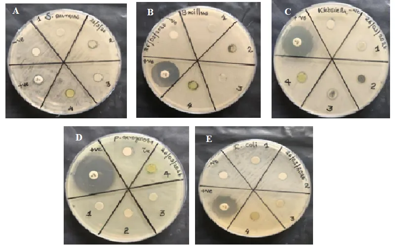
Figure 3. Antibacterial activity of seaweed extracts against different bacterial pathogens (A) Staphylococcus aureus, (B) Bacillus subtilis, (C) Klebsiella pneumoniae, (D) Pseudomonas aeruginosa, and (E) Escherichia coli. Wells 1–4 represent seaweed extracts, (+ve) indicates the positive control (ciprofloxacin), and (−ve) indicates the negative control (methanol).
Cytotoxicity of Crude Seaweed Extracts
The ethyl acetate extracts of six seaweed species, Grateloupia lithophila, Ulva fasciata, Gracilaria corticata, Ulva lactuca, Gelidium micropterum and Chaetomorpha antennina, were evaluated for cytotoxicity by MTT reduction assay against CHO-K1 cells. The results showed that none of the tested extracts showed ≥50% cytotoxicity (Table 3 and Figure 4). The highest cell viability was exhibited by Grateloupia lithophila and Gracilaria corticata with cell viability of 82.55% and 82.38%, respectively, corresponding to cytotoxicity values of 17.45 ± 2.4% and 17.62 ± 1.4%. Similarly, Ulva fasciata showed a cell viability of 75.13% with a cytotoxicity of 24.87 ± 1.4%. These species, Grateloupia lithophila, Gracilaria corticata, and Ulva fasciata, were considered non-cytotoxic according to ISO 10993-5 guidelines. Chaetomorpha antennina showed the lowest cell viability (53.82%) and the highest cytotoxicity (46.18 ± 1.7%), indicating a moderate level of cytotoxicity. The crude extracts of Ulva lactuca and Gelidium micropterum showed cell viability of 68.08% and 68.91%, respectively, with cytotoxicity values of 31.92 ± 3.7% and 31.09 ± 3.5%. These species were considered slightly cytotoxic. All the extracts showed varying degrees of cytotoxicity in CHO-K1 cells characterised by cell rounding, shrinkage, detachment and reduced cell density compared to the control, which retained normal spindle-shaped morphology and confluency (Figure 5). Among the six species tested, the species Grateloupia lithophila, Ulva fasciata, and Gracilaria corticata showed good biocompatibility with minimal adverse effect on CHO-K1 cells.
Table 3. Cytotoxic effects of selected seaweed extracts on CHO-K1 cells.
|
Seaweed species |
Cell Viability (%) |
Cytotoxicity (%) |
Interpretation |
|
Grateloupia lithophila |
82.55 |
17.45±2.4 |
Non cytotoxic |
|
Ulva fasciata |
75.13 |
24.87±1.4 |
Non cytotoxic |
|
Gracilaria corticata |
82.38 |
17.62±1.4 |
Non cytotoxic |
|
Chaetomorpha antennina |
53.82 |
46.18±1.7 |
Moderately cytotoxic |
|
Ulva lactuca |
68.08 |
31.92±3.7 |
Slightly cytotoxic |
|
Gelidium microsporium |
68.91 |
31.09±3.5 |
Slightly cytotoxic |

Figure 4. Cytotoxicity (%) of crude extract of different seaweed species on CHO-K1 cells.
Figure 5. Cytotoxic effects of selected seaweed extracts on CHO-K1 cells. A. Grateloupia lithophila, B. Ulva fasciata, C. Gracilaria corticata, D. Chaetomorpha antennina, E. Ulva lactuca, F. Gelidium micropterum and G. Untreated CHO-K1 cells. Scale bar = 150 μm.
DISCUSSION
Marine macroalgae are an important source of bioactive compounds with pharmaceutical potential. Several secondary metabolites like polysaccharides, alkaloids, fatty acids, terpenoids, phlorotannins, halogenated compounds, and pigments, with a broad spectrum of biological activities, have been reported from seaweeds (Pérez et al., 2016; Shannon and Abu-Ghannam, 2016; Biris-Dorhoi et al., 2020).
In the present study, ethyl acetate extracts of selected seaweed species were tested for antimicrobial activity against five human pathogens: Staphylococcus aureus, Bacillus subtilis, Escherichia coli, Pseudomonas aeruginosa, and Klebsiella pneumoniae. None of the extracts showed significant antimicrobial activity against the tested pathogens. Similar observations have been reported in many other studies; despite the presence of biologically active secondary metabolites, crude seaweed extracts exhibited little or no antibacterial activity against some of the tested pathogens (Pérez et al., 2016). This might be due to differences in seaweed species, seasonal variation, geographical location, environmental parameters such as temperature, salinity, light, and nutrients, the extraction solvent, and the concentration of active compounds in the crude extract (Trigui et al., 2013; Stabili et al., 2014; Biris-Dorhoi et al., 2020). The polarity and structure of algal antimicrobial compounds vary greatly, so the use of different extraction solvents with varying polarity would aid in effective extraction of both the polar and non-polar compounds from the seaweeds (Cox et al., 2014; Amorim et al., 2017).
In the advancement of drug development from natural resources, it is essential to determine the cytotoxicity and biocompatibility of the natural compounds (Harvey et al., 2015). Therefore, it is important to screen the extracts with common methods like the MTT assay. In the present study, seaweed extracts showed different levels of cytotoxic effects in CHO-K1 cells. Crude extracts from Grateloupia lithophila, Ulva fasciata, and Gracilaria corticata were found to be non-cytotoxic. Ulva lactuca and Gelidium micropterum are slightly cytotoxic, and Chaetomorpha antennina is moderately cytotoxic. The decreased cell viability with Chaetomorpha antennina may be associated with the presence of sulphated compounds, flavonoids, phenolics and terpenoids (Cotas et al., 2020); specifically, phenolic compounds can induce apoptosis due to their pro-oxidant and antioxidant activities (Lomartire et al., 2021). The presence of excessive amounts of these kinds of secondary metabolites in the crude extract may impart cytotoxicity on normal cells; therefore, cytotoxic evaluation and dose optimisation are important in therapeutic applications (Gunasekaran et al., 2017; Lomartire et al., 2021).
The extraction yield was different for all six seaweed isolates, indicating variations in the amount of extractable secondary metabolites in each species. The highest yield was obtained from Gracilaria corticata, and the lowest yield was obtained from Gelidium micropterum, followed by Ulva lactuca. There was no correlation found between the extraction yield and the cytotoxicity. Gracilaria corticata showed low cytotoxicity but with high yield. For Gelidium micropterum and Ulva lactuca, the yield was low, but they were slightly cytotoxic. In the case of Chaetomorpha antennina, the yield was relatively high, and it also showed high cytotoxicity. Overall, the study shows that the cytotoxic effect cannot be determined from the extraction yield alone. The most cytotoxic strain, Chaetomorpha antennina, requires further evaluation for anticancer properties and the strains Grateloupia lithophila, Ulva fasciata, and Gracilaria corticata have minimum cytotoxicity and high extraction yield warranties additional research for further nutraceutical and pharmaceutical applications.
CONCLUSION
The present study evaluated the antimicrobial activity and cytotoxicity of ethyl acetate extracts of six seaweed species collected from Kannur coast, Kerala. None of the crude extracts showed antimicrobial activity against the test pathogens, which revealed that the antimicrobial activity of seaweed extracts is greatly influenced by the seasonal, geographical and environmental factors. Additionally, the effective extraction of secondary metabolites from seaweeds requires multiple solvent extractions with different polarities. The cytotoxicity assessment using CHO-K1 cells showed that Gracilaria corticata is the least cytotoxic with high extraction yield, making it a valuable candidate for further pharmaceutical and nutraceutical evaluation. The moderate cytotoxicity exhibited by Chaetomorpha antennina suggests its importance in future anticancer studies with the isolation and characterisation of active metabolites.
REFERENCES


Arya K V, Adhithya P, Namitha R, Ayana N, Jisha Kumaran, Evaluation of Antimicrobial Potential and Cytotoxicity of Selected Marine Seaweed Extracts, Int. J. of Pharm. Sci., 2026, Vol 4, Issue 7, 1116-1124, https://doi.org/10.5281/zenodo.21217791
 10.5281/zenodo.21217791