

We use cookies to ensure our website works properly and to personalise your experience. Cookies policy
1,2 Student, Department of Pharmaceutics, Government College of Pharmacy, Karad, Maharashtra,415124.
3Associate Professor, Department of Pharmaceutics, Government College of Pharmacy, Karad, Maharashtra, 415124
Exosomes are small extracellular vesicles released by cells that play an important role in intercellular communication by transporting proteins, lipids, nucleic acids, and other bioactive molecules. Their unique biological properties, biocompatibility, and ability to transfer functional cargo to recipient cells have attracted considerable attention in diagnostics, drug delivery, and therapeutic applications. This review provides a comprehensive overview of exosomes, including their classification, biogenesis, biological composition, isolation, characterization, and storage methods. Particular emphasis is placed on their emerging role as diagnostic and therapeutic tools in dermatological disorders, cancer, and various immunopathological conditions. The potential of exosomes as non-invasive biomarkers and their application in liquid biopsy are also discussed. In addition, plant-derived extracellular vesicles (PDEVs) are reviewed as an emerging alternative delivery platform, with attention to their biological characteristics, therapeutic potential, and advantages over conventional mammalian extracellular vesicles. The review further explores the involvement of exosomes in the immunopathology of neurodegenerative, respiratory, cardiovascular, renal, digestive, and endocrine diseases, highlighting their role in disease progression and intercellular signalling. Despite their considerable potential, challenges related to isolation, purification, characterization, storage, standardization, scalability, safety, and clinical translation remain. Overall, exosomes and PDEVs represent promising platforms for biomarker discovery, targeted drug delivery, and therapeutic intervention, although further standardized research and well-designed clinical studies are required to establish their clinical applicability.
Extracellular vesicles (EVs) are membrane-enclosed structures released by cells that can carry protein, lipid, and nucleic acid cargos to recipient cells. Initially regarded as cellular debris, EVs are now recognized to play key roles in intercellular communication and are of great interest as diagnostic and therapeutic tools for various diseases. The ability of EVs to shield and carry specific cargo from the cells of origin has contributed to their increased interest in basic and clinical research. [1-3]
Historical development of exosome research
Early observations leading to the discovery of exosomes date back decades before the term exosome was introduced. As early as 1967, Wolf described platelet dust as small particles present in human plasma. [1] The discovery of exosomes was closely linked to the study of reticulocyte maturation. Harding et al. demonstrated in 1983 that transferrin receptors were internalized and recycled in rat reticulocytes via endocytosis, whereas electron microscopy of these cells revealed vesicles containing transferrin receptors. [4,5] In 1987, Johnstone et al. demonstrated that vesicles were released from reticulocytes during their maturation and suggested the term exosome for these structures. [6]
Early descriptions of exosomes and other EVs primarily focused on their role in removing membrane receptors and other components during endocytosis and recycling. The view of exosomes as garbage bags changed when it was discovered that they contain bioactive.
molecules, such as proteins, lipids, and RNAs, that can modulate the functions of recipient cells. In particular, the ability of exosomes to transfer functional RNAs has opened up new opportunities for research. [2,3,7]
From cell to cellular waste to intercellular communication
Exosomes are formed from the endosomal system during inward budding of the limiting endosomal membrane, resulting in the formation of intraluminal vesicles (ILVs). Exosomes are released when multivesicular bodies (MVBs) fuse with the plasma membrane, and intraluminal vesicles are released into the extracellular space. This pathway clearly distinguishes exosomes from other EVs, which are formed by the outward budding of the plasma membrane. [7,8]
The importance of exosomes is underscored by the diverse range of bioactive molecules that can be incorporated into their cargo. Exosomes contain membrane- and cytosolic proteins, lipids, messenger RNA, microRNA, and other non-coding RNAs that regulate gene expression and physiological processes in recipient cells. In addition, exosomes can carry various metabolites and affect cellular signalling pathways, leading to changes in metabolism and other physiological functions. Moreover, recent studies have provided evidence that the incorporation of specific RNA species into exosomes may be regulated at the level of individual cells. [9,10]
Furthermore, the ability of exosomes to deliver specific cargo to recipient cells makes them important players in disease pathogenesis. For example, tumor-derived exosomes can modulate the tumor microenvironment and affect distant sites, participating in all stages of tumor development and progression. [11,12]
Exosomes as diagnostic and therapeutic candidates
The close link between the molecular content of exosomes and the functional state of cells has prompted extensive efforts to harness exosomes for diagnostic purposes. The ability to detect exosomes in accessible biofluids and the potential of their molecular content as disease biomarkers have led to their extensive preclinical investigation. Simultaneously, the ability of exosomes to affect the functions of recipient cells makes them attractive vehicles for the delivery of nucleic acids, proteins, and other therapeutics. [3,12,13]
However, the use of exosomes for practical applications is hampered by their heterogeneity and the limitations of current approaches for their isolation and characterization. Exosome preparations typically contain a mixture of different EVs and other extracellular particles, and their protein content often overlaps with that of other EV fractions. Moreover, many exosome preparations lack consistent cargo molecules that are traditionally used as markers. These challenges highlight the importance of developing more reliable approaches for exosome isolation and characterization in the future. [14]
Current perspective
Overall, the initial recognition of exosomes as cellular garbage bags has been replaced by a broader view of these structures as key modulators of intercellular communication. The current understanding of the biogenesis, cargo composition, and physiological and pathological functions of exosomes has advanced due to numerous studies using modern electron microscopy, flow cytometry, next-generation RNA sequencing, lipidomic, and proteomic approaches. However, despite rapid progress in the field, there are still many challenges in the isolation, identification, and characterization of exosomes. [8,14]
The latest recommendations from the International Society for Extracellular Vesicles (ISEV) suggest using the term exosomes with caution. According to the MISEV2023 guidelines, the term exosome should be used only for EVs with a demonstrated endosomal origin. The designation of small extracellular vesicles (sEVs) should be used for EVs of unknown origin. [15] Thus, the current perspective on exosomes and other EVs highlights the importance of caution and careful consideration when interpreting exosome-related data.
Overall, the journey from the initial description of exosomes as garbage bags from reticulocytes has led to a better understanding of the biological roles of EVs and the opportunities for their use as biomarkers and therapeutic agents in various diseases. Further studies are required to fully harness the potential of membrane-enclosed structures. The classification and characterization of exosomes is depicted in fig.1[8,15–19] and the comparison of synthetic nanoparticles, mammalian EV’s and PDEV’s depicted in fig.2.[19–31]
CLASSIFICATION AND CHARACTERIZATION OF EXOSOME
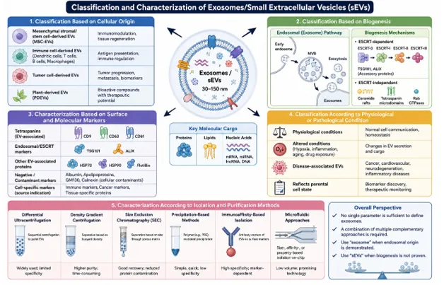
Figure 1: Classification and characterization of exosomes/small extracellular vesicles (sEVs).
COMPARISON OF SYNTHETIC NANOPARTICLES, MAMMALIAN EV’S AND PDEVS
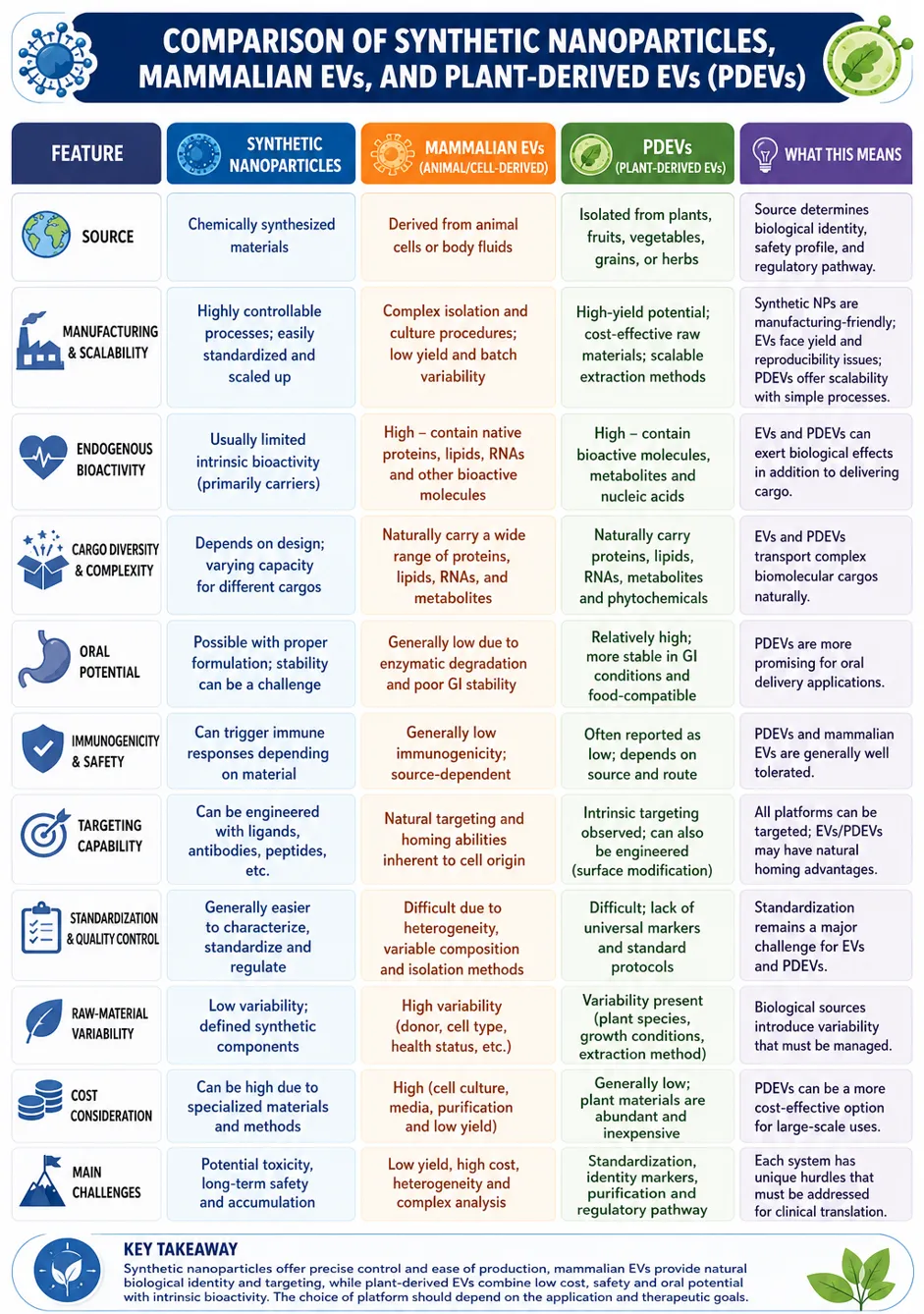
Figure 2: Comparison of synthetic nanoparticles, mammalian EV’s and PDEVs
BIOGENESIS OF EXOXOMES
Exosome biogenesis primarily occurs through the endosomal route. As shown in fig. 3 it starts with the inward budding of the plasma membrane to form endosomes. Early endosomes mature into late endosomes and multivesicular bodies (MVBs) [32–35]. During MVB biogenesis, the limiting membrane invaginates to produce intraluminal vesicles (ILVs) with specific protein, lipid, and nucleic acid content [33,36]. This process involves the endosomal sorting complex required for transport (ESCRT), including ESCRT-0, -I, -II, and -III, and ESCRT-independent mechanisms such as tetraspanins, ceramide, and other lipids [36–39]. MVBs deliver their ILVs to lysosomes for degradation or to the cell surface for secretion [33,36]. This trafficking is mediated by small guanosine triphosphatases of the Rab family, including Rab27a and 27b, which control MVB docking and fusion with the plasma membrane [40,41]. Once released, ILVs are referred to as exosomes [32,34,42]. Nevertheless, according to the MISEV2023 recommendations, it is mandatory to demonstrate the endosomal origin of the vesicles because, otherwise, they should be defined as small extracellular vesicles (sEVs) [43,44].
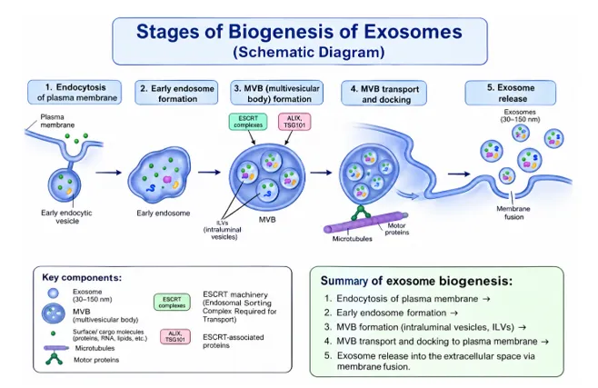
Figure 3: stages of biogenesis of exosomes
EXOSOME ISOLATION METHODS
Complete overview of isolation methods of exosome is represented in table.1
Table 1: exosome isolation methods
|
Isolation method |
Principle and procedure |
Advantages |
Limitations |
Suitable applications |
|
1. Differential ultracentrifugation (dUC) |
Sequential centrifugation removes cells and debris at lower speeds, followed by high-speed ultracentrifugation to sediment small EVs. The final pellet was washed and resuspended in PBS [49–51]. |
Widely established, relatively high recovery, suitable for large sample volumes, and does not require affinity reagents. |
This method is time-consuming, requires an ultracentrifuge, and may cause vesicle aggregation/deformation. In addition, protein and other particle contamination may remain [49–52]. |
Cell culture medium, plasma, serum, and large-volume research samples. |
|
2. Density-gradient ultracentrifugation (DGUC) |
EVs are separated according to their buoyant density using sucrose or iodixanol gradients. Vesicles migrate to their characteristic density regions and are collected as separate fractions [53–56]. |
Higher purity than conventional dUC; better separation from soluble proteins and other particles; useful for complex samples. |
It is laborious, lengthy, and technically demanding, and the recovery may be lower because of the multiple processing steps. |
Proteomics, functional studies, and samples that require high-purity EV preparations. |
|
3. Sucrose cushion ultracentrifugation |
A sample is layered over a concentrated sucrose cushion and subjected to ultracentrifugation. EVs pass through/collect at the cushion, whereas soluble contaminants remain separated [57,58]. |
Can reduce non-vesicular contamination; is comparatively simple; may improve recovery and vesicle quality compared with direct dUC. |
Requires ultracentrifugation; sucrose must be removed before downstream applications; yield and purity strongly depend on the protocol conditions. |
Cell culture supernatants and biological fluid EV enrichment |
|
4. Size-exclusion chromatography (SEC) |
EVs were separated according to their hydrodynamic size using porous chromatographic beads. Larger EVs elute earlier, whereas smaller proteins enter the pores and elute later [59–61]. |
Gentle method that preserves vesicle integrity, is rapid and reproducible, and removes many soluble proteins. |
Limited sample-loading capacity; EV subtypes are not specifically separated; dilution of the EV fraction may occur. |
Plasma, serum, and samples were intended for functional or molecular analyses. |
|
5. Ultrafiltration (UF) |
Membrane filters with defined molecular weight or pore size cut-offs retain EVs, whereas smaller molecules and soluble proteins pass through [59,62]. |
Rapid; requires relatively small equipment; concentrates large sample volumes; useful before additional purification. |
Membrane fouling, vesicle retention, and possible deformation can reduce recovery, and purity alone may be insufficient. |
Concentration of EVs before SEC or other purification methods |
|
6. Polymer precipitation |
Polymers such as polyethylene glycol (PEG) reduce solubility and promote vesicle precipitation, which are subsequently recovered by low-speed centrifugation [46,63]. |
It is simple, inexpensive, does not require specialized equipment, and is suitable for processing many samples. |
Poor specificity: Proteins and other non-vesicular materials may co-precipitate, thereby reducing purity [46,63]. |
Preliminary enrichment and high-throughput studies, where maximum purity is not essential. |
|
7. Immunoaffinity capture |
Antibodies immobilized on magnetic beads or other matrices recognize specific EV surface markers, such as CD9, CD63, CD81, or disease-associated antigens [64–66]. |
High specificity allows for the enrichment of selected EV subpopulations and is useful for biomarker studies. |
Expensive; antibody-dependent; limited processing capacity; does not capture EVs lacking selected markers. |
Disease-specific biomarker studies and isolation of defined EV populations |
|
8. Microfluidic isolation |
Miniaturized devices separate EVs based on size, surface affinity, charge, or a combination of these physical properties [67,68]. |
Small sample requirement, rapid processing, and potential for automation and point-of-care applications. |
Device fabrication and optimization can be complex, and limited standardization and scalability remain challenging. |
Clinical diagnostics and small-volume biological samples |
STORAGE METHODS OF EXOSOMES
Proper storage of exosomes/small extracellular vesicles (sEVs) is critical for preserving their size distribution, membrane morphology, cargo content, concentration, and biological functions. The storage temperature, duration, buffer composition, and number of freeze–thaw cycles influence the quality of the isolated EVs [69–72].
The following are the storage recommendations.
BIOLOGICAL COMPOSITION OF EXOSOMES
Exosomes are small extracellular vesicles surrounded by a lipid bilayer and are enriched with biologically active proteins, lipids, nucleic acids, and metabolites. The composition of these vesicles is highly dependent on the cells of origin and their physiological or pathological state, which allows exosomes to be used as biomarkers of cellular function [79–83].
Protein Composition
Proteins constitute an essential component of exosomes, both in the membrane and in the internalized form. Exosomes contain tetraspanins, such as CD9, CD63, and CD81; heat-shock proteins, including HSP70 and HSP90; TSG101; ALIX; Rab proteins; integrins; annexins; and various other enzymes and ion channels. These proteins participate in a wide array of critical cellular functions, including maintaining membrane integrity, facilitating vesicular trafficking, recognizing target cells, and regulating exosome function [79,83,89,90]. For instance, exosomes possess cell-specific integrins, adhesion molecules, and cytoskeleton-associated proteins that enhance their cellular uptake through interactions with target cell receptors [90,95–98].
Lipid Composition
Similar to other cell membranes, exosome membranes are composed of cholesterol, sphingomyelin, ceramide, phosphatidylserine, and other lipids. Most of these lipids are involved in membrane stabilization and vesicular trafficking, facilitating exosome formation and uptake by recipient cells [79,83,99–102]. Specifically, the sphingomyelinase-mediated ceramide signalling pathway has been implicated in the regulation of exosome-mediated intercellular communication [103–108]. Prostaglandins and leukotrienes are among the lipids found in exosomes that play critical roles in the regulation of inflammation [79,111,112].
Nucleic Acids
Exosomes contain various RNAs, including mRNA, microRNA (miRNA), long non-coding RNA (lncRNA), circRNA, transfer RNA (tRNA), ribosomal RNA (rRNA), small nuclear RNA (snRNA), small nucleolar RNA (snoRNA), and PIWI-interacting RNA (piRNA) [79,81,114–117]. miRNAs are transported to target cells within exosomes, where they regulate distinct cellular processes by binding to specific messenger RNA molecules [114,118]. Notably, several types of RNAs, including lncRNAs and circRNAs, have been shown to modulate distinct cellular functions [115–121]. Apart from RNAs, exosomes also contain various types of DNA, although their content is highly variable depending on the cell of origin [79,123,124].
Other Bioactive Components
In addition to lipids, proteins, and nucleic acids, exosomes contain metabolites and amino acids that can originate from host cells or the environment. Moreover, exosomes can carry a variety of bioactive molecules, including cytokines, chemokines, growth factors, and extracellular vesicle RNA. All these diverse molecular components allow exosomes to participate in numerous physiological and pathological processes [79,80,125]. Consequently, exosome functionality is determined by the combination of diverse molecular components present in them, rather than individual molecules [80,83]. Biological composition of exosomes are described in the table.2
Table 2: Biological Components Of Exosomes
|
Component |
Major examples |
Principal significance |
|
Proteins |
CD9, CD63, CD81, ALIX, TSG101, HSP70/90, Rab |
Recognition, trafficking, signalling and uptake |
|
Lipids |
Cholesterol, sphingolipids, PS, PA |
Membrane stability, curvature and cell interaction |
|
RNAs |
miRNA, mRNA, lncRNA, circRNA, tRNA |
Gene regulation and intercellular communication |
|
DNA |
Genomic and mitochondrial DNA |
Potential genetic signalling |
|
Metabolites |
Amino acids and metabolic intermediates |
Metabolic and immunological regulation |
|
Plant EV components |
PA, PC, PE, MGDG, DGDG, PI |
Distinct membrane properties and bioactivity |
EXOSOMES AND PLANT-DERIVED EXTRACELLULAR VESICLES AS DRUG DELIVERY SYSTEMS
Overview
Exosomes are nanosized extracellular vesicles (EVs) that represent the link between donor and target cells by carrying biologically active information from the former to the latter. Their ability to deliver complex cargo, phospholipid membrane composition, nano size dimensions, and specific targeting potential makes them highly efficient carriers for targeted drug delivery (TDD). Plant-derived EVs (PDEVs) or plant exosome-like nanovesicles (PELNVs) are promising nanocarriers because they can carry various bioactive phytochemical compounds and endogenous biomolecules of plant origin. As naturally produced carriers, PDEVs offer the possibility of combined conventional drug plus cell communication factor delivery to specific sites of action, which is of great interest in pharmaceutical science [126,127]. Unlike artificial nanoparticles, which usually lack endogenous metabolic activity, plant EVs have the ability to provide an additional effect owing to their endogenous nature, as they contain various metabolites, lipids, proteins, and RNAs that may have a pharmacological impact. Additionally, membrane properties protect their contents from external factors, while their physicochemical properties ensure their penetration into cells [126,129]. Due to their biocompatibility, high drug targeting potential, natural origin, and opportunity for large-scale production, such nanocarriers seem to be a great source for innovative pharmaceutical production which is illustrated in fig,4 [126,129]
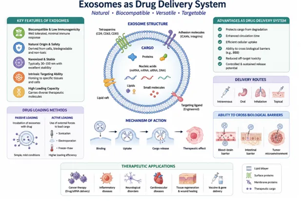
Figure 4: Exosomes And PDEV’s As Drug Delivery System
Drug-Loading Mechanisms[126,129]
Therapeutic Molecules Delivered by the Nanocarriers
Targeted Delivery[126,133]
Oral and Barrier Delivery Mechanisms[126]
The ability of PDEVs to resist the harsh conditions of the gastrointestinal tract allows for the oral administration of therapeutics, including PDEVs. In addition, these vesicles can cross various biological barriers to access specific tissues, including the blood-brain barrier.
Major Advantages [126-133]
Major Disadvantages [126-134]
Exosomes and PDEV are promising natural delivery vehicles that can deliver various types of pharmaceuticals and biological molecules while potentially providing additional therapeutic benefits owing to their endogenous nature. The major challenge associated with these vesicles is the need to optimize loading, targeting, and isolation techniques, as well as extensive preclinical trials to assess their safety, optimal pharmacokinetic profiles, and treatment regimens [126,128,133,134].
DIAGNOSTIC APPLICATIONS OF EXOSOMES IN DERMATOLOGY
Exosomes are small extracellular vesicles that are naturally secreted from cells under physiological and pathological conditions. Their membrane lipid bilayer structure contains bioactive cargos, such as proteins, lipids, DNA, and diverse types of RNA, which can provide indicative information about their cells of origin. Therefore, exosomes have become promising research targets for disease diagnosis, prognosis, and treatment response monitoring. As previously reported, due to their relative stability in biological fluids and the informative nature of their biochemical cargo, exosomes can serve as highly valuable biomarkers for both minimally invasive or liquid biopsy approaches [135-138].
Exosomes as Emerging Biomarkers in Dermatology
In dermatology, exosomes may serve as valuable biomarkers for inflammatory, autoimmune, pigmentary, and malignant skin diseases. The content of exosomes can differ depending on the state of the disease, thus contributing to the discrimination between healthy and diseased states. Nevertheless, the practical application of exosomes in dermatology remains limited and requires extensive clinical evaluation [139,140].
Exosomal Nucleic Acids as Biomarkers
Exosomal miRNAs are one of the most studied classes of RNA species because of their involvement in the regulation of inflammation, immunity, proliferation, apoptosis, melanogenesis, and tissue repair processes. Therefore, their altered expression patterns can serve as informative biomarkers for disease-associated transcriptomic changes. Other classes of RNA that can be used as biomarkers include mRNAs, long non-coding RNAs, circular RNAs, and tRNA-derived fragments [137,142].
Psoriasis
Psoriasis is a complex multifactorial inflammatory disease involving cross-talk among keratinocytes, dendritic cells, and T lymphocytes, where exosomes have been implicated in this process by carrying messenger molecules involved in inflammatory cascades. Consequently, exosomal RNA/proteins have been suggested as potential biomarkers for psoriasis and psoriatic arthritis. However, their clinical implementation is hampered by interindividual heterogeneity and possible treatment effects [143,144].
Atopic Dermatitis
Atopic dermatitis is characterized by impaired epidermal barrier function and immune dysregulation. Episomal RNAs may serve as biomarkers for these processes. Specifically, in patients with paediatric atopic dermatitis, decreased levels of transcripts of plasma exosomal tRFs, including tRF-28-QSZ34KRQ590K, have been reported. The diagnostic accuracy of this RNA species was supported by ROC analysis; however, the link between atopic dermatitis and tRFs was postulated based on a small patient cohort, and further validation is required [145].
Vitiligo
Exosomes may also play a role in identifying biomarkers of vitiligo, in which oxidative stress, melanocyte damage, and immune activation are vital pathogenic factors. Exosomes derived from oxidatively stressed keratinocytes have been reported to carry miR-31-3p, which enhances melanocyte injury and increases CD8+ T cell cytotoxicity in vivo, supporting the pathogenic role of exosomal miRNAs in vitiligo [146].However, their diagnostic and prognostic values in patients with vitiligo remain unclear [146].
Melanoma And Other Cutaneous Malignancies
Genomic research on tumour cell exosomes has established that they contain biologically relevant information about oncogenic transformation, tumour development, metastasis formation, immunological interactions, and treatment response. Consequently, the RNA species (miRNA), proteins, and other molecules present in tumour cell exosomes have been suggested as potential biomarkers for melanoma and other skin cancer types. Exosome biomarker testing may be used as a non-invasive test for detecting and assessing the response to therapy of malignant tumours but cannot substitute diagnostic histopathology and requires further clinical investigations [139,140].
Sweat-Derived Exosomes and Non-Invasive Diagnosis
Sweat is an attractive source of skin-associated extracellular vesicles because of the ease of its non-invasive isolation. Proteomic analysis of human sweat-derived exosomes revealed the presence of 1,062 different proteins, among which are proteins and antimicrobial peptides involved in skin immunity [141]. Although the study described above did not identify disease-specific biomarkers, it nevertheless showed the potential utility of sweat exosomes in the diagnosis of inflammatory and infectious skin diseases. Analysis of exosomal RNA content may further extrapolate this approach [141].
Exosome-Based Liquid Biopsy
Exosome-based liquid biopsy refers to the analysis of molecules contained in extracellular vesicles isolated from fluid samples, preferably blood or sweat, for diagnostic purposes. This non-invasive procedure is particularly attractive in dermatology, where skin biopsies are often associated with patient discomfort. For inflammatory conditions, liquid biopsy has the potential to enable the assessment of inflammatory status and treatment efficacy by characterizing disease-associated exosomal content, whereas in skin cancer, it may serve to monitor tumorigenic activity. The key to success in this regard is the identification of relevant biomolecules characteristic of a particular disease [136,139,140].
Potential Biomarker Classes
Several exosomal components can act as biomarkers.
Utilisation of Exosomes in Cancer
Exosomes are membrane vesicles of endocytic origin, containing various proteins, mRNA, miRNA, circular RNA, DNA, and other biomolecules released from cells. Due to their accessibility in biofluids such as blood and urine, they can serve as valuable cancer biomarkers for diagnosis and liquid biopsy [147,148,149]. Since exosomal content reflects changes in tumour cells, exosomes can help predict cancer development and progression, determine prognosis, and monitor therapy.
Since exosomal protein content can be used as a biomarker, it is of great interest to evaluate the role of various proteins in cancer. Thus, an increase in the number of CD63-positive vesicles was associated with melanoma [150], while CD151, CD171, and tetraspanin-8 proteins were linked with lung cancer [151,152]. In pancreatic cancer, GPC1-positive exosomes have been studied [153], while urinary exosomal proteins have been suggested to be used as potential bladder and prostate cancer biomarkers [154]. However, to predict cancer, it is necessary to validate particular proteins and study their concentrations in detail.
Exosomal miRNAs are promising biomarkers because they are protected by the lipid layer of the exosomes. They can be used to predict cancer development or to monitor its progress. For instance, miR-21 and miR-1246 levels are linked to breast and oesophageal cancers, while miR-141 and miR-375 are associated with prostate cancer [147-149,155]. Recent studies have combined exosome biomarkers with microfluidics, spectroscopy, and artificial intelligence to develop highly sensitive liquid biopsy tests for cancer screening [156,157]. In general, exosomes have great potential as non-invasive biomarkers for diagnosing and predicting cancer, monitoring therapy, and assessing prognosis of cancer. However, it is necessary to conduct more high-level studies and clinical trials for this technology to be used in practice [158].
Table 3: diagnostic application of exosomes in cancer
|
Cancer |
Candidate exosomal marker |
Specimen |
Diagnostic significance |
|
Lung cancer |
CD151, CD171, Tetraspanin-8 |
Serum |
Potential NSCLC biomarker |
|
Exosomal miRNA profile |
Circulating blood |
Potential screening approach |
|
|
Breast cancer |
miR-21, miR-1246 |
Plasma/serum |
Tumour-associated biomarker |
|
Colorectal cancer |
miR-638 |
Circulating exosomes |
Candidate diagnostic marker |
|
Pancreatic cancer |
GPC1-positive exosomes |
Blood |
Early-detection candidate |
|
Exosomal SORL1 |
Blood/plasma |
Microfluidic diagnostic platform |
|
|
Prostate cancer |
Survivin |
Plasma |
Early-detection candidate |
|
PCA3, TMPRSS2:ERG |
Urine |
Non-invasive molecular detection |
|
|
miR-141, miR-375 |
Serum/plasma |
Diagnosis/progression |
|
|
Melanoma |
CD63, caveolin-1, TYRP2, VLA-4, HSP70/HSP90 |
Plasma |
Tumour-associated biomarkers |
|
Glioblastoma |
miR-21, miR-320, miR-574-3p, RNU6-1 |
Exosomes |
Diagnostic/molecular characterization |
|
ESCC |
miR-21, miR-1246 |
Serum |
Diagnosis/prognosis |
|
Bladder cancer |
EGFR-related proteins, Gsα, resistin, RAI3 |
Urine |
Candidate urinary biomarkers |
THERAPEUTIC APPLICATIONS
The Role of Exosomes in Immunopathology
Exosomes are a type of cell vesicle that carries proteins, nucleic acids, and lipids, which can be transferred from one cell to another. The contents and properties of exosomes are regulated by the state of the cells from which they originate and affect the functionality of recipient cells upon uptake [158,159,160]. Thus, acting as regulators, exosomes modulate both immune responses and the processes of pathological development. In addition, exosomes enable homotypic (recipient and donor cells of the same type) and heterotypic (recipient and donor cells of different types) cell communication, as well as communication between organs via the bloodstream [161-163]. Moreover, due to the possibility of carrying specific cargo molecules, exosomes can be used not only as information carriers but also as drug delivery vehicles for targeted treatment. Therefore, exosomes are multifunctional regulators of both healthy and pathological immunity with great therapeutic potential for use in exosome-based treatments [158,163].
Exosomes In Immunopathology of Neurodegenerative Diseases
Neurodegenerative disorders are characterized by a pathogenic link between the dysregulated immune system and malfunctioning of the nervous system. For instance, exosomes seem to play a role in amyloid-β (Aβ) pathogenesis and neuroinflammation associated with Alzheimer’s disease (AD). Astrocyte-derived exosomes carrying complement proteins may contribute to neuronal damage, whereas exosomal miR-146a is implicated in the dysregulation of microglial response to Aβ and pro-inflammatory stimulation [164, 165]. Similarly to AD, exosomes are considered to participate in the mechanism of Parkinson’s disease (PD). Exosomes derived from astrocytes, neurons, microglia, macrophages, and other cells mediate communication between these cells and may propagate pathological processes. Specifically, inflammatory macrophage-derived exosomes enriched with miR-155-5p may trigger glial cells, whereas exosomes carrying alpha-synuclein may amplify neuroinflammation and neurodegeneration [166-170].
Exosomal Immunopathology in The Respiratory Tract
The respiratory tract is characterized by a complex network of communication between epithelial cells, endothelial cells, macrophages, neutrophils, and other cells via exosomes. Exosomes may carry inflammatory proteins and regulatory RNAs that affect lung inflammation and tissue injury.
Acute Lung Injury and Acute Respiratory Distress Syndrome
During acute lung injury (ALI) and acute respiratory distress syndrome (ARDS), pro-inflammatory signalling by exosomes, as well as disruption of the alveolar-capillary barrier, leads to increased permeability. In particular, endothelial extracellular vesicles may mediate monocyte polarization into inflammatory macrophages and facilitate pathological neutrophil trafficking [169,170]. Alveolar epithelial cell exosomes may carry miR-92a-3p, which can promote macrophage activation and pulmonary inflammation. Particulate matter may also modify the content of exosomal miRNAs, while tenascin-C-enriched exosomes may enhance inflammation by polarizing macrophages towards the M1 phenotype [171,172]. Altogether, these data suggest that exosomes play a critical role in sustaining inflammatory responses in the lungs during ALI or ARDS.
Exosomes In Asthma
Exosomes are involved in a variety of pro-inflammatory processes in asthma, ranging from epithelial-immune cell crosstalk to the promotion of airway remodelling. In particular, exosomes derived from Mycoplasma pneumoniae-infected macrophages may activate TLR2/NF- κB/JNK signalling pathways in macrophages and T cells [173]. At the same time, exosomes from patients with chronic airway inflammation are capable of modulating monocytes, NK cells, and neutrophils. Exosomal signalling can also be dysregulated by environmental factors, including exposure to house-dust mites and particulate matter, leading to enhanced airway inflammation. Thus, exosomes are critically involved in immune-cell activation and contribute to the development of chronic airway inflammation in asthma.
Exosomes In COPD
Exosomes are key players in the development and progression of chronic obstructive pulmonary disease (COPD). Enhanced levels of circulating exosomes are often reported in patients with COPD, and these vesicles may carry specific miRNAs that contribute to systemic inflammation. For instance, airway epithelial-cell-derived exosomes can mediate M1 macrophage polarization via miR-125a-5p and miR-221-3p [174,175]. In contrast, cigarette smoke can promote the release of exosomes from epithelial cells and neutrophils that carry inflammatory Wnt5a protein, potentially propagating the inflammatory response to other tissues [176].
The Role of Exosomal Immunopathology in The Systemic Circulation
Exosomes participate in communication between cardiomyocytes, endothelial cells, macrophages, fibroblasts, and immune cells. Acting via local and systemic routes, they modulate cardiovascular inflammation, fibrosis, vasculature function, and tissue remodelling [177].
Heart Failure and Cardiac Remodelling
Exosomes participate in the development of myocardial inflammation, fibrosis, hypertrophy, and remodelling in heart failure. For instance, fibroblast-derived exosomes enriched with miR-21a-5p contribute to cardiac fibrosis, whereas cardiomyocyte stress promotes the release of exosomes carrying inflammatory cargo. Notably, following myocardial ischemia-reperfusion injury, cardiomyocyte-derived exosomal miR-155-5p can activate macrophages and trigger inflammatory signalling. Thus, exosomes play a paracrine role in the development of cardiac damage and remodelling.
Atherosclerosis
Exosomes carry bioactive molecules that modulate endothelial cell dysfunction, macrophage activation, vascular smooth-muscle cell proliferation, and vascular inflammation. For example, obesity- and nicotine-induced exosomal miRNAs can promote endothelial inflammation and smooth-muscle cell proliferation. Moreover, macrophage-derived exosomes may exacerbate inflammatory responses and neutrophil extracellular trap formation, linking metabolic disturbances with vascular inflammation. Overall, exosomes are implicated in atherogenesis and could serve as biomarkers or therapeutic targets.
Exosomal Immunopathology in the Urinary System
The kidney hosts numerous cell types, including epithelial and endothelial cells of the urinary tract, which communicate with each other via extracellular vesicles. Urinary exosomes reflect the functional state of the kidney and participate in inflammatory signalling.
Acute Kidney Injury
In acute kidney injury (AKI), tubular epithelial cells release exosomes containing pro-inflammatory messenger RNA (mRNA). For instance, exosomal CCL2 mRNA can activate macrophages and promote tubulointerstitial inflammation, while exosomal miR-23a and miR-19b-3p enhance macrophage activation and M1 polarization [178-180]. Thus, AKI induces the release of pro-inflammatory exosomes that modulate immune cell function and worsen kidney injury.
Chronic Kidney Disease
Chronic kidney disease (CKD) is associated with enhanced tubular injury and dysfunction as well as dysregulated inflammatory response. Exosomes derived from tubules or immune cells could carry profibrotic and inflammatory cargo, exacerbating CKD progression. For instance, tubule-derived exosomes could activate fibroblasts and promote extracellular matrix remodelling, whereas macrophage-derived exosomes could facilitate the recruitment of inflammatory cells into the kidney. Additionally, high-phosphate diet could alter macrophage exosomal RNA content and promote vascular calcification in CKD [181]. Altogether, exosomes are regulators of renal inflammation and could serve as promising biomarkers of CKD.
Exosomal Immunopathology in the Digestive System
The gastrointestinal tract and liver represent a complex network of immunologically active cells, including epithelial cells, macrophages, hepatocytes, endothelium, and other cell types. Exosomes link the intestine and the liver, participating in the regulation of intestinal immunity, inflammatory responses, and metabolic homeostasis.
Inflammatory Bowel Disease
Inflammatory bowel disease (IBD) is characterized by deregulated intestinal immune responses causing mucosal damage and chronic intestinal inflammation. Exosomes from inflamed intestine contain bioactive molecules that modulate epithelial-cell and immune-cell functions. For instance, macrophage-derived exosomal miR-223 could suppress intestinal barrier function, whereas IBD-associated exosomes from epithelial cells and neutrophils worsened colonic inflammation [182]. Altogether, defective exosome-mediated communication promotes intestinal damage in IBD.
Non-Alcoholic Fatty Liver Disease
Non-alcoholic fatty liver disease (NAFLD) is associated with lipid accumulation in hepatocytes and chronic liver inflammation. Exosomes link NAFLD with systemic inflammation by transporting bioactive molecules between hepatocytes, macrophages, vascular endothelium, and other cells. For instance, lipotoxic hepatocytes could release exosomes enriched with miR-9-5p that promote M1 macrophage polarization. Cell-cell communication via integrin β1+ containing extracellular vesicles enhances monocyte adhesion to the liver endothelium and contributes to liver inflammation. Additionally, ceramide-enriched extracellular vesicles from hepatocytes could promote macrophage recruitment to the liver. Moreover, liver-derived exosomes could affect the function of distant tissues, such as the intestine and endocrine system, fine-tuning glucose metabolism and other physiological processes.
Exosomal Immunopathology in the Endocrine System
Exosomes bridge endocrine and immune systems, affecting β-cell survival, insulin secretion, and autoimmune response. Their implication in the development of diabetes has been described for both type 1 and type 2 diabetes.
Diabetes Mellitus
Extracellular vesicles from β-cells carry autoantigens and contribute to the development of autoimmunity in type 1 diabetes [183]. In type 2 diabetes (T2D), adipose-derived exosomes enhance monocyte activation and promote macrophage polarization into pro-inflammatory M1 subtype, thus driving insulin resistance. Exosomes from pancreatic islets could affect immune responses via TLR4/MyD88-dependent signalling.
Autoimmune Thyroid Disease
Autoimmune thyroid diseases are associated with deregulated immune response against thyrocytes. The role of exosomes in this pathology has been scarcely studied; however, recent data highlight their potential as serological biomarkers. In Hashimoto’s thyroiditis, T-cell-derived exosomes carrying miR-142-3p contribute to the suppression of regulatory T cells and thyrocyte destruction. Overall, deregulated exosome-mediated communication exacerbates autoimmunity in the thyroid.
Integrated Mechanistic Perspective of Exosomal Immunopathology
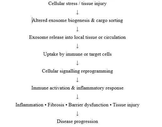
Injured tissue → circulating exosomes → distant organs → immune-cell activation → systemic inflammation
CHALLENGES AND LIMITATIONS OF EXOSOMES AND PLANT-DERIVED EXTRACELLULAR VESICLES
Despite the anticipated breakthrough in biomarker discovery and drug delivery, exosomes and other extracellular vesicles (EVs) have limitations that hinder their clinical exploitation. These include biological heterogeneity, inadequate isolation and purification techniques, low production yields, challenges in standardization, stability, targeting, safety, and regulatory issues [184–187]. Below are the challenges associated with mammalian exosomes and plant extracellular vesicles (PDEVs).
Challenges of Mammalian Exosomes
Heterogeneity and characterization: They possess diverse sizes, content, cellular origins, and biological properties that limit their diagnostic and therapeutic potency. In addition, there is no standardized characterization technique due to the lack of unique markers [184, 185].
Isolation, purification, and yield: Exosome isolation techniques such as differential centrifugation, density-gradient centrifugation, precipitation, size-based isolation, and immunoaffinity capture vary in terms of simplicity, scalability, purity, and copurification of contaminants [184]. The yield is low mainly due to their dependence on cell-culture techniques, which are considered to be too laborious [187].
Stability and standardization: Several factors including cell origin, culture conditions, isolation procedures, storage, and characterization impact their stability, which limits standardization [186].
Cargo loading and targeting: The cargo of most exosomes is randomly determined depending on their origin. Although this can be manipulated, the process is not well understood and may affect their stability, targeting, and function [185]. In addition, poor control of biodistribution limits their diagnostic and therapeutic applications [187].
Safety and regulatory constraints: There is a risk of triggering adverse effects since EVs contain numerous bioactive molecules [186]. In addition, standardization and potency testing at good manufacturing practice (GMP) levels are required to demonstrate efficacy and safety before clinical translation [187].
Challenges of Plant-Derived Extracellular Vesicles
PDEVs are promising therapeutics with diverse sources; however, their translation is hindered by heterogeneity, copurification of unwanted metabolites or toxins, safety concerns, targeting limitations, and standardization issues [188]. Their isolation is complicated by variable composition due to differences in plant species, plant tissues, and production systems. In addition, some PDEVs preparations may lack sufficient safety evaluations, including unknown effects of plant-specific metabolites and toxins such as mycotoxins. Targeting and biodistribution to specific tissues and cells need to be investigated due to the lack of standardized isolation and characterization technologies. Moreover, their storage, stability, and pharmacokinetic properties are poorly characterized [188]. Therefore, standardization of PDEVs production procedures, quality control measures, and regulatory guidelines are needed.
Generally, the challenges facing exosome and PDEVs therapeutics include the need for standardization of manufacturer and preparation protocols, improved isolation and characterization techniques, optimization of targeting, cargo loading, stability and safety assessment, and appropriate regulatory guidelines.
FUTURE PERSPECTIVE
Extracellular vesicles (EVs), particularly plant-derived extracellular vesicles (PDEVs), represent a promising platform for the development of next-generation drug delivery systems. Their biocompatibility, biological activity, and ability to carry diverse therapeutic molecules provide opportunities for targeted and personalized delivery. Future research should focus on establishing standardized methods for PDEV isolation, purification, characterization, and large-scale production to improve reproducibility and facilitate clinical translation. Greater understanding of their cellular uptake, biodistribution, pharmacokinetics, and molecular mechanisms is also required. Advances in bioengineering may further enhance their targeting ability, cargo loading efficiency, and therapeutic performance. In addition, systematic evaluation of long-term safety, immunogenicity, stability, and interactions with biological systems will be essential. Integration of PDEVs with modern therapeutic approaches, including nucleic acid delivery and combination therapy, may expand their clinical applications. Overall, multidisciplinary research and well-designed clinical studies will be crucial for translating the potential of PDEVs from laboratory research into practical therapeutic applications.
CONCLUSION
Extracellular vesicles have emerged as important biological carriers capable of transferring proteins, lipids, nucleic acids, and other bioactive molecules between cells. Among the different EV platforms, plant-derived extracellular vesicles have attracted increasing attention because of their biocompatibility, natural origin, relative accessibility, and potential for therapeutic delivery. Their intrinsic biological properties, together with their capacity to transport diverse cargos, make them attractive candidates for applications in drug delivery, cancer therapy, tissue regeneration, inflammation, and other therapeutic areas. However, despite considerable progress, several challenges remain before their full pharmaceutical potential can be realized. Variations in isolation and purification methods, limited standardization of characterization techniques, uncertainty regarding mechanisms of cellular uptake, stability, biodistribution, and insufficient long-term safety data continue to restrict clinical translation. Future efforts should therefore emphasize standardized manufacturing, rigorous characterization, scalable production, and comprehensive preclinical and clinical evaluation. Advances in engineering and cargo-loading strategies may further improve their therapeutic efficiency and targeting capability. Overall, PDEVs represent a promising and developing nanomedicine platform, but stronger mechanistic evidence, regulatory standardization, and clinical validation are required to establish their safety, reproducibility, and therapeutic value.
REFERENCES

Raj Jagtap*, Yash Gandhi, Kranti Deshmukh, Akanksha Tattu, Dr. Sadhana Shahi, Exosomes And Plant Derived Extracellular Vesicles (Pdev’s): Pioneering The Transition From Cellular Messengers To Therapeutic Innovations, Int. J. of Pharm. Sci., 2026, Vol 4, Issue 9, 510-541. https://doi.org/ 10.5281/zenodo.22305639
 10.5281/zenodo.22305639