

We use cookies to ensure our website works properly and to personalise your experience. Cookies policy
Narasaraopeta Institute of Pharmaceutical Sciences, Narasaraopet, Palnadu, Andhra Pradesh, India 522601.
Lipid nanoparticles (LNPs) have become one of the most successful platforms for modern drug delivery due to their biocompatibility, biodegradability, high drug loading capacity and ability to deliver both hydrophilic and hydrophobic therapeutics. Their clinical importance was established with FDA approval of Doxil (PEGylated liposomal doxorubicin) and further highlighted by the mRNA, COVID-19 vaccines developed by Pfizer biotech and moderna. This review presents a comprehensive overview of major lipid based nanocarriers including liposomes, lipid nano emlusions, solid lipid Nano particles (SLNS), nanostructured lipid carriers (NLCs) and lipoid polymer hybrid nanoparticles. The structural features, advantages and limitations of each system are discussed. Various preparation techniques such as hot and cold high pressure homogenization, solvent emulsification along with mechanisms of drug encapsulation, protection from degradation, cellular up via membrane fusion and endocytosis, endosomal escape and targeted release.
Numerous drug delivery system have been developed for protecting active ingredients, improving drug efficacy and directing site – specific drug delivery [1]. Nanoparticle have been extensively investigated for drug delivery for decode [1,2]. Lipid based nanoparticles such as liposomes, solid lipid nanoparticles (SLNs) and nanostructured lipid carriers (NLCs) have demonstrated gremedous clinical success in delivering both hydrophobic and hydrophilic therapeutics [3]. The first FDA- approved nanodrug. Doxil is a doxorubicin (DOX) loaded PEGylated liposomes for treating breast cancer, ovarian cancer, and other solid tumors [4,5]. Compared to free DOX, the PEGylated liposomal doxorubicin Doxil offers several benefits including dramatic reduction of cardiotoxicity, prolonged retention time in human plasma and passively targeted drug delivery to tumors by taking advantages of the enhanced permeability and retention (EPR) effected [6].
Lipid nanoparticle drug delivery systems were considered from the beginning of the 19th century by professor R.H. Miller from GERMANY and professor M. Gascon from ITALY [7,8]. These nanoparticles are manufactured from solid or mixture of solid and liquid lipid and stabilized by emulsifiers. Lipids used in these nano particles are biocompatible and completely tolerated by the body like triglycerides, fatty acids, steroids and waxes. In addition, using combination of emulsifiers could stabilize the formulation more efficiently. Lipid nano particles have many advantages in comparison of other particulate system such as ease of largescale production [9]. The most important parameters in lipid nano particles, characterization are particle size and size distribution, geta potential, polymorphism, degree of crystallinity, drug loading, entrapment efficiency, and drug release. These are three different types of lipid nanoparticles, homogenous drug lipid matrix, drug enriched core and drug enriched cell. Probably the most important reasons of using lipid nanoparticles as a suitable alternative of previous polymeric nanoparticle are the ease of largescale production and their low toxicity potential [7].
LNPs a key component in the successful COVID-19 vaccines developed by Modern and Pfizer/Bio nanotic, are going recognition for their role in mRNA delivery. Their advantages, such as CDR, target delivery, reduced cellular degradation and improved efficacy, have played a crucial role in delivery drugs inside target cells [10].
LNPs are small spherical vesicle composed of lipids. They consist of form main components. Ionizable lipid, PEGylated lipid, phospholipids and cholesterol [10].
CLASSIFICATION OF LNPS:
LNPs can be categorized into five sub groups. Liposomes, lipid nano emulsion, solid lipid nano particles, nano structured lipid carriers, and lipid polymer hybrid nano particles. This section offers an outline of the structural elements, physicochemical characteristics, computational modeling studies, and clinical applications of these LNP subgroups.
Liposomes first discovered in 1965 by Bangham and Colleagues, are self assembling nanosized lipid vesicles comprising one or more concentric phospholipid bilayer that enclose discrete aqueous spaces [11]. Liposomes can be synthesized into unilamellar or multilamellar vesicles with sizes varying from 20 to 1000nm, depending on the specific formulations and synthesis procedure [12]. The core-shell nanostructure of liposomes makes them suitable for loading both hydrophilic and hydrophobic molecules. Normally, hydrophobic drugs are encapsulated into lipophilic bilayers of the shell and hydrophilic drugs are to be entrapped into aqueous phase of the core [13]. For the liposomal doxorubicin Doxil, all the encapsulated Dox drugs in the aqueous phase of the core and they are in the form of aggregated or crystalline (Dox) 2 so4 salt, which is loaded into aqueous core driven by the transmembrane ammonium ion gradient [14]. Computational pro drug design methodology has been applied to explore various active pharmaceutical ingredients for optimizing drug encapsulation and release from liposomes [15]. Liposomes are widely recognized as a versatile drug delivery system which advantages such as rapid absorption, improved drug bio availability, reduced toxicity and protection against hydrolysis and oxidation [16]. Various methods have been developed for making liposomes including electro formation method, microfluidic-based methods, thin-film hydration etc…[17].
Nano emulsions are another type of LNP that consist of spherical biphasic liquid droplets ranging in size from 50 to 500nm [18]. They are composed of an internally dispersed oil phase covered by an external continuous phase. Nano emulsions have a colloidal structure that enables solubilization and encapsulation of hydrophobic drugs, reducing adverse effects[19]. These are mainly used for topical drug delivery but have potential for other modes of administration like intra venous, ocular, intra nasal and oral delivery [20,21]. Nano emulsions fined applications in the food industry as flavoring, coloring, nutraceutical, or preservative agents [22]. These surfactants, whether ionic or nonionic prevent droplet aggregation through electrostatic repulsion or steric hindrance, hydration and thermal fluctuation interactions [23,24]. In summary, these stimulations aid in developing efficient and stable nano emulsion formulation for drug delivery, cosmetics and food technology [25]. In clinical applications the aqueous dispersion medium is adopted, LNEs i.e, nanodroplets of oil dispersed in water, are the most investigated and employed. LNEs can be directly employed for active delivery and targeting as drug delivery systems or can act as a templet for the preparation of polymeric NPS and LNCs [26,27].
Solid lipid nanoparticles are colloidal delivery carriers in which the drug is incorporated into solid lipid matrix. The lipid remains solid at both room and physiological temperature [28]. SLNs are introduced around 1991 as an alternative to conventional colloidal carriers such as emulsions, liposomes and polymeric nanoparticles [29]. An solid lipid nanoparticles mainly consist of solid lipid matrix containing the drug and a surfactant/emulsifier layer that stabilizes the nanoparticles. SLNs consist of a core of solid lipids with (high MP) that posses a highly ordered crystalline structure, where the encapsulated compound is integrated into the lipid matrix [30]. Surrounding the particles, SLNs feature an external layer composed of emulsifiers, either alone or in combination, which stabilize the structure [31]. SLNs carrying several therapeutic agents including antioxidants, anticancer agents, nucleic acids, antibiotics, cytokines and other hydro phobic drugs have currently been examined in clinical trails [32]. Using computational modeling specifically MD stimulations, offers valuable insights into the physicochemical properties and behavior of SLNs at the molecular level [33]. From the pharmaceutical aspects, SLNs have both advantages and disadvantages compared to other types of drug carriers. SLNs offer several technical advantages such as drug protection from chemical and enzymatic degradation, improved physical stability [34,35,36]. They also have great potential for various routes of administration [37].
SLNs were presented has an avant-garde alternative to LNEs, liposomes, and polymeric NPs, However, problems related to the crystalline lipid core, such as low drug loading, drug exclusion, and slow drug release, lead to the development. In 1999 of a new generation of LCNPs the so called NLCs [38,39]. In these particles, the matrix is composed not only of one solid lipid, but of a blend of solid and liquid lipids [40]. The main advantage of NLCs, when compared to SLNs is an increased loading capacity of actives. Which was first show for retinol [41]. This structural characteristic follows for higher drug loading capacity and improved drug release kinetics compared to SLNs [42,43]. The ration between solid lipid (ex: cetyl palmitate) liquid oil ( ex: caprylic triglyceride) and surfactants (ex: polysorbate 80) place a critical role in determining the entrapment efficient and stability of therapeutic agents in vivo[44] they are biocompatible and biodegradable making them ideal drug carriers, NLCs have shown promise in increasing oral drug availability and have been studied in clinical trials for various pharmaceuticals, including COVID-19 m RNA vaccines, anticancer agents, antioxidants, and antiviral agents[45].They also hold potential for gene therapy, chemotherapy, and applications in the food and cosmetic industry[46].
Lipid polymer hybrid nanoparticles(LPNs) are another major type of lipid nanoparticles that combines the advantages of lipids and polymers for various biomedical applications[47].LPNs have polymer cores that contain therapeutic substances and lipid\lipid PEG shells has a stealth coating for improved in vivo circulation[48].This unique structural composition offers optimal biocompatibility and physical stability, making them an ideal vehicle for drug delivery [49].LPLs have been successfully used to encapsulate various pharmaceuticals, including nucleic acids for sustained released and improved stability. Density functional theory (DET) stimulations, one of the interaction mechanism between the anticancer drug doxorubicin and the polymers with in LNPs [50].
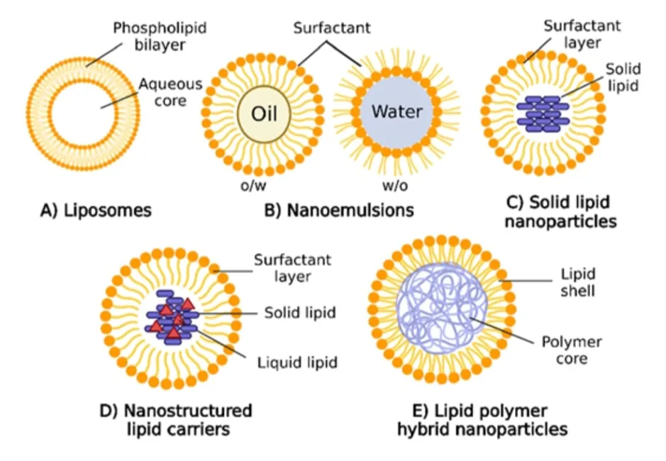
Fig:1 Different types of LNPs used in drug delivery system
METHODS OF LIPID NANOPARTICLES PREPARATION:
Lipid nanoparticles could be prepared by different methods such as hot and cold high pressure homogenization solvent emulsification/ evaporation. Micro emulsification formation technique and ultrasonic solvent emulsification. Larga scale productions mainly obtained by high pressure homogenization technique [51].
High pressure homogenization technique:
In this method, lipid phase is heated up to 90°c under 3 cycles of high pressure homogenizer at 5 × 10⁷ pa. finally, the obtained oil in water emulsion is cooled down to room temperature and solidified SLNs or NLCs [52].
In this method, the melted lipid phase is cooled to solidify and the ground to form lipid nanoparticles. Obtain lipid microparticles are dispersed in cool aqueous phase containing surfactants to form pre suspension. Then the pre suspension is homogenized under 5 cycles of high pressure homogenizer at room temperature and pressure of 1.5 × 10⁸ pa [53].
In this method, lipid phase is dissolved in an organic solvent such as acetone (organic phase). Then organic phase is added to the aqueous phase (surfactant solution in water) under continuous stirring at 70 – 80°c. The stirring will be continued until the organic phase is completely evaporated. The obtained nano emulsion is cooled (below 5°c) to solidify lipid nanoparticles [54].
In this method, lipids are melted at appropriate temperature and aqueous phase containing surfactants are heated up to some temperature. Then the aqueous phase will be added to the melted lipids under stirring at some temperature. The hot oil in water microemulsion is dispersed in cold water at 1:5 ratio to solidify lipid nanoparticles [55].
In this method, lipid phase is dissolved in an organic solvent such as dichloromethane and heated up to 50°c. then aqueous phase containing surfactants and emulsification is heated up to the some temperature. After particle evaporation of dichloromethane. The aqueous phase is added to the organic phase under stirring at 50°c obtained emulsion is sonicated for appropriate time and finally cooled in an both to solidify lipid nanoparticles [56].
MECHANISM:
lipid nanoparticles are the effective drug delivery system. The main mechanism that enable the encapsulation, protection, cellular uptake and target releases the therapeutic agents. The encapsulation of lipid nanoparticles are including hydrophilic, hydrophobic and nucleic acids, with in their lipid bilayer or core material. The encapsulation payload from enzymatic degradation and improves stability and bioavailability. Lipid nanoparticles are interact with lipid membrane fusion and endocytosis pathway. A critical step effective drug delivery in lipid nanoparticles are escaped from endosomal compartments into the cytoplasm. This is facilitated with ionizable and cationic lipids that become positive charge in their environment. Endosomal membrane are release the cargo safely inside the cell.
Lipid nanoparticles are surface modified with antibodies, peptides or aptamers that are binding to receptors over expressed on their targeted cells for active targeting. Polyethylene glycol are modified on lipid nanoparticles surface of steric stabilization and immune system, though polyethylene glycol can be designed to certain environment for time release. Passive targeting through the enhanced permeability and retention (EPR) can effect in cancer therapeutics, where lipid nanoparticles accumulate preferentially in tumor tissues due to leaky vasculature and poor lymphatic drainage.
Together, these are enable to protect therapeutic agents, cellular internalization, escape in transcellular trafficking barriers and delivery selective site and minimize therapeutic efficacy and minimize off- target effects optimize the surface chemistry and ligand conjugation is thus critical to tailor lipid nanoparticles for specific drug delivery challenges [57].
CHARACTERIZATION OF LNPS:
Proper characterization of LNPs is critical not only far investigating the synthesis of LNPs but also for controlling their quality to meet the requirement of various applications. A few important properties of LNPs need to be carefully characterized including particle size and drug release, surface charge, entrapment efficiency, crystallinity.
Particle size is a critical parameter for nanoparticle drug delivery applications. The average particle size of LNPs is normally between 100 and 400nm, and LNPs with particle size ranging from 10 to about 150nm are desirable for systemic drug delivery via intravenous injection [58]. The extent of particle size distribution with a range of 0 to 1 and PPI of less than 0.2 is after consider as narrow size distribution alters most of studies sep PPI, value less than 0.3 as the upper limit. Normally particle size and PPI can be determined by dynamic light scattering (DLS). Which measures the intensity differences of fluctuated light due to the motion of particles [59]. Size of nanoparticles can be determined by several methods such as photon-correlation spectroments (PCS) TEM, scanning electron microscopy (SEM). SEM combined with energy dispersion. X-ray spectroscopy, scanned probe microscopy and Fraunhofer diffraction.
Normally drug release from the LNPs is mainly controlled by two process, namely biodegradation and diffusion. Invitro studies are vary useful to predict the in vivo behaviors of drug loaded LNPs. Invitro drug release studies simulating in vivo behaviors of drug loaded LNPs. Invitro drug release are often conducted in phosphate buffered saline (PBS) or simulated body fluids using side-by-side diffusion cells with biological or article membrane like reverse dialysis sacs, ultracentrifugation, and ultrafiltration. Then uv-spectroscopy or HPLC is used to analyses the drug release profile. Many factors influence the invitro release profiles of LNPs including drug loading, drug location, particle size, size distribution, degree of crystallinity, lipid content types of release medium, morphologies, surfactants applied and preparation techniques [58].
The measurement of the zetapotential allows predictions about the storage stability of colloidal dispersions at high zeta potential particle aggregation is less likely to occur, due to electrical repulsions. Zetapotential measurement also helps in designing dosage form with reduced RES uptake. In order to reduce RES uptake, the surface of the particle should be hydrophilic [60].
The pH of the formulation influences the zetapotential. Addition oh high electrolyte concentrations causes compression of diffusion layer, leading to decrease in zetapotential value. Schwarz et.al. prepared SLNs by incorporating tetracaine and etomidate and studied the effect of drug loading on physical properties of SLNs and showed that incorporation of drugs has no or little effect on particles size and zetapotential compared to drug free SLNs dispersion [61].
The entrapment efficiency of the system can be determined by measuring the concentration of free drug in the dispersion medium. Ultrafiltration can be employed using consort this consists of filter membrane (molecular weight cut-off 20000 dalton) at the base of the sample recovery chamber. The sample is placed in the outer chamber and subjected for centrifugation. So that aqueous phase moves into the sample recovery chamber through filter membrane. Analyzing drug concentration in an aqueous phase, gives entrapment efficiency [62].
It is very important to determine the crystallinity of LNP components. Because the lipids and drugs encapsulated may undergo polymorphic transformation during storage, leading to drugs expulsion and instability [63]. Lipid crystallinity also strongly affects drug incoparation and drug release. DSC and X-ray diffractometry (XRD) are often used for the investigation of structure, content, and size of lipid crystalline [64]. DSC measures heat capacity changes of the tested samples during the warming up a cooling down process, and peaks are observed at different phase transition temperature. DSC is a simple quick, and intuitive way to establish the degree of LNPs crystallinity through enthalpy change. The main drawback of DSC is that it is a destructive method. Because heat is applied [65]. The nondestructive XRD indicates the crystallographic structure by measuring the intensity and angle of X-ray scattered through the tested sample. It usually DSC and XRD can be combined to analyze the atom-to-atom distance of LNPs, there by understanding the composition of LNPs [58].
IN VIVO FATE OF LNPS VIA DIFFERENT ADMINISTRATION ROUTES:
The in vivo fate, in a broad sense, primarily involves four processes: absorption, distribution, metabolism and clearance. Different routes of administration significantly influence the absorption phase, as each route encounters unique sets of barriers. These include mechanical barriers (ex: mucociliary clarence in the respiratory tract, treat turnover and nasolacrimal drainage), biochemical barriers (ex: alveolar macrophages and dendritic cells) and permeability related barriers (ex: mucus, epithelial layer) [66]. Once LNPs overcome the absorption barrier and enter the blood stream, their distribution, clearence and metabolic process are similar to those of intravenous injected LNPs [67].
Oral administration is a widely used and convenient drug delivery route. Know for its high patient compliance [68] upon gastrointestinal administration, nanocarrier can follow two possible trajectors: they can be absorbed into the systemic circulation or excreted through feces or other pathways [69]. Some absorption occurs in the gastrointestinal tract through paracellular transport, endocytosis and uptake by immune cells [70]. After gastric emptying, LNPs are transported to the small intestine, where they undergo lipase-mediated breakdown.
Research suggests that nanocarriers within the size range of 50 to 200nm can be effectively absorbed through payers patches in the small intestine, effectively bypassing metabolism [71]. For ex, song et.al. designed LNP loaded with 12-22 encoding, mRNA for inflammatory bowel diseases [72].
LNPs have attracted attention as potential inhalation formulations, with their capacity to stabilize the drugs and facilitate effective drug accumulation in the lungs showing potential for addressing respiratory diseases [73]. During inhalation exposure, nanoscale particles face two competing process: non-absorptive clarence and absorption [74]. Developing LNPs for inhalation demands careful consideration of factors such as particle size, charge and surfaces properties to optimize their behavior with in the respiratory systems. Thus enhancing pulmonary targeting, minimizing systemic side effects and amplifying therapeutic outcomes [75]. The versatility of LNPs and their potential to encapsulate various types of drugs make them a potent tool for improving the treatment of respiratory diseases via the inhalation administration route.
LNPs also shows promising potential in transdermal administration, a route that involves delivering drugs through the skin for systemic absorption. This approach offers distinct advantages, including non-invasiveness, sustained release of bypassing first-pass metabolism. LNPs have garnered attention for their ability to enhance transdermal drug delivery by encapsulating both hydrophilic and hydrophobic compounds, facilitating their penetration through the skins lipid-rich stratum corneum [76]. Transdermal absorption must penetrate the stratum corneum, epidermis and dermis. Nanoparticles are absorbed through the lymphatic system and lymph nodes in the skin [77]. LNPs especially those with size below 600nm are considerable capable of penetrating the skin [78]. Reports suggest the nanoparticles of approximately 20nm accumulate exhibit time – dependent follicular permeability. These finding highlight the potential of LNPs to increase transdermal drug delivery [79].
Local injection refers to the administration of drugs into a specific, localized area of the body, allowing for their locally or having the potential to diffuse or enter the bloodstream producing systemic therapeutic outcomes. LNPs via local injection are primarily absorbed through the lymphatic system before entering the systemic circulation [80] for on intradermal (ID), intramuscular (IM) and subcutaneous (SC) injections are commonly utilized for LNP vaccine administration. These routes capitalize resident and recruited angle. Presenting cell present in the skin and muscle [81]. The COVID-19 vaccine serves as a typical illustration of a vaccine that exerts systemic effects following IM injections [82]. For local injections the size of LNPs also impact lymphatic absorption. Our previous studies indicated that after IM injections of LNPs with sizes of 100nm, 200nm and 300nm. Smaller nanoparticles were move rapidly absorbed into the systemic circulation. In conclusion localized injection routes provide a targeted approach for drug delivery. Allowing drugs to act locally while potentially yielding systemic effects. Which depends on LNP design schemes [80].
intravenous injections (IV) of LNP formulation involves administrating these nanoparticles directly into the bloodstream through a vein. This route of administration allow for rapid systemic distribution of the encapsulated drug or therapeutic agents. The size of LNPs is a vital parameter that drug circulation half-life. It primarily influence the phenomenon of optimization protein absorption amount and interactions [83]. They reported that liposomes within the range of 100 to 200nm were detected in the systemic circulation at 60%. Where at nanoparticles smaller than 250nm accounted for 20% of the plasma count.
The hydrophobicity of the nanoparticle surface effects the quantity and the characteristics of the bound protein thus affecting their circulation kinetics. We have examined the in vivo fate of LNPs based on their administration routes. Each route has unique physiological barriers and benefits for targeting specific tissues. Selecting the proper route for each medical indication and optimization. LNP design can greatly enhance drug delivery efficiency and therapeutic outcomes [84].
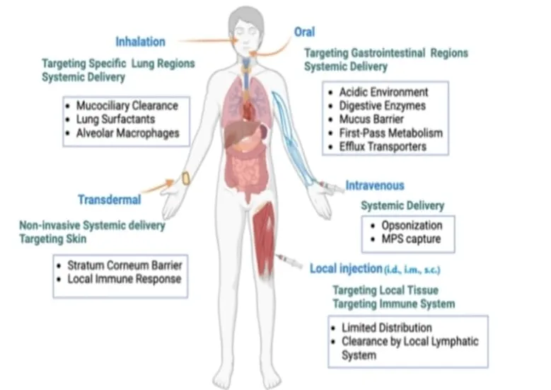
Fig:2 Biological barriers and targeting regions by various administration routes of LNPs.
CURRENT CHALLENGES AND FUTURE PERSPECTIVES:
Current challenges:
Lipid nanoparticles (LNP) drug delivery faces several significant challenges that influence their effectiveness and clinical translation [85]. A primary concentration is the stability of LNP formulations during storage and in vivo circulation. Lipid crystallization and polymorphic transitions can lead to structural changes, resulting in drug leakage and reduced encapsulation efficiency [86]. This instability is exacerbated by environmental factors such as temperature fluctuation, PH variations, and enzymatic degradation, which are particularly problematic for oral and system delivery routes [87]. overcoming biological barriers, especially the blood-brain barrier, remains a major hurdle due to its selective permeability, requiring sophisticated targeting strategies and nanoparticle engineering to enable CNS delivery. Another major challenge lies in balancing PEGylation, which extends circulation time by reducing immune clearance but can simultaneously hinder cellular uptake and endosomal escape, thus impairing therapeutic efficacy [88]. Designing PEG molecules that can be reversibly shed or optimized in density and chain length is essential to strike this balance. Toxicity and immunogenicity of LNP components. Particularly cationic lipids, also pose safety concerns. While ionizable lipids reduce toxicity compared to permanently charged cationic lipids, immune activation and complement system engagement must be carefully managed to avoid adverse reactions[89]. Manufacturing and reproducibility issues further complicate clinical applications. Large scale prodution requires stringenent control of particles size, composition, and batch consistency, as minor variations can significantly affect pharmacokinetic and bio activity. Advanced fabrication techniques such as microfluidics have improved process precision but cost and scale remain limiting factors [90].
Future perspectives:
The future of lipid nanoparticle (LNP) drug delivery is poised for transformative advancements driven by innovations in targeting, therapeutic, and regulatory evolution [91]. Improved targeting is expected through sophisticated ligand and receptor engineering, enabling highly selective delivery to specific cell types or tissues beyond the liver. This precision targeting will rely on advancements in lipid chemistry, ligand conjugation techniques, and molecular recognition strategies, potentially aided by artificial intelligence to optimize ligand selection and nanoparticles formulation [92].
Integration of LPs with cutting-edge therapies such as CRISPR-Cas gene editing represents a major frontier. Next-generation LNPs are being designed to enhance the efficiency of genome editing tools, facilitating safe and effective delivery of gene editors to target cells for treatment of genetic disorders and personalized medicine applications [93,94]. These developments include engineering of ionizable lipids with optimized stereochemistry for reduced toxicity and enhanced endosomal escape, as well as modular LNP platforms tailored for multi-functional payload delivery [95]. Enhanced functionality of future LNPs will also be achieved through stimuli-responsive lipids enabling controlled and on-demand drug release, multi-ligand targeting for complex diseases, and formulations, that improve pharmacokinetics ang biodistribution [96]. Advances in scalable, sustained manufacturing processes, including continuous flow technologies and AI-driven quality control, will aid in overcoming current commercialization barriers [97].
APPLICATIONS:
Drug loaded LNPs have been used for other drug delivery applications:
Cancer therapy:
Nanoparticles, particularly pH-sensitive polymers, play a crucial role in cancer therapy. Their ability to target tumors, which have on acidic environment is well established. The increased vascular pores and the EPR effect also make tumors accessible through passive targeting. Several FDA-approved nanomedicines, such as DOXIFIL and ONIVYDE are PEG-functionalized liposomal nanoparticle design for various cancer treatments [98].
Gene therapy:
Gene therapy is widely used to treat or prevent genetics and acquired diseases like cancer, cystic fibrosis and diabetes. Gene therapy offers long-lasting treatment as it directly targets gene instead of downstream proteins. Recently the FDA approved. A cell based gene therapy that treats patients with sickle cell enemia. Lipid based nanoparticles closely resemble cell membrane structure and allow the encapsulating and delivery of mRNA molecules. LNPs encapsulating mRNA was the most recent FDA-approved vaccine for the COVID-19 pandemic [99].
Target delivery:
The reaction of excess vascular permeability factors and the development of aberrant blood arteries within the tumor tissue cause the enhanced permeability and retention (EPR) effect. The ability of liposomal drug delivery system (DDS) to selectively target cancer cella and enrich tumors regions while reducing toxicity to normal tissues and side effects is mostly due to absence of lymphatic drainage (). It is not able that although the EPR effect is the foundation of the active targeting technique, through these modified molecules, the internalization process of liposomes with in tumor cell can be further enhanced. There by improving the targeting and therapeutic effect of the drug [100].
Combination therapy:
LNP can also be used in combination with chemotherapy. Drugs photothermal therapy drugs or immunotherapy drug to achieve multi model therapy.
The use of LNPs in the field of chemotherapy:
LNPs can keep medications from down and increase how long they stay in the body LNP reduces side effects by accumulating at the tumor site through regulated release for ex: chistoson – coated nano lipid carriers (NLCs) significantly enhance skin permeability, cellular uptake, and cytotoxicity of breast cancer cell when tetra hydro curcumin (THC) is delivered [101].
Application of LNP in photothermal therapy and photodynamic therapy:
As a carrier of photo sensitizer LNPs enhance cell permeability and targeted delivery of photo sensiting molecule improving the therapeutic effect. For ex: gold- BODIPY co-ordination liposomal nano composites (LABs) effectively solve the changes faced by traditional photo thermal agents. LAB significantly increased the level of intracellular reactive oxygen species and induced apotosis under lasser irradiation. In addition, cetuximab conjugated chitosan based iron oxide nanoparticle (cet-LINPs) combine the advantages of chemotherapy and photothermal therapy [101].
Application of LNPs in immunotherapy:
LNPs enable precise targeted delivery to tumor cells by encapsulating tumor antigens or immunostimulants, improving the bioavailability of drugs and improving treatment outcomes. For ex heterocyclic LNPs are capable of efficient delivery of oncology vaccines and STAT 3 si RNAs. By activating the STING pathway. It induces the production of type of interferons. Which in form promotes dendric cell maturation and antigen presentation of attenuates. The immunosuppressive tumor microenvironment [102]. In addition the U-101-LNP/1L-2F mRNA formulation releases 1L-2F at a tumor site via MMP-14-mediated cleavage, significantly enhancing antitumor activity while reducing systemic toxicity [103].
ADVANTAGES OF LNPS:
LNPs offer numerous advantages that make them attractive carriers or modern drug delivery applications.
They are composed of biocompatible and biodegradable lipids. Which minimize toxicity and improve patient safety.
LNPs enhance the stability of encapsulated drugs by protecting them from chemical and enzymatic degradation.
This nanoscale size improve drug solubility and bioavailability leading to better therapeutic outcomes.
They provide controlled and sustained drug release, reducing dosing frequency and improving patient compliance.
Lipid nanoparticles can also be engineered for site specific targeting enabling the selective delivery to disease tissue, while minimizing adverse effects on healthy cells [104].
LIMITATIONS:
Despite their numerous benefits. Lipid nanoparticles have certain limitations:
Physical instability during long term storage may result in changes in particle size and drug release characteristics:
Aggregation of nanoparticles can occur due to inadequate stabilization, affecting formulation performances.
Drug leakage during storage remains a concern, particularly for certain lipid nanoparticles [105].
CONCLUSION:
Lipid nanoparticles are promising drug delivery systems that improve drug solubility, stability, bioavailability, and control release of drug. Their ability to protect therapeutic molecules from degradation and facilitate targeted delivery can enhance drug accumulation at the desired site while decreasing unwanted exposure to healthy tissues. Lipid nanoparticles also used in a delivering a wide rang of therapeutic agents such as small molecules, proteins, peptides, nucleic acid and vaccines. By using different types of lipid nanoparticles are including liposomes, lipid nano emulsions, solid lipid nano emulsions etc.… over all, lipid nanoparticles have emerged as an innovative plat form for improving drug delivery.
REFERENCES

P. Adi Narayana Reddy, Dr. J. N. Suresh Kumar, G. Gopi Raju, S. Vignesh, G. Pavani, K. Harshita, U. Ravi Teja, Ch. Sai Charan, Lipid Nanoparticles as Versatile Plat Forms for Drug Delivery: From Design to Clinical Translation, Int. J. of Pharm. Sci., 2026, Vol 4, Issue 10, 2155-2171. https://doi.org/10.5281/zenodo.23375981
 10.5281/zenodo.23375981