

We use cookies to ensure our website works properly and to personalise your experience. Cookies policy
Department of Pharmaceutics, Sri Vijay Vidyalaya College of Pharmacy, Nallampalli, Dharmapuri, Tamil Nadu, India 636807
Background: Nimodipine is a highly lipophilic dihydropyridine calcium-channel blocker with established clinical use for improving neurological outcome after aneurysmal subarachnoid haemorrhage. Its poor aqueous solubility creates a formulation challenge for aqueous parenteral delivery. The present work investigated a laboratory-scale injectable formulation using a PEG 400–ethanol cosolvent system and evaluated its physicochemical and preliminary in vitro characteristics. Methods: Preformulation studies included appearance, organoleptic characteristics, solubility, pH, FTIR compatibility, pH-dependent stability and moisture content. Ten formulations (F1–F10) containing 10 mg nimodipine were prepared by systematically varying PEG 400, ethanol and Polysorbate 80 while maintaining the total cosolvent volume at 40 mL. Formulations were evaluated for appearance, clarity, pH, drug content, viscosity, osmolality, sterility testing, particulate matter and preliminary stability. Selected biological assessments included MTT cell viability, DPPH radical-scavenging activity, calcium-influx inhibition and ex vivo goat-corneal permeation. Results: Nimodipine showed very limited aqueous solubility and improved solubility in PEG 400 and ethanol. FTIR spectra showed retention of characteristic drug bands in the physical mixture. F6, containing 25 mL PEG 400, 15 mL ethanol and 0.45% v/v Polysorbate 80 per 100 mL, showed pH 6.85 ± 0.02, drug content 99.62 ± 0.18%, viscosity 5.28 ± 0.04 cP and osmolality 296 ± 2 mOsm/kg. No microbial growth was observed in the soybean–casein digest medium test performed for F6. One-month storage at 2–8 °C showed minimal change, whereas increased temperature produced marked pH drift and turbidity. Conclusion: The PEG 400–ethanol approach produced a clear laboratory-scale nimodipine formulation with favorable short-term physicochemical characteristics. F6 was selected for further evaluation, but the one-month stability findings indicate a need for extended stability, complete compendial sterility testing, container-closure compatibility, extractables/leachables assessment and in vivo safety before any clinical or commercial application. The biological experiments are exploratory and do not establish efficacy for glaucoma or epilepsy.
Nimodipine is a 1,4-dihydropyridine calcium-channel blocker characterized by high lipophilicity and a preferential cerebrovascular effect. Current product information identifies nimodipine as a therapy for neurological deficits associated with aneurysmal subarachnoid haemorrhage (aSAH). Oral products are widely used, while sterile infusion formulations are also authorised in several jurisdictions for the same clinical setting. The established clinical indication should be distinguished from experimental investigations of nimodipine in other neurological or ophthalmic conditions.
The formulation problem is driven primarily by the drug’s very low aqueous solubility. An injectable solution must maintain the drug in solution during manufacture, storage and administration while meeting requirements for sterility, particulate control, chemical stability, container compatibility and safe administration. Published formulation research has therefore explored cosolvents, micellar systems, nanoemulsions and other delivery platforms.
Interest in ophthalmic delivery has also generated preclinical evidence for topical nimodipine formulations. Such work is relevant to formulation science but does not establish nimodipine as an approved glaucoma treatment. Similarly, experimental studies of calcium-channel modulation in epilepsy do not establish nimodipine as an approved antiseizure medicine. The present work therefore treats glaucoma- and epilepsy-related findings as research context rather than therapeutic indications.
1.1 Rationale for formulation development
For a poorly water-soluble molecule, cosolvency is a straightforward approach in which a water-miscible organic solvent is combined with the aqueous vehicle to increase apparent solubility. PEG 400 is widely used as a pharmaceutical cosolvent, while ethanol can substantially increase the solubility of lipophilic compounds. Polysorbate 80 may further support wetting and solubilization. Chelation and antioxidant protection can be incorporated when justified by formulation-development studies.
The formulation strategy evaluated here used PEG 400 and ethanol as the principal variables, with Polysorbate 80, disodium EDTA and sodium metabisulfite maintained at controlled levels. The purpose was not to reproduce a marketed product exactly, but to identify a composition that provided acceptable solution characteristics at laboratory scale.
1.2 Objectives
To characterize the physical and physicochemical properties of nimodipine relevant to injectable formulation development.
To improve apparent aqueous solubility using a PEG 400–ethanol cosolvent system.
To prepare and compare ten trial formulations with systematically varied cosolvent and surfactant levels.
To evaluate appearance, clarity, pH, drug content, viscosity, osmolality, particulate matter and microbiological quality.
To investigate short-term stability under refrigerated, intermediate and accelerated conditions.
To perform exploratory in vitro biological and ex vivo corneal-permeation assessments of the selected formulation.
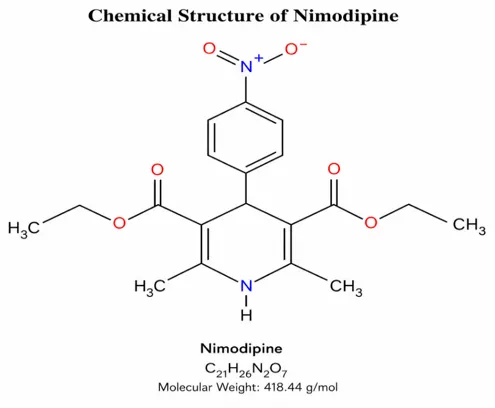
Figure 1. Chemical structure of nimodipine.
2. BACKGROUND AND LITERATURE REVIEW
2.1 Glaucoma and neuroprotection
Glaucoma is a group of progressive optic neuropathies characterized by retinal ganglion-cell loss, optic-nerve damage and corresponding visual-field impairment. Elevated intraocular pressure is the principal modifiable risk factor, but disease progression can occur despite pressure control. This has stimulated research into neuroprotective approaches directed at mechanisms such as excitotoxicity, oxidative stress, mitochondrial dysfunction and impaired ocular perfusion.
Nimodipine has been investigated in this context because L-type calcium-channel blockade can alter calcium influx and vascular tone. A 2017 study evaluated topical nimodipine formulations containing cyclodextrin complexes and reported improved ocular delivery characteristics and an intraocular-pressure-lowering response in experimental models. These findings support continued formulation research but remain preclinical evidence.
2.2 Epilepsy and calcium signalling
Epilepsy is a neurological disorder characterized by a persistent predisposition to epileptic seizures. Abnormal neuronal excitability and altered ion-channel activity contribute to seizure generation and propagation. Calcium signalling is one component of neuronal excitability, providing a mechanistic basis for experimental investigation of calcium-channel modulators. However, clinical management of epilepsy relies on established antiseizure therapies, and nimodipine is not an approved first-line antiseizure drug.
2.3 Nimodipine delivery systems
The formulation literature demonstrates several strategies for overcoming nimodipine’s low aqueous solubility. Song and colleagues developed nimodipine-loaded mixed micelles and reported markedly increased apparent solubility, compatibility with infusion media and reduced vascular irritation compared with conventional nimodipine solution. Other investigators have explored nanoemulsions, polymeric micelles, liposomes and ophthalmic formulations. Collectively, these studies show that delivery-system design can substantially influence solubility, release, tissue exposure and tolerability.
2.4 Clinical formulation context
Authorised Nimotop infusion products provide an important benchmark for parenteral formulation science. Current product information describes a sterile 0.02% nimodipine solution containing 10 mg in 50 mL and an ethanol-containing vehicle, with administration through a controlled continuous-infusion system. Product information also identifies compatibility restrictions, light sensitivity and clinically important hypotension risks. These characteristics highlight why an experimental formulation should not be considered interchangeable with an authorised product.
The present laboratory formulation contains 10 mg nimodipine in a final volume of 100 mL and therefore differs in concentration and composition from authorised infusion products. It should be considered an investigational laboratory formulation requiring comprehensive quality and safety evaluation.
3. MATERIALS
3.1 Active pharmaceutical ingredient and excipients
|
Material |
Grade/ quality |
Function |
|
Nimodipine |
USP/IP grade |
Active pharmaceutical ingredient |
|
PEG 400 |
USP/NF |
Primary cosolvent |
|
Ethanol |
AR grade |
Cosolvent |
|
Polysorbate 80 |
USP/NF |
Surfactant/solubilizing aid |
|
Water for Injection |
IP/sterile |
Aqueous vehicle |
|
Disodium EDTA |
AR/USP |
Chelating agent |
|
Sodium metabisulfite |
AR |
Antioxidant |
|
Sodium hydroxide |
AR |
pH adjustment |
|
Hydrochloric acid |
AR |
pH adjustment |
|
Nitrogen gas |
Medical grade |
Oxygen displacement/ purging |
3.2 Equipment
|
Equipment |
Application |
|
Analytical balance |
Accurate weighing |
|
Magnetic stirrer/hot plate |
Dissolution and controlled mixing |
|
Digital pH meter |
pH measurement |
|
Ultrasonicator |
Degassing and dissolution support |
|
Water bath |
Controlled heating |
|
Laminar-air-flow cabinet |
Aseptic handling |
|
0.22 µm membrane filtration unit |
Sterile filtration |
|
UV–Visible spectrophotometer |
Drug-content estimation |
|
Viscometer |
Viscosity measurement |
|
Osmometer |
Osmolality determination |
|
Incubator/culture facilities |
Microbiological testing |
|
Particle counter |
Sub-visible particulate assessment |
3.3 Formulation composition
Nimodipine was maintained at 10 mg in every 100 mL formulation. PEG 400 was increased from 15 to 33 mL, ethanol was decreased from 25 to 7 mL, and the combined cosolvent volume was kept constant at 40 mL. Polysorbate 80 was increased from 0.20 to 0.65% v/v. Disodium EDTA (10 mg) and sodium metabisulfite (20 mg) were kept constant. Sodium hydroxide or hydrochloric acid was used as required for pH adjustment and Water for Injection was used to make up the final volume.
|
Formulation |
PEG 400 (mL) |
Ethanol (mL) |
Polysorbate 80 (% v/v) |
|
F1 |
15 |
25 |
0.20 |
|
F2 |
17 |
23 |
0.25 |
|
F3 |
19 |
21 |
0.30 |
|
F4 |
21 |
19 |
0.35 |
|
F5 |
23 |
17 |
0.40 |
|
F6 |
25 |
15 |
0.45 |
|
F7 |
27 |
13 |
0.50 |
|
F8 |
29 |
11 |
0.55 |
|
F9 |
31 |
9 |
0.60 |
|
F10 |
33 |
7 |
0.65 |
4. METHODS
4.1 Preformulation studies
The preformulation programme was designed to establish baseline characteristics and identify formulation variables likely to affect solubility and stability.
|
Study |
Procedure/ measurement |
Purpose |
|
Appearance |
Visual examination under normal illumination |
Identity and physical quality |
|
Organoleptic properties |
Colour, odour, texture and physical state |
Preliminary characterization |
|
Solubility |
10 mg sample in 10 mL test medium; equilibrium at 25 ± 2 °C |
Selection of cosolvent system |
|
Melting point |
Capillary method |
Identity/purity assessment |
|
pH |
Calibrated digital pH meter |
Formulation compatibility |
|
Partition coefficient |
Shake-flask approach |
Lipophilicity assessment |
|
FTIR compatibility |
API and physical mixture spectra |
Drug–excipient compatibility |
|
pH stability |
Observation in pH 1.2, 4.5, 6.8 and 7.4 media |
pH-dependent physical stability |
|
Moisture content |
Loss-on-drying |
Moisture assessment |
4.2 Solubility assessment
Approximately 10 mg nimodipine was added separately to distilled water, PEG 400, ethanol and phosphate buffer pH 6.8. Each mixture was stirred for 30 min at 25 ± 2 °C and visually assessed for complete dissolution or undissolved material. The observations were used qualitatively to guide cosolvent selection.
4.3 Drug–excipient compatibility by FTIR
FTIR spectra of nimodipine and the physical mixture with selected excipients were compared. Characteristic absorption bands were recorded and examined for disappearance, major displacement or new bands suggestive of a chemical interaction.
4.4 Formulation preparation
Nimodipine was dissolved in the PEG 400–ethanol cosolvent system under continuous stirring. Polysorbate 80, disodium EDTA and sodium metabisulfite were incorporated according to the formulation composition. The pH was adjusted with dilute sodium hydroxide or hydrochloric acid. Water for Injection was added to final volume. The solution was passed through a 0.22 µm membrane under aseptic conditions, filled into sterile amber glass vials, nitrogen-purged and closed with sterile stoppers and aluminium seals.
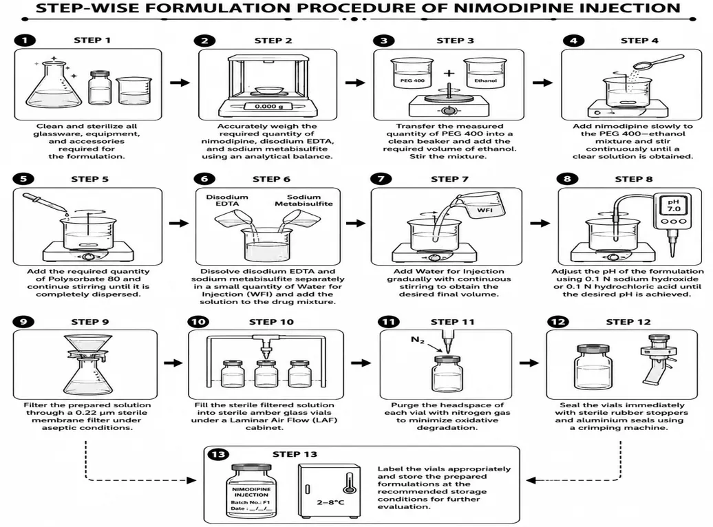
Figure 2. Schematic workflow for preparation and evaluation of the nimodipine injectable formulation
4.5 Post-formulation evaluation
4.5.1 Appearance and clarity
Filled containers were examined against light and dark backgrounds after gentle rotation. Colour, turbidity, precipitation, fibres, visible particles and container-closure defects were recorded.
4.5.2 pH, drug content, viscosity and osmolality
pH was measured in triplicate using a calibrated digital pH meter. Nimodipine content was estimated by the specified UV method and expressed as mean ± SD (n=3). Viscosity was measured using a rotational viscometer and reported in centipoise. Osmolality was measured in mOsm/kg and used to assess the osmotic characteristics of each formulation.
4.5.3 Sterility and particulate matter
The selected formulation F6 was subjected to a soybean–casein digest medium test under aseptic conditions, with an uninoculated control. Visible particulate matter was examined visually, and sub-visible particles were assessed by light obscuration.
The reported microbiological observation is limited to the test actually performed; it should not be interpreted as a complete compendial sterility validation unless all applicable USP <71> requirements are fulfilled.
4.5.4 Stability study
F6 was stored for one month under 2–8 °C, 25 ± 2 °C/60 ± 5% RH and 40 ± 2 °C/75 ± 5% RH. Appearance, pH and visual inspection were recorded. The study represents a preliminary short-term stability assessment rather than a shelf-life study. ICH Q1A(R2) describes longer data requirements for formal stability programmes, particularly when a shelf life is proposed.
4.6 Exploratory biological evaluations
Cell viability was assessed by MTT at 10–250 µg/mL. DPPH radical-scavenging activity was measured at 100–1000 µg/mL. Calcium influx was assessed using a fluorescence-based assay with Fluo-4 AM, and inhibition was calculated relative to stimulated control. Ex vivo corneal permeation was studied using freshly excised goat cornea mounted in a modified Franz diffusion cell for 120 min.
4.7 Data analysis
Results are presented as mean ± SD for triplicate measurements where stated. The formulation selected for further work was identified from the combined pattern of clarity, pH, drug content, viscosity, osmolality, preliminary stability and biological observations. No claim of clinical efficacy was inferred from these in vitro or ex vivo findings.
5. RESULTS
5.1 Preformulation characterization
Nimodipine was observed as a crystalline powder with a white to pale-yellow appearance. No visible foreign matter or discoloration was observed. In the solubility assessment, undissolved particles remained in water and phosphate buffer pH 6.8, whereas clear solutions were obtained in PEG 400 and ethanol. These observations supported the use of a cosolvent-based strategy.
|
Solvent |
Observation |
Qualitative solubility |
|
Distilled water |
Undissolved particles |
Very slightly soluble |
|
PEG 400 |
Clear solution |
Freely soluble |
|
Ethanol |
Clear solution |
Soluble |
|
Phosphate buffer pH 6.8 |
Slight turbidity/undissolved particles |
Very slightly soluble |
5.2 pH stability profile
|
Medium |
0 h |
6 h |
12 h |
24 h |
|
pH 1.2 |
Clear/slightly turbid; no precipitation |
No significant change |
No significant change |
No significant change |
|
pH 4.5 |
Clear; no precipitation |
Clear |
Clear |
Clear |
|
pH 6.8 |
Slightly turbid; no precipitation |
No significant change |
No significant change |
No significant change |
|
pH 7.4 |
Slightly turbid; no precipitation |
Slight change in clarity |
Slight change in clarity |
Slight turbidity |
5.3 FTIR compatibility
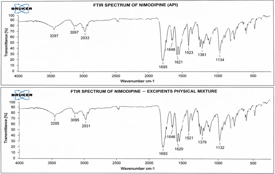
Figure 3. FTIR spectra of nimodipine and the drug–excipient physical mixture.
The characteristic bands of nimodipine were retained in the physical mixture with small shifts of approximately 0–2 cm⁻¹. No major disappearance or new prominent band was reported, supporting physical compatibility of the tested components under the conditions evaluated.
5.4 Optimization of formulation composition
The ten formulations were designed to examine the effect of a controlled shift from ethanol-rich to PEG 400-rich cosolvent conditions while progressively increasing Polysorbate 80. All formulations produced clear, colourless and homogeneous solutions without visible particulate matter. This established that the experimental composition range was capable of maintaining nimodipine in solution at the tested concentration.
|
F |
PEG 400 (mL) |
Ethanol (mL) |
PS80 (% v/v) |
pH |
Drug content (%) |
|
F1 |
15 |
25 |
0.2 |
6.42 ± 0.03 |
98.42 ± 0.38 |
|
F2 |
17 |
23 |
0.25 |
6.55 ± 0.04 |
97.86 ± 0.42 |
|
F3 |
19 |
21 |
0.3 |
6.68 ± 0.03 |
98.74 ± 0.31 |
|
F4 |
21 |
19 |
0.35 |
6.48 ± 0.05 |
99.18 ± 0.27 |
|
F5 |
23 |
17 |
0.4 |
6.72 ± 0.02 |
99.35 ± 0.24 |
|
F6 |
25 |
15 |
0.45 |
6.85 ± 0.02 |
99.62 ± 0.18 |
|
F7 |
27 |
13 |
0.5 |
6.63 ± 0.04 |
98.94 ± 0.29 |
|
F8 |
29 |
11 |
0.55 |
6.51 ± 0.03 |
98.28 ± 0.35 |
|
F9 |
31 |
9 |
0.6 |
6.76 ± 0.03 |
99.28 ± 0.22 |
|
F10 |
33 |
7 |
0.65 |
6.80 ± 0.04 |
99.48 ± 0.20 |
F6 showed a pH of 6.85 ± 0.02 and the highest measured drug content (99.62 ± 0.18%). Its composition provided an intermediate PEG 400–ethanol balance rather than an extreme of either cosolvent. On the combined physicochemical results, F6 was carried forward for additional evaluation.
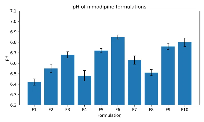
Figure 4. pH of F1–F10 formulations (mean ± SD, n=3).
5.5 Physicochemical evaluation of F1–F10
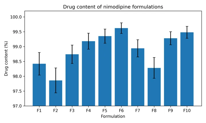
Figure 5. Drug content of F1–F10 formulations (mean ± SD, n=3).
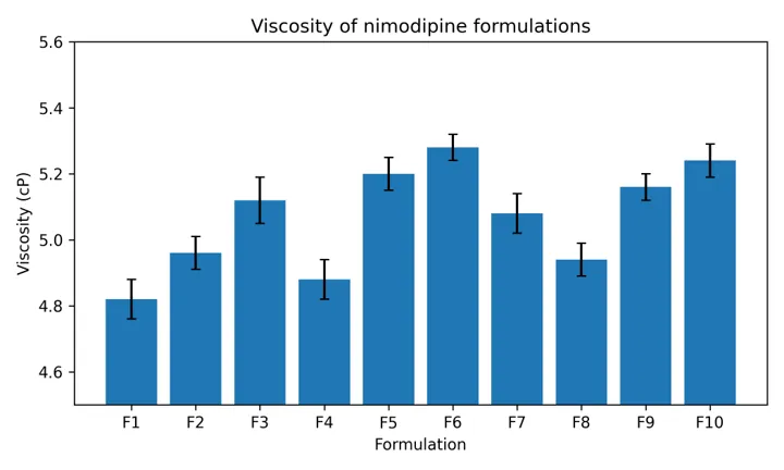
Figure 6. Viscosity of F1–F10 formulations (mean ± SD, n=3).
Drug content ranged from 97.86 ± 0.42% to 99.62 ± 0.18%. Viscosity ranged from 4.82 ± 0.06 to 5.28 ± 0.04 cP. The variation was modest across the series, indicating that changing the cosolvent ratio did not produce an extreme increase in viscosity within the tested range.
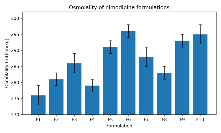
Figure 7. Osmolality of F1–F10 formulations (mean ± SD, n=3).
Osmolality values ranged from 276 ± 3 to 296 ± 2 mOsm/kg. F6 showed 296 ± 2 mOsm/kg. These values indicate relatively similar osmotic characteristics among the formulations, although a formal parenteral acceptability assessment should consider the intended route, product volume, administration rate and applicable pharmacopoeial requirements.
5.6 Microbiological and particulate quality of F6
|
Test |
Result |
Interpretation |
|
Appearance |
Clear, colourless |
No visible precipitation or phase separation |
|
Visible particulate matter |
None observed |
No visible particles detected |
|
SCDM sterility test |
No turbidity or visible growth |
No growth detected under test conditions |
|
Sub-visible particles ≥10 µm |
18 particles/container |
Within the applicable reported limit |
|
Sub-visible particles ≥25 µm |
2 particles/container |
Within the applicable reported limit |
No microbial growth or turbidity was observed in the soybean–casein digest medium test of F6. The formulation was also free of visible particles, while the reported sub-visible particle counts were within the applicable limits used during evaluation. Because the reported microbiological work does not document the complete set of media, controls and incubation details required for a formal compendial sterility test, the result is appropriately described as no growth detected in the performed test rather than as definitive proof of sterility.
5.7 Preliminary stability
|
Condition |
Time |
Appearance |
pH |
Visual finding |
|
2–8 °C |
Initial |
Clear, colourless |
6.85 ± 0.02 |
No visible particles |
|
2–8 °C |
1 month |
Clear, colourless |
6.84 ± 0.02 |
No visible particles |
|
25 ± 2 °C/60 ± 5% RH |
Initial |
Clear, colourless |
6.85 ± 0.02 |
No visible particles |
|
25 ± 2 °C/60 ± 5% RH |
1 month |
Clear, colourless |
7.80 ± 0.03 |
No visible particles |
|
40 ± 2 °C/75 ± 5% RH |
Initial |
Clear, colourless |
6.85 ± 0.02 |
No visible particles |
|
40 ± 2 °C/75 ± 5% RH |
1 month |
Turbid |
7.99 ± 0.03 |
Small particles observed |
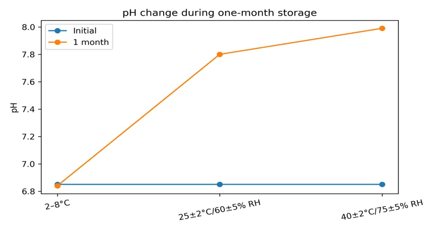
Figure 8. Change in pH of F6 during one-month storage.
5.8 Exploratory biological evaluation
5.8.1 MTT cell-viability assay
|
Nimodipine concentration (µg/mL) |
Cell viability (%) |
Cytotoxicity (%) |
|
10 |
97.8 ± 1.2 |
2.2 |
|
25 |
96.4 ± 1.4 |
3.6 |
|
50 |
94.7 ± 1.6 |
5.3 |
|
100 |
92.1 ± 1.8 |
7.9 |
|
250 |
88.6 ± 2.1 |
11.4 |
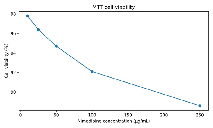
Figure 9. MTT cell viability across the tested nimodipine concentrations.
Cell viability decreased progressively as concentration increased, but remained above 80% across the reported concentration range. Under the assay conditions, the results indicate limited cytotoxicity within the tested range. The identity of the cell line, exposure duration and detailed assay controls should be reported in a full biological-methods section for independent reproduction.
5.8.2 DPPH radical-scavenging assay
|
Concentration (µg/mL) |
Scavenging activity (%) |
|
100 |
9.8 |
|
250 |
23.5 |
|
500 |
46.1 |
|
750 |
68.2 |
|
1000 |
90.7 |
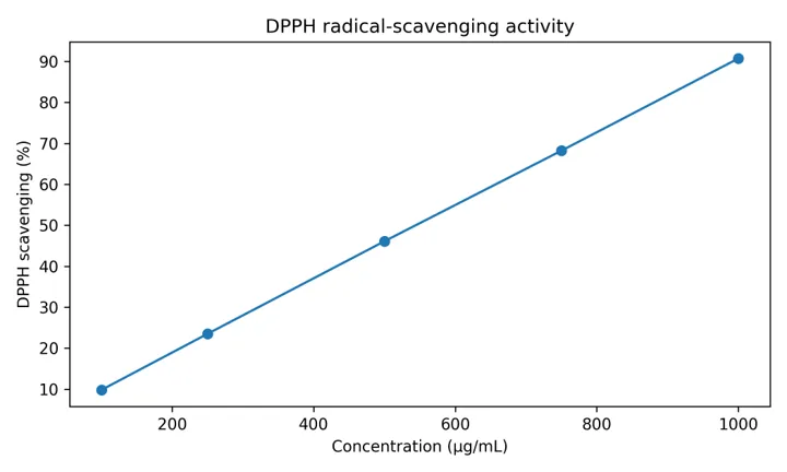
Figure 10. DPPH radical-scavenging activity of the tested sample.
The DPPH response increased with concentration, reaching 90.7% at 1000 µg/mL. The reported IC50 was approximately 546 µg/mL. Because antioxidant activity is not a validated clinical endpoint for injectable nimodipine, this result should be interpreted as an exploratory chemical-assay observation rather than evidence of therapeutic antioxidant efficacy.
5.8.3 Calcium-influx inhibition
|
Concentration (µg/mL) |
Inhibition (%) |
Mean ± SD |
|
10 |
18.0 |
18.00 ± 2.10 |
|
30 |
39.0 |
39.00 ± 2.45 |
|
100 |
58.0 |
58.00 ± 2.18 |
|
300 |
72.0 |
72.00 ± 1.95 |
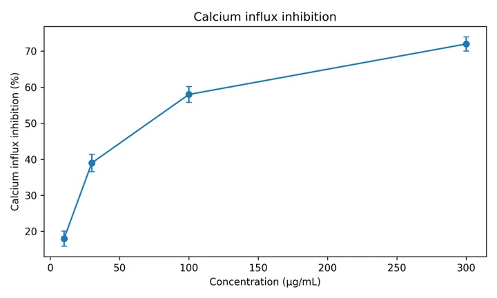
Figure 11. Concentration-dependent inhibition of calcium influx by the tested sample.
Calcium-influx inhibition increased from 18.00 ± 2.10% at 10 µg/mL to 72.00 ± 1.95% at 300 µg/mL. The positive control showed 69.00 ± 1.96% inhibition in the reported experiment. The concentration-dependent pattern is consistent with calcium-channel-related activity, although the assay does not establish clinical antiseizure or neuroprotective efficacy.
5.8.4 Ex vivo goat-corneal permeation
|
Time (min) |
Amount permeated (mg) |
Permeation (%) |
Papp (×10⁻⁶ cm/s) |
|
30 |
0.38 ± 0.02 |
7.60 ± 0.40 |
— |
|
60 |
0.82 ± 0.03 |
16.40 ± 0.60 |
— |
|
90 |
1.24 ± 0.04 |
24.80 ± 0.80 |
— |
|
120 |
1.68 ± 0.05 |
33.60 ± 1.00 |
7.84 ± 0.23 |
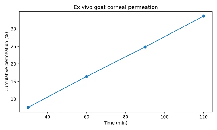
Figure 12. Ex vivo cumulative corneal permeation of the tested F6 preparation.
Permeation increased progressively over 120 min, reaching 33.60 ± 1.00% with a reported apparent permeability coefficient of 7.84 ± 0.23 × 10⁻⁶ cm/s. This experiment provides preliminary evidence of corneal transport under ex vivo conditions. It should not be interpreted as proof that an injectable formulation is suitable for ophthalmic administration.
6. DISCUSSION
The preformulation findings establish the principal formulation challenge: nimodipine is poorly soluble in aqueous media but dissolves readily in PEG 400 and ethanol. This behaviour is consistent with the high lipophilicity of the molecule and supports the use of cosolvency as an initial formulation strategy. The progressive changes in PEG 400, ethanol and Polysorbate 80 allowed the formulation space to be explored without changing the total cosolvent volume.
All ten formulations were visually clear and homogeneous. This is important for an injectable solution because visible precipitation or particulate contamination would be unacceptable. However, visual clarity alone cannot establish chemical stability or physical stability over the product lifetime. The stability study demonstrates this distinction: F6 remained clear under refrigeration for one month, while the formulation showed substantial pH drift at 25 °C and turbidity with small particles at 40 °C/75% RH.
The pH behaviour is particularly important. F6 changed from 6.85 to 7.80 at 25 °C and to 7.99 with turbidity at 40 °C/75% RH. These changes indicate that the formulation requires further investigation of degradation pathways, buffer capacity, packaging interactions and temperature sensitivity. A one-month study cannot support a commercial shelf-life claim.
The physicochemical profile of F6 was favourable within the tested series. Drug content was 99.62 ± 0.18%, viscosity was 5.28 ± 0.04 cP and osmolality was 296 ± 2 mOsm/kg. These values are useful for comparative formulation screening, but injectable-product suitability also depends on sterility assurance, endotoxin control, visible and sub-visible particles, container closure integrity, compatibility with administration sets, extractables/leachables, pH, degradation products and systemic tolerability.
The formulation composition differs from authorised Nimotop infusion products. Current product information describes a 10 mg/50 mL sterile infusion solution with ethanol and macrogol 400, together with specific administration and compatibility requirements. The experimental formulation contains 10 mg/100 mL and adds Polysorbate 80, disodium EDTA and sodium metabisulfite. Therefore, direct equivalence with a marketed product should not be claimed.
The biological experiments provide supportive mechanistic observations but should be interpreted cautiously. The MTT results indicate preserved cell viability over the tested range. DPPH activity increased with concentration, and calcium-influx inhibition showed a concentration-dependent response. These assays can support early formulation research but cannot establish efficacy in glaucoma or epilepsy. The corneal-permeation experiment is similarly exploratory and does not convert an injectable preparation into an ophthalmic dosage form.
7. QUALITY AND REGULATORY CONSIDERATIONS
7.1 Sterile product development
A parenteral formulation requires a higher level of control than a conventional oral dosage form. Development should address bioburden, endotoxins, sterility assurance, aseptic processing or validated terminal sterilization where feasible, particulate matter, container-closure integrity, compatibility with the intended administration system, and stability of the active and excipients. The current results represent an early laboratory evaluation and do not replace formal sterile-product qualification.
7.2 Container and administration compatibility
Authorised nimodipine infusion information identifies compatibility restrictions with certain plastics and requires controlled administration equipment. Accordingly, future work should include systematic compatibility testing with the intended vial, stopper, syringe, tubing and infusion set. Adsorption, leachables, precipitation during dilution and light exposure should be investigated before clinical translation.
7.3 Stability programme
The observed pH drift and turbidity under elevated-temperature storage demonstrate why a formal stability programme is essential. Long-term and accelerated studies should be designed according to the intended storage condition, with appropriate time points and validated analytical methods. Chemical assay, related substances/degradation products, pH, appearance, particulate matter and container-closure integrity should be monitored.
7.4 Biological evidence
The current biological studies should be classified as preliminary. A future programme should specify the cell line, passage number, test concentration basis, vehicle controls, positive controls, exposure time, replicate structure and statistical analysis. For ocular research, corneal integrity, tissue viability and drug recovery should be documented. For neurological applications, pharmacokinetic exposure and validated disease-relevant models would be needed.
7.5 Statistical considerations
The reported results are primarily descriptive and are based on triplicate measurements. Future optimization should use a predefined experimental design with statistical modelling rather than selecting a formulation from individual highest values. A quality-by-design approach can identify critical material attributes and critical process parameters and establish a design space supported by experimental evidence.
7.6 Clinical and regulatory positioning
Nimodipine is clinically established for aneurysmal subarachnoid haemorrhage, whereas use in glaucoma and epilepsy remains investigational. The present formulation should therefore be positioned as a preclinical pharmaceutical-development study. No conclusion regarding clinical efficacy, clinical safety or suitability for human administration should be drawn from the current data.
8. CONCLUSION
A laboratory-scale nimodipine injectable formulation was developed using PEG 400 and ethanol as the principal cosolvents, with Polysorbate 80, disodium EDTA and sodium metabisulfite as supporting excipients. Preformulation studies demonstrated poor aqueous solubility and improved solubilization in PEG 400 and ethanol. FTIR evaluation showed retention of characteristic nimodipine bands in the tested physical mixture.
Among the ten trial formulations, F6 contained 25 mL PEG 400, 15 mL ethanol and 0.45% v/v Polysorbate 80 per 100 mL and was selected for further evaluation based on its overall physicochemical profile. F6 showed pH 6.85 ± 0.02, drug content 99.62 ± 0.18%, viscosity 5.28 ± 0.04 cP and osmolality 296 ± 2 mOsm/kg. No microbial growth was observed in the SCDM test performed, and no visible particulate matter was detected.
The preliminary stability findings were informative: F6 remained clear with minimal pH change at 2–8 °C over one month, whereas marked pH changes occurred at 25 °C and 40 °C/75% RH, with turbidity and small particles observed at the highest temperature. Accordingly, the formulation cannot be described as stable for long-term storage on the basis of the current dataset.
The exploratory MTT, DPPH, calcium-influx and ex vivo corneal-permeation studies generated concentration- or time-dependent responses. These observations support further investigation but do not demonstrate clinical efficacy or establish a therapeutic role for injectable nimodipine in glaucoma or epilepsy.
9. LIMITATIONS
10. FUTURE WORK
Future development should prioritize validated assay and related-substance methods, complete sterile-product quality assessment, container and administration compatibility, formal stability studies, endotoxin testing, validated aseptic processing, pharmacokinetic characterization and appropriately designed preclinical safety studies. If glaucoma remains a target, a dedicated ophthalmic formulation should be developed rather than assuming that an injectable composition is suitable for ocular administration.
DECLARATIONS
Ethics Statement
No live-animal experiment was conducted in the present study. The ex vivo corneal permeation experiment was performed using post-mortem goat corneal tissue. The tissue was obtained and used in accordance with applicable institutional and local requirements governing the collection and use of post-mortem animal tissues.
Consent for Publication
Not applicable.
Funding
The authors received no external funding or financial support for the present study.
Conflict of Interest
The authors declare that there are no financial or non-financial conflicts of interest related to the present work.
Data Availability
All numerical data generated and analyzed in this study are presented in the tables and figures of the manuscript. Additional data may be made available by the corresponding author upon reasonable request, subject to applicable institutional requirements.
Author Contributions
All authors contributed to the conception and design of the study, formulation development, experimental work, data analysis, interpretation of results, manuscript preparation, critical revision of the manuscript, and approval of the final version.
ACKNOWLEDGEMENTS
The authors would like to acknowledge the support and technical assistance provided by the respective laboratory and institutional facilities during the conduct of this study.
REFERENCES

P. Senthilkumar, A. Vasanthan, Development and In Vitro Evaluation of a Nimodipine Injectable Formulation: Preformulation, Optimization and Preliminary Biological Characterization, Int. J. of Pharm. Sci., 2026, Vol 4, Issue 10, 2260-2277. https://doi.org/10.5281/zenodo.23377341
 10.5281/zenodo.23377341