Diabetes remains a massive global health hurdle, and while nature offers powerful tools like Jamun (Syzygium cumini) leaves, turning those leaves into reliable medicine is tricky. The raw extracts often break down too quickly in the body or aren't absorbed well.To solve this, our study developed a specialized "slow-release" system. We used a gentle technique called ionotropic gelation to tuck the leaf’s natural antioxidants—specifically its phenolic acids and flavonoids—inside tiny sodium alginate microbeadsWhy this matters:• Protection: These microbeads act like a protective shield, keeping the active ingredients safe from harsh stomach acids.• Precision: Instead of a sudden spike, the beads release the extract gradually.• Effectiveness: The formulation successfully blocked ?-amylase and ?-glucosidase (the enzymes responsible for sugar spikes) while keeping its full antioxidant power intact.Ultimately, this isn't just about better chemistry; it’s about creating a smarter, plant-based supplement that is easier for patients to take and more effective at managing blood sugar over the long term
Diabetes mellitus has emerged as one of the most pressing metabolic challenges of the modern era. At its core, the condition involves a persistent state of hyperglycemia, where the body’s ability to manage blood sugar is compromised—either because the pancreas fails to produce sufficient insulin or because cells become resistant to its effects.
The stakes of poor management are incredibly high. Over time, erratic glucose levels can trigger a cascade of systemic damage, potentially leading to renal complications, vision loss, and cardiovascular instability. While lifestyle shifts remain the first line of defense for prediabetics, maintaining these habits over a lifetime is notoriously difficult. This creates a critical gap in care, driving the search for plant-derived nutraceuticals that offer a safer, more sustainable way to maintain glycemic balance without the harsh side effects often linked to long-term synthetic drug use. (Ahmad Raza, 2017)
2. SYZYGIUM CUMINI: A BOTANICAL POWERHOUSE
Known widely as Jamun , Syzygium cumini has been a cornerstone of traditional healing systems like Ayurveda for centuries. Its therapeutic value lies in its complex phytochemicals, which is packed with anthocyanins, flavonoids, and phenolic acids. These act as natural antioxidants that shield the body from the oxidative stress often exacerbated by diabetes.
While the fruit itself supports healthy lipid levels, the seeds and leaves are the true heroes in glucose regulation. They possess unique compounds that help stimulate pancreatic function and improve how the body processes sugar. By leveraging this "green" pharmacy, we can find holistic ways to address the root causes of metabolic imbalance.(Pooja Patel, 2023)
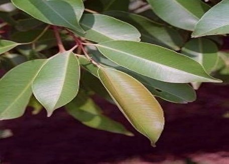
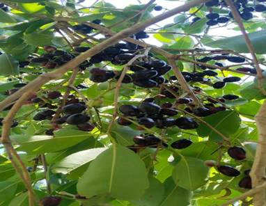
Fig.no.01 Fig.no.02
Jamun leaves Jamun Fruits
3. PRECISION DELIVERY VIA MICROBEADS
The challenge with herbal extracts is ensuring they actually reach the bloodstream in a stable, effective form. This is where microbead technology changes the game.
By encapsulating Syzygium cumini extracts within a protective polymer matrix—such as sodium alginate—we create a sophisticated delivery vehicle.
These microscopic spheres (typically ranging from 0.5 to 1000 µm) offer several strategic advantages:
Sustained Action: Instead of a sudden spike, the medication is released gradually, keeping blood sugar levels steady over a longer window.
Protection & Absorption: The beads shield the delicate plant compounds from being destroyed by stomach acid, ensuring higher bioavailability once they reach the intestines.
Targeted Efficiency: By controlling exactly where and how the extract is released, we can maximize the healing impact while minimizing any systemic strain on the body. (Umesh D. Shivhare, 2013) (Mullaicharam Bhupathyraaj *, 2021)
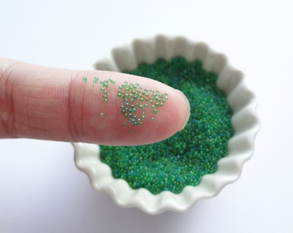
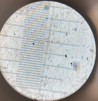
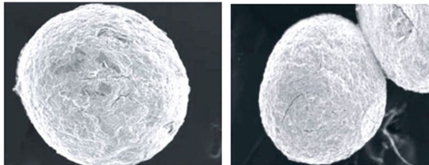
Fig.no.03 Fig.no.04
Extarct Microbeads
MATERIALS AND METHODS
A single leaf matters when pulling together a clean batch of Syzygium cumini extract - each one checked, rinsed under slow running water. Instead of rushing drying, folks wait near open windows where air moves softly, letting green fade without baking out fragile elements such as flavonoids. After seven quiet days, crunch sets in; that signals it is time to crush them down by hand until bits turn rough and ready. With more edges exposed, soaking pulls deeper into what was once whole foliage.
A few days soaking in 70 percent ethanol or methanol usually kicks off the process, although a Soxhlet setup works better for stronger results. Afterward, what's left gets pushed through a filter, then spun in a rotary evaporator under 45 °C to strip away the solvent, ending with a rich dry residue best chilled for durability. From there, turning that thick liquid into tiny dissolvable beads relies on ionic gelation, forming small biodegradable carriers.
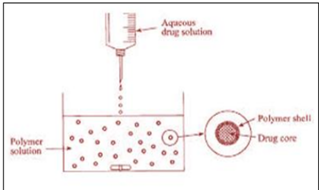
This begins with a mix made by dissolving sodium alginate in distilled water, forming a transparent, viscous liquid. Into this goes the Syzygium cumini extract, ensuring key components spread evenly through the blend. Such a mixture becomes what's called the "internal phase," set for transformation shortly after. Shape forms when droplets of this solution fall from a syringe into a small cup filled with calcium chloride. Every time a droplet meets the solution, calcium ions swap places with sodium along the alginate strands, forming bonds that turn it into a firm, water-resistant gel called calcium alginate. Half an hour to one full hour passes while the beads set properly before being strained out. Rinsing follows, sweeping away leftover particles floating free. Then drying takes over - either under open air or via freeze-drying - to finish crafting the tiny plant-based spheres (Mullaicharam Bhupathyraaj *, 2021)
Fig.no.05
Ionotropic Gelation Method
RESULTS
How Jamun May Help with Diabetes
Diabetes keeps showing up more often around the world, now touching about six out of every hundred people. Most cases - nearly nine in ten - are Type II, where blood sugar stays high over time. Though doctors usually recommend changes in daily habits along with medicines, interest is shifting toward natural options that help control glucose levels. One such option is Jamun, a fruit used widely in India's healing practices for generations. Before insulin came into play, it was already being used across parts of Europe to manage symptoms linked to elevated sugar. Its role has lasted through decades, standing apart from modern pills yet still relevant today. (Pooja Patel, 2023)
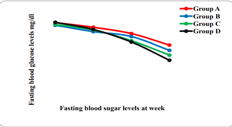
Fresh evidence from lab tests on Wistar rats and rabbits - made diabetic with alloxan or streptozotocin - shows Jamun extract brings down blood sugar. While some experiments used high doses, others found results even at low levels. Because responses varied by species, findings still point in one direction. Though animal data isn’t human proof, patterns here stand out clearly. Each trial followed strict measurement routines without skipping controls. From start to finish, glucose drops were tracked with care. Not every marker shifted, yet the main trend stayed strong (Ahmad Raza, 2017)
From 50 to 400 mg/kg, alcoholic, ethanolic, and ethyl acetate seed extracts kept lowering blood sugar. Rats fed seeds showed as much as a 65% drop in glucose, researchers like Shankar found. (Ahmad Raza, 2017)
Fresh evidence points one way - the gooey part inside the fruit may help lower blood sugar by about 9 to 15 percent. Yet, tucked away, the seed turns out stronger effects when tested under similar conditions. (Ahmad Raza, 2017)
Jamun helps control blood sugar mainly by acting on the pancreas. Studies show it boosts insulin release from beta cells found in the pancreas. In animal tests involving mild and more advanced diabetes, insulin went up between 26.3% and 44%. That rise appears tied directly to how Jamun interacts with those cells. The stronger the condition, the clearer the response became under observation. (Ahmad Raza, 2017)
Altogether, the results point to Jamun packing strong natural compounds that help control blood sugar and insulin levels. Because it has worked well in both traditional use and modern tests, it can play a supportive role in handling high glucose and excess insulin. What stands out is how long it has been trusted across practices. Not every plant earns that kind of lasting footprint. (Ahmad Raza, 2017)
Fig.no.06 (Ahmad Raza, 2017)
Fig.no.07 (Umesh D. Shivhare, 2013)
CONCLUSION
Every now and then, something small catches the eye - Syzygium cumini might just put a brake on diabetes using what nature already made. Lab work keeps adding up: chemicals hiding in its leaves and seeds team up, each boosting the other. Bit by bit, results hint that these plant parts react predictably when tested behind glass walls. Some studies pass right by it, true - still, patterns form where sugar levels shift through repeated tests
From the moment they’re released, phenolic acids work alongside flavonoids to block oxidation. Moving quickly, these natural compounds jump into action when damage begins, slowing it down right away. Before things escalate, they’re already cutting the risk.
Lately, research has pointed to anthocyanins playing a role in healing pancreatic beta cells, while also making the body respond more clearly to insulin. Evidence isn’t complete, yet signs show these substances take part in fixing metabolism. As cells begin renewing, blood sugar handling tends to get smoother too. One clue after another builds up - simple plant colors may quietly guide how diabetes develops. What was once overlooked now seems central. Finding each piece takes effort. Though progress feels small, moments like this push curiosity further into how plants shape outcomes.
Here’s something different: some substances don’t just hide high blood sugar - they alter how glucose flows in the body. Instead of changing just one thing, they affect multiple phases together. Inside cells, changes start to appear without warning. Far from stopping a single reaction, they influence many parts of the process at once. Data suggests these agents shift how cells handle excess sugar across several levels. Blood sugar levels hold more stable during daylight hours because of shifts in bodily energy flow. These actions reshape movement within the system, rather than covering up symptoms.
The Microbead Edge Found in Studies
Some days, plant-based solutions fail since guts absorb so little. Wrapped in a shield made of seaweed and crushed shellfish, they survive stomach acids more easily. That idea outperforms most past methods tried until today.
This method works well, research shows. What makes it tick is where the real difference lies. Its form matters, contributing in quiet ways. Built like this, performance climbs without fuss. Alignment shifts everything when timing clicks into place. Favorable conditions line up quietly. Yet results keep showing the same pattern
Inside the jamun plant, tiny beads guard delicate components, studies find they withstand stomach juices. When hit by tough conditions, these miniature casings act like armor. Even as acids tear apart most compounds in digestion, the outer layer holds firm. Evidence shows consistent shielding during stomach transit, letting key parts reach further intact. What counts is not just safety but how well the core remains untouched.
Steady blood counts stick around through therapy, research finds, because tiny beads dribble out medication slowly. Release happens in small waves, not a flood, data showed. Tests pointed to lasting effects when doses spread out over time instead of hitting hard up front.
REFERENCES
Ahmad Raza, M. S.-U.-H. (2017). Jamun (Syzygium cumini) seed and fruit extract attenuate hyperglycemia in diabetic. Asian Pacific Journal of Tropical Biomedicine, 4.
ullaicharam Bhupathyraaj *, A. A. (2021). FORMULATION OF MICRO BEADS: A REVIEW. International Journal of Pharmaceutical Sciences and Research, 103.
Pooja Patel, R. F. (2023). BIOCHEMICAL INVESTIGATION AND PHYSICOCHEMICAL STUDY ON JAMUN (SYZYGIUM CUMINI LINN.) LEAVES-A PROMISING ANTIDIABETIC DRUG. World Journal of Pharmaceutical Research, 10.
Umesh D. Shivhare, V. B. (2013). Preparation of Microbeads by different Techniques and. 288.
Swami S.B, Thakor N.S.J, Patil M.M, Haldankar P.M. Jamun (Syzygium Cumini (L.)). A review of its food and medicinal uses, 2012; 3: 1100-1117.
Hasan S.M, Zulfiqar S, Sajid T, Hassan S.K, Ibrahim A, Majeed A, Hassan H. Jamun: A multifunctional medicinal plant. Journals of life sciences, 2018; 2(4): 284-290.
Dagadhair A.C, Pakhare K. N, Todmal A.K, Andhale R.M. Jamun (Syzygium cumini) Skeels. A traditional therapeutic tree and its processed food products. International Journal of Pure and Applied Bioscience, 2017; 5(5): 1202-1209.
Nadeem A, Mohammad N, Husain K.M. Medicinal potential of Jamun (Syzygium Cumini linn). A review. Journal of drug delivery and Therapeutics, 2019; 9(5): 175-180.
Ruhil P, Arora R, Kumar V. A review article on preliminary screening and pharmacological activities of Jamun (Syzygium Cumini). Indian Journal of Research, 2018; 7(1): 129-130. Anjali V, Sindhu G, Girish C. A review on pharmacology and phytochemistry of Syzygium Cumini. Indian Journal of Pharmaceutical and Biological Research (IJPBR), 2017; 5(4): 24-28.
Jahan A, Reddy M.J.M. Syzygium Cumini: A plant with high medicinal and nutritional value. The International Journal of Botany and Research (IJBR), 2018; 8(6): 1-4.
Marufa M.K, Das P.C, Iqbal A. utilization of Jamun seed powder is composite cake formulation. The Journal of Bangladesh agricultural university, 2019; 17(4): 599-605.
Anonymous, The Ayurvedic Pharmacopoeia of India, Government of India, Ministry of Health & Family Welfare, Department of Indian systems of medicine & Homoeopathy, New Delhi Part, 1989; 1(I).
Quality standards of Indian Medicinal Plant (published by Indian Council of Medical Research, V. Ramalingaswami Bhawan, Ansari Nagar, New Delhi. 15th edn, 2017; 198-208.
Baravkar AA, Kale RN, Patil RN, Sawant SD Pharmaceutical and biological evaluation of formulated cream of methanolic extract of Acacia nilotica leaves. Res. J. Pharm. Technol., 2008; 1(4): 481-483.
Kaur K, Michael H, Arora S, Harkonen P, Kumar S In vitro bioactivity-guided fractionation and characterization of polyphenolic inhibitory fractions from Acacia nilotica (L.) Willd. ex Del. J. Ethnopharmacol., 2005; 99: 353-630.
Venugopal G, Srinivasa R C, Rao N B, Rao V B, Rao V T, Rao A P Pharmacognostic and Preliminary Phytochemical Evaluation of Ocimum basilicum L. var. pilosum (Willd.) Benth. and O. tenuiflorum var. CIM-AYU, International Journal of Pharmacognosy and Phytochemical Research, 2015; 7(3): 519-526.
Mukherjee PK Quality Control of Herbal Drugs- an Approach to Evaluation of Botanicals, 1st edn, Business Horizons, New Delhi, 2002; 695.
Tripathi M, Sikarwar R L S, Tiwari A & Dwivedi N, Pharmacognostical identification of ingredients in Laghulai curna Indian J Tradit Knowle, 2015; 14(4): 531-536.
Anonymous Protocol for Testing of Ayurvedic, Siddha & Unani Medicines. Governmentof India, Department of AYUSH, Ministry of Health & Family Welfare, Pharmacopoeial Laboratory for Indian Medicines Ghaziabad, 2007. Mitra R and Mehrotra S, Pharmacognostical studies on Priyal: fruits and seeds of Buchanania lanzan Spreng. (Family: Anacardiaceae) Bull Bot Surv India, 1980; 22: 68-76.
Choudhary N, Siddiqui MB and Khatoon S, Pharmacognostic evaluation of Tinospora cordifolia Willd.) Miers and identification of biomarkers, Indian J Tradit Knowle, 2014; 13(3): 543-540.
Sholapur Hasan Pasha N & Patil Basanagouda M, Pharmacognostical and phytochemicalinvestigations on the bark of Moringa oleifera Lam. Indian J Nat Prod Resour, 2013;4(1): 96-101.
Tripathi M and Sikarwar R L S Pharmacognostic standardization of stem bark of Erythrina variegate L. Journal of the Indian Botanical Society, 2014; 93(3&4): 248-253.
Kokate CK, Practical Pharmacognosy Is ted., Vallabh Prakashan, New Delhi, 2006.
Anonymous Laboratory guide for the analysis of Ayurveda and Siddha formulations, published by Central Council for Research in Ayurveda and Siddha, Department of AYUSH, Ministry of Health and family Welfare, Government of India, New Delhi, 2010; 83-87.
Tripathi M and Sikarwar R L S Pharmacognostic Studies on Plaksa (Ficus virens Ait.) Stem Bark Indian Journal of Natural Products and Resources, 2015; 6(1): 27-32.
Tiwari A, Tripathi M and Dwivedi N, Standardization of Ayurvedic compound formulation Kutajastaka Kvatha Curna J. Trop. Med. Plants, 2015; 15: 1-13.
Williams and Wilkins.Noida:B.I. Publications, 2000,903-904.
Lachman L, Liberman H, Kanig J. The Theory and Practice of Industrial Pharmacy. 3rd ed.,Mumbai:Varghese Publishing House, 1986, p. 430.
Gibaldi M, Parrier D. Biopharmaceutics and clinical Pharmacokinetics.Philadelphia:Lea and Febiger 3rd ed., Vol 15, ,1984,64-82.
Belyaeva E, Valle D.D., Neufeld R.J. Ponceleta D. Newapproach to the formulation of hydrogel beads by emulsification/thermal gelation using a static mixer. Chem Eng Sci 2004; 59(2): 2913–20.
Badarinath A.V., Reddy J.R. Mallikarjuna R. K.,.Alagusundaram M, Gnanaprakash K., Chetty M.S. Formulation and Characterization of Alginate Microbeads of Flurbiprofen by Ionotropic Gelation Techniques. Int J Chem Tech Res 2010;2(1):361-367.
Reddy T., Tammishetty S. Formulation of barium chloride crosslinked beads of carboxymethyl guar gum. J Applied Poly Sci 2001; 82(7): 3084-3090.
Bopanna R., Kulkarni R.V., Setty C.M. Carboxymethylcellulose-aluminium hydrogel microbeads for prolong release of Simvastatin. Acta Pharm Sci 2010; 52(2): 137-143.
Elzatahry A.A., Soliman E.A. Evaluation of alginate- chitosan bioadhesive beads as a drug delivery system for the controlled release of Theophylline, J Apppl Poly Sci 2009; 111(34): 2452-2459.
Gursoy A., Cevik S. Sustained release properties of alginate microsphere and tableted microsphere of diclofenac sodium, J Microencapsul 2000; 17(4): 565-575.
Joseph P, Loyd V, Allen and Remington: The science and practice of pharmacy, lippincott williams and wilkins. Edition 22nd 2013; 1: 759-04.
Michael E, Aulton, Kevin MG and Taylor: Aulton’s pharmaceutics, the design and manufacture of medicines. International Elsevier Publication Editi 4th 2013; 465-50.
Da SP, Diniz MM and De Jong G: Chitosan-alginate beads as encapsulating agents for Yarrowia lipolytica lipase: Morphological, physico-chemical and kinetic characteristics. Int J of Biol Macro 2019; 139: 621-30.
Kota RK and Gande S: Development and characterization of alginatemicrospheres containing olmesartan by ionotropic gelation method. International Journal of Pharmaceutical Sciences and Drug Research 2018; 10(4): 335-41.
Bilal M, Rasheed T, Iqbal HMN, Li C, Hu H and Zhang X: Development of silver nanoparticles loaded chitosan- alginate constructs with biomedical potentialities.International Journal of Biological Macromolecules 2017; 105(1): 393-400.
Gierszewska MJ, Ostrowska-Czubenko and Chrzanowska E: pH-responsive chitosan/alginate polyelectrolyte complex membranes reinforced by tripolyphosphate.European Polymer Journal 2018; 101: 282-90.
Zakaria L, Wong TW and Anuar NK: Enhancing sustained drug release property of chitosan in spheroids through crosslinking reaction and coacervation. Powder Technology 2019; 354: 815-21.
Ling K, Wu H, Neish AS and Champion JA: “Alginate/chitosan microparticles for gastric passage and intestinal release of therapeutic protein nanoparticles. Journal of Controlled Release 2019; 295: 174-86.
Sorasitthiyanukarn FN, Muangnoi C, Ratnatilaka Na Bhuket P, Rojsitthisak P and Rojsitthisak P: Chitosan alginate nanoparticles as a promising approach for oral
delivery of curcumin diglutaric acid for cancer treatment. Materials Science and Engineering C 2018; 93: 178-90. 11. Ways TMM, Lau WM and Khutoryanskiy V: Chitosan and its derivatives for application in mucoadhesive drug delivery system. Polymers 2018; 10: 267.
Thakur G, Singh A and Singh I: Chitosan-Montmorillonite polymer composite: Formulation and evaluation of sustained release tablets of aceclofenac. Sci Ph 2016; 84: 603-17.
Dhas P: Mechanism of drug release, In Slide Share net. Health and Medicine 2018; 1-15.
Bidyut Das and Juti Rani Devi: Microparticulate drug delivary system- a review. WJPLS 2016; 2(6): 243-58.
Giri TK: Alginate containing nanoarchitectonics for improved cancer therapy. NASDDT 2016; 20: 565-88.
Victor Badron and Pavel Gurikov: A continous approch to the emulsion gelation method for the production of aerogel microparticle. CSPEA 2019; 566: 58-69.
Wong RSH and Doudou K: Effect of drug loading method and drug physicochemical properties on the material and drug release properties of poly (ethylene oxide) hydrogels for transdermal delivery. Polymers 2017; 9(7): 286-02.
Aluani D, Tzankova V and Kondeva-Burdina M: “?valuation of biocompatibility and antioxidant efficiency of chitosan-alginate nanoparticles loaded with quercetin.Int J of Biological Macromolecules 2017; 103: 771-82.
Bhanja S, Sudhakar M, Neelima V and Roy H: Develop-ment and evaluation of mucoadhesive microspheres ofIrbesartan. Int J Pharm Res Health Science 2013; 1: 17-26.
Kumar A, Mahajan S and Bhandari N: Microspheres: a review. World J Pharm Pharm Sci 2017; 6: 724-40.
Giri TK: Alginate containing nanoarchitectonics for improved cancer therapy. Nanoarchitectonics for Smart Delivery and Drug Targeting 2016; 566-98.
Umesh D, Shivhare, Vijay B, Mathur, Chandrashekhar G, Shrivastava, Vivek I and Ramteke: Preparation of microbeads by different techniques and study of their influence on evaluation parameters. J Adv Pharm Edu & Res 2013; 3(3): 279-88.
Saxena C, Vats S, Easwari TS and Shaukla VK: Floating microsphere: a novel approach used to develop gastroretentive drug delivery system. International Journal for Pharmaceutical Research Scholars 2014; 2(3): 699-16.
Meghna KS, Krishna MP, Giridas S, Sreelakshmi C and Vijayakumar B: Microsphere a drug delivery system- a review. Int J Novel Trends Pharm Sci 2017; 7: 104.
Sagar SK: Formulation and evaluation of microspheres with aceclofenac. WJPMR 2016; 2(4): 181-87.
Unagolla JM and Jayasuriya AC: Drug transport mechanisms and in-vitro release kinetics of vancomycin encapsulated chitosan-alginate polyelectrolyte micro-particles as a controlled drug delivery system. EJPS 2018; 114: 199-09.
Sherina VM, Santhi K and Sajeeth CI: Formulation and evaluation of sodium alginate microbeads as a carrier for the controlled release nifedipine. IJPCS 2012; 1: 699-10.
Sarvan K, Ramu S and Ishwarya M: Floating microspheres: a promising drug delivery. International J of Pharmacy and Pharmaceut Research 2017; 11(1): 375-88.
Farah FH: Magnetic microsphere: a novel drug delivery system. J of Ana and Pharmac Research 2016; 3(5):1-10.
Danaei M, Dehghankhold M, Ataei S, Hasanzadeh Davarani F, Javanmard R, Dokhani A, Khorasani S and Mozafari MR: Impact of particle size and polydispersity index on the clinical applications of lipidic nanocarrier systems. Pharmaceutics 2018; 10(2): 57.
Krishna A and Jyothika M: A review on microcapsules. Canadian Journal of Plant and Science 2015; 4(2): 26-33.
Anuradha K, Bangal P and Madhavendra S: Structural characterization and water turbidity removing efficacy of Portulaca mucilage alginate (PMA) beads. Desalination and Water Treatment 2016; 57 (10): 4386-97.
Singh MK, Prajapati SK, Mahor A, Rajput N and Singh R: Chitosan: a novel excipient in pharmaceutical formulation:A Review. IJPSR 2011; 2(9): 2266-77.
Patel KK, Tripathi M and Pandey N: Alginate lyase immobilized chitosan nanoparticles of ciprofloxacin for the improved antimicrobial activity against the biofilm associated mucoid P. aeruginosa infection in cystic fibrosis. International J of Pharmaceutics 2019; 56: 30-42.
Awasthi R, Kulkarni GT, Ramana MV, Pinto TD, Kikuchi IS, Ghisleni DD, de Souza Braga M, De Bank P and Dua K: Dual crosslinked pectin alginate network as sustained release hydrophilic matrix for repaglinide. Int J Biological Macromolecules 2017; 97: 721-32.
Prakash U and Sharma V: Role Of xanthan gum (Xanthomonascampestris) in gastroretentive drug delivery system: An Overview. Int Res J Pharm 2013; 4(4): 35-38.
Lv X, Zhang W, Liu Y, Zhao Y, Zhang J and Hou M: “Hygroscopicity modulation of hydrogels based oncarboxymethyl chitosan/Alginate polyelectrolyte complexes and its application as pH- sensitive delivery system. Carbohydrate Polymers 2018; 198: 86-93.\
Dhote V, Dhote K and Mishra DK: Formulation and Characterization of Microbeads as a Carrier for the Controlled Release of Rioprostil. Asian Journal of Pharmacy and Pharmacology 2015; 1(1): 27-32
Nayak AK, Khatua S, Hasnain MS, Sen K.K, Development of diclofenac sodium-loaded alginate-PVP K 30 microbeads using central composite design. Daru 2011;19(5): 356-66.
Shefrin S, Sreelaxmi CS, Vishnu V and Sreeja CN: Enzymosomes: a rising effectual tool for targeted drug delivery system. Int J Appl Pharm 2017; 9: 1-9.
Revathy BM, Lakshmi VS, Aishwarya MU, Keerthana R and Sreeja CN: Porphysomes: a paradigm shift in targeted drug delivery. Int J Appl Pharm 2018; 10: 1-6.
Bashir S, Development of Sustained-Release Microbeads of Nifedipine and In-vitro Characterization, Trop J Pharm Res 2014; 13(4): 505-510.
Segale L, Giovannelli L, Mannina P and Pattarino F: Calcium alginate and calcium alginate-chitosan beads containing celecoxib solubilized in a self-emulsifying phase. Scientifica 2016; 50: 6.
Nayak K and Mishra MK: Formulation and Evaluation of ibuprofen loaded agar microbeads. International Journal of Advanced Pharmaceutics 2017; 7(1): 11-15.
Reference
Ahmad Raza, M. S.-U.-H. (2017). Jamun (Syzygium cumini) seed and fruit extract attenuate hyperglycemia in diabetic. Asian Pacific Journal of Tropical Biomedicine, 4.
ullaicharam Bhupathyraaj *, A. A. (2021). FORMULATION OF MICRO BEADS: A REVIEW. International Journal of Pharmaceutical Sciences and Research, 103.
Pooja Patel, R. F. (2023). BIOCHEMICAL INVESTIGATION AND PHYSICOCHEMICAL STUDY ON JAMUN (SYZYGIUM CUMINI LINN.) LEAVES-A PROMISING ANTIDIABETIC DRUG. World Journal of Pharmaceutical Research, 10.
Umesh D. Shivhare, V. B. (2013). Preparation of Microbeads by different Techniques and. 288.
Swami S.B, Thakor N.S.J, Patil M.M, Haldankar P.M. Jamun (Syzygium Cumini (L.)). A review of its food and medicinal uses, 2012; 3: 1100-1117.
Hasan S.M, Zulfiqar S, Sajid T, Hassan S.K, Ibrahim A, Majeed A, Hassan H. Jamun: A multifunctional medicinal plant. Journals of life sciences, 2018; 2(4): 284-290.
Dagadhair A.C, Pakhare K. N, Todmal A.K, Andhale R.M. Jamun (Syzygium cumini) Skeels. A traditional therapeutic tree and its processed food products. International Journal of Pure and Applied Bioscience, 2017; 5(5): 1202-1209.
Nadeem A, Mohammad N, Husain K.M. Medicinal potential of Jamun (Syzygium Cumini linn). A review. Journal of drug delivery and Therapeutics, 2019; 9(5): 175-180.
Ruhil P, Arora R, Kumar V. A review article on preliminary screening and pharmacological activities of Jamun (Syzygium Cumini). Indian Journal of Research, 2018; 7(1): 129-130. Anjali V, Sindhu G, Girish C. A review on pharmacology and phytochemistry of Syzygium Cumini. Indian Journal of Pharmaceutical and Biological Research (IJPBR), 2017; 5(4): 24-28.
Jahan A, Reddy M.J.M. Syzygium Cumini: A plant with high medicinal and nutritional value. The International Journal of Botany and Research (IJBR), 2018; 8(6): 1-4.
Marufa M.K, Das P.C, Iqbal A. utilization of Jamun seed powder is composite cake formulation. The Journal of Bangladesh agricultural university, 2019; 17(4): 599-605.
Anonymous, The Ayurvedic Pharmacopoeia of India, Government of India, Ministry of Health & Family Welfare, Department of Indian systems of medicine & Homoeopathy, New Delhi Part, 1989; 1(I).
Quality standards of Indian Medicinal Plant (published by Indian Council of Medical Research, V. Ramalingaswami Bhawan, Ansari Nagar, New Delhi. 15th edn, 2017; 198-208.
Baravkar AA, Kale RN, Patil RN, Sawant SD Pharmaceutical and biological evaluation of formulated cream of methanolic extract of Acacia nilotica leaves. Res. J. Pharm. Technol., 2008; 1(4): 481-483.
Kaur K, Michael H, Arora S, Harkonen P, Kumar S In vitro bioactivity-guided fractionation and characterization of polyphenolic inhibitory fractions from Acacia nilotica (L.) Willd. ex Del. J. Ethnopharmacol., 2005; 99: 353-630.
Venugopal G, Srinivasa R C, Rao N B, Rao V B, Rao V T, Rao A P Pharmacognostic and Preliminary Phytochemical Evaluation of Ocimum basilicum L. var. pilosum (Willd.) Benth. and O. tenuiflorum var. CIM-AYU, International Journal of Pharmacognosy and Phytochemical Research, 2015; 7(3): 519-526.
Mukherjee PK Quality Control of Herbal Drugs- an Approach to Evaluation of Botanicals, 1st edn, Business Horizons, New Delhi, 2002; 695.
Tripathi M, Sikarwar R L S, Tiwari A & Dwivedi N, Pharmacognostical identification of ingredients in Laghulai curna Indian J Tradit Knowle, 2015; 14(4): 531-536.
Anonymous Protocol for Testing of Ayurvedic, Siddha & Unani Medicines. Governmentof India, Department of AYUSH, Ministry of Health & Family Welfare, Pharmacopoeial Laboratory for Indian Medicines Ghaziabad, 2007. Mitra R and Mehrotra S, Pharmacognostical studies on Priyal: fruits and seeds of Buchanania lanzan Spreng. (Family: Anacardiaceae) Bull Bot Surv India, 1980; 22: 68-76.
Choudhary N, Siddiqui MB and Khatoon S, Pharmacognostic evaluation of Tinospora cordifolia Willd.) Miers and identification of biomarkers, Indian J Tradit Knowle, 2014; 13(3): 543-540.
Sholapur Hasan Pasha N & Patil Basanagouda M, Pharmacognostical and phytochemicalinvestigations on the bark of Moringa oleifera Lam. Indian J Nat Prod Resour, 2013;4(1): 96-101.
Tripathi M and Sikarwar R L S Pharmacognostic standardization of stem bark of Erythrina variegate L. Journal of the Indian Botanical Society, 2014; 93(3&4): 248-253.
Kokate CK, Practical Pharmacognosy Is ted., Vallabh Prakashan, New Delhi, 2006.
Anonymous Laboratory guide for the analysis of Ayurveda and Siddha formulations, published by Central Council for Research in Ayurveda and Siddha, Department of AYUSH, Ministry of Health and family Welfare, Government of India, New Delhi, 2010; 83-87.
Tripathi M and Sikarwar R L S Pharmacognostic Studies on Plaksa (Ficus virens Ait.) Stem Bark Indian Journal of Natural Products and Resources, 2015; 6(1): 27-32.
Tiwari A, Tripathi M and Dwivedi N, Standardization of Ayurvedic compound formulation Kutajastaka Kvatha Curna J. Trop. Med. Plants, 2015; 15: 1-13.
Williams and Wilkins.Noida:B.I. Publications, 2000,903-904.
Lachman L, Liberman H, Kanig J. The Theory and Practice of Industrial Pharmacy. 3rd ed.,Mumbai:Varghese Publishing House, 1986, p. 430.
Gibaldi M, Parrier D. Biopharmaceutics and clinical Pharmacokinetics.Philadelphia:Lea and Febiger 3rd ed., Vol 15, ,1984,64-82.
Belyaeva E, Valle D.D., Neufeld R.J. Ponceleta D. Newapproach to the formulation of hydrogel beads by emulsification/thermal gelation using a static mixer. Chem Eng Sci 2004; 59(2): 2913–20.
Badarinath A.V., Reddy J.R. Mallikarjuna R. K.,.Alagusundaram M, Gnanaprakash K., Chetty M.S. Formulation and Characterization of Alginate Microbeads of Flurbiprofen by Ionotropic Gelation Techniques. Int J Chem Tech Res 2010;2(1):361-367.
Reddy T., Tammishetty S. Formulation of barium chloride crosslinked beads of carboxymethyl guar gum. J Applied Poly Sci 2001; 82(7): 3084-3090.
Bopanna R., Kulkarni R.V., Setty C.M. Carboxymethylcellulose-aluminium hydrogel microbeads for prolong release of Simvastatin. Acta Pharm Sci 2010; 52(2): 137-143.
Elzatahry A.A., Soliman E.A. Evaluation of alginate- chitosan bioadhesive beads as a drug delivery system for the controlled release of Theophylline, J Apppl Poly Sci 2009; 111(34): 2452-2459.
Gursoy A., Cevik S. Sustained release properties of alginate microsphere and tableted microsphere of diclofenac sodium, J Microencapsul 2000; 17(4): 565-575.
Joseph P, Loyd V, Allen and Remington: The science and practice of pharmacy, lippincott williams and wilkins. Edition 22nd 2013; 1: 759-04.
Michael E, Aulton, Kevin MG and Taylor: Aulton’s pharmaceutics, the design and manufacture of medicines. International Elsevier Publication Editi 4th 2013; 465-50.
Da SP, Diniz MM and De Jong G: Chitosan-alginate beads as encapsulating agents for Yarrowia lipolytica lipase: Morphological, physico-chemical and kinetic characteristics. Int J of Biol Macro 2019; 139: 621-30.
Kota RK and Gande S: Development and characterization of alginatemicrospheres containing olmesartan by ionotropic gelation method. International Journal of Pharmaceutical Sciences and Drug Research 2018; 10(4): 335-41.
Bilal M, Rasheed T, Iqbal HMN, Li C, Hu H and Zhang X: Development of silver nanoparticles loaded chitosan- alginate constructs with biomedical potentialities.International Journal of Biological Macromolecules 2017; 105(1): 393-400.
Gierszewska MJ, Ostrowska-Czubenko and Chrzanowska E: pH-responsive chitosan/alginate polyelectrolyte complex membranes reinforced by tripolyphosphate.European Polymer Journal 2018; 101: 282-90.
Zakaria L, Wong TW and Anuar NK: Enhancing sustained drug release property of chitosan in spheroids through crosslinking reaction and coacervation. Powder Technology 2019; 354: 815-21.
Ling K, Wu H, Neish AS and Champion JA: “Alginate/chitosan microparticles for gastric passage and intestinal release of therapeutic protein nanoparticles. Journal of Controlled Release 2019; 295: 174-86.
Sorasitthiyanukarn FN, Muangnoi C, Ratnatilaka Na Bhuket P, Rojsitthisak P and Rojsitthisak P: Chitosan alginate nanoparticles as a promising approach for oral
delivery of curcumin diglutaric acid for cancer treatment. Materials Science and Engineering C 2018; 93: 178-90. 11. Ways TMM, Lau WM and Khutoryanskiy V: Chitosan and its derivatives for application in mucoadhesive drug delivery system. Polymers 2018; 10: 267.
Thakur G, Singh A and Singh I: Chitosan-Montmorillonite polymer composite: Formulation and evaluation of sustained release tablets of aceclofenac. Sci Ph 2016; 84: 603-17.
Dhas P: Mechanism of drug release, In Slide Share net. Health and Medicine 2018; 1-15.
Bidyut Das and Juti Rani Devi: Microparticulate drug delivary system- a review. WJPLS 2016; 2(6): 243-58.
Giri TK: Alginate containing nanoarchitectonics for improved cancer therapy. NASDDT 2016; 20: 565-88.
Victor Badron and Pavel Gurikov: A continous approch to the emulsion gelation method for the production of aerogel microparticle. CSPEA 2019; 566: 58-69.
Wong RSH and Doudou K: Effect of drug loading method and drug physicochemical properties on the material and drug release properties of poly (ethylene oxide) hydrogels for transdermal delivery. Polymers 2017; 9(7): 286-02.
Aluani D, Tzankova V and Kondeva-Burdina M: “?valuation of biocompatibility and antioxidant efficiency of chitosan-alginate nanoparticles loaded with quercetin.Int J of Biological Macromolecules 2017; 103: 771-82.
Bhanja S, Sudhakar M, Neelima V and Roy H: Develop-ment and evaluation of mucoadhesive microspheres ofIrbesartan. Int J Pharm Res Health Science 2013; 1: 17-26.
Kumar A, Mahajan S and Bhandari N: Microspheres: a review. World J Pharm Pharm Sci 2017; 6: 724-40.
Giri TK: Alginate containing nanoarchitectonics for improved cancer therapy. Nanoarchitectonics for Smart Delivery and Drug Targeting 2016; 566-98.
Umesh D, Shivhare, Vijay B, Mathur, Chandrashekhar G, Shrivastava, Vivek I and Ramteke: Preparation of microbeads by different techniques and study of their influence on evaluation parameters. J Adv Pharm Edu & Res 2013; 3(3): 279-88.
Saxena C, Vats S, Easwari TS and Shaukla VK: Floating microsphere: a novel approach used to develop gastroretentive drug delivery system. International Journal for Pharmaceutical Research Scholars 2014; 2(3): 699-16.
Meghna KS, Krishna MP, Giridas S, Sreelakshmi C and Vijayakumar B: Microsphere a drug delivery system- a review. Int J Novel Trends Pharm Sci 2017; 7: 104.
Sagar SK: Formulation and evaluation of microspheres with aceclofenac. WJPMR 2016; 2(4): 181-87.
Unagolla JM and Jayasuriya AC: Drug transport mechanisms and in-vitro release kinetics of vancomycin encapsulated chitosan-alginate polyelectrolyte micro-particles as a controlled drug delivery system. EJPS 2018; 114: 199-09.
Sherina VM, Santhi K and Sajeeth CI: Formulation and evaluation of sodium alginate microbeads as a carrier for the controlled release nifedipine. IJPCS 2012; 1: 699-10.
Sarvan K, Ramu S and Ishwarya M: Floating microspheres: a promising drug delivery. International J of Pharmacy and Pharmaceut Research 2017; 11(1): 375-88.
Farah FH: Magnetic microsphere: a novel drug delivery system. J of Ana and Pharmac Research 2016; 3(5):1-10.
Danaei M, Dehghankhold M, Ataei S, Hasanzadeh Davarani F, Javanmard R, Dokhani A, Khorasani S and Mozafari MR: Impact of particle size and polydispersity index on the clinical applications of lipidic nanocarrier systems. Pharmaceutics 2018; 10(2): 57.
Krishna A and Jyothika M: A review on microcapsules. Canadian Journal of Plant and Science 2015; 4(2): 26-33.
Anuradha K, Bangal P and Madhavendra S: Structural characterization and water turbidity removing efficacy of Portulaca mucilage alginate (PMA) beads. Desalination and Water Treatment 2016; 57 (10): 4386-97.
Singh MK, Prajapati SK, Mahor A, Rajput N and Singh R: Chitosan: a novel excipient in pharmaceutical formulation:A Review. IJPSR 2011; 2(9): 2266-77.
Patel KK, Tripathi M and Pandey N: Alginate lyase immobilized chitosan nanoparticles of ciprofloxacin for the improved antimicrobial activity against the biofilm associated mucoid P. aeruginosa infection in cystic fibrosis. International J of Pharmaceutics 2019; 56: 30-42.
Awasthi R, Kulkarni GT, Ramana MV, Pinto TD, Kikuchi IS, Ghisleni DD, de Souza Braga M, De Bank P and Dua K: Dual crosslinked pectin alginate network as sustained release hydrophilic matrix for repaglinide. Int J Biological Macromolecules 2017; 97: 721-32.
Prakash U and Sharma V: Role Of xanthan gum (Xanthomonascampestris) in gastroretentive drug delivery system: An Overview. Int Res J Pharm 2013; 4(4): 35-38.
Lv X, Zhang W, Liu Y, Zhao Y, Zhang J and Hou M: “Hygroscopicity modulation of hydrogels based oncarboxymethyl chitosan/Alginate polyelectrolyte complexes and its application as pH- sensitive delivery system. Carbohydrate Polymers 2018; 198: 86-93.\
Dhote V, Dhote K and Mishra DK: Formulation and Characterization of Microbeads as a Carrier for the Controlled Release of Rioprostil. Asian Journal of Pharmacy and Pharmacology 2015; 1(1): 27-32
Nayak AK, Khatua S, Hasnain MS, Sen K.K, Development of diclofenac sodium-loaded alginate-PVP K 30 microbeads using central composite design. Daru 2011;19(5): 356-66.
Shefrin S, Sreelaxmi CS, Vishnu V and Sreeja CN: Enzymosomes: a rising effectual tool for targeted drug delivery system. Int J Appl Pharm 2017; 9: 1-9.
Revathy BM, Lakshmi VS, Aishwarya MU, Keerthana R and Sreeja CN: Porphysomes: a paradigm shift in targeted drug delivery. Int J Appl Pharm 2018; 10: 1-6.
Bashir S, Development of Sustained-Release Microbeads of Nifedipine and In-vitro Characterization, Trop J Pharm Res 2014; 13(4): 505-510.
Segale L, Giovannelli L, Mannina P and Pattarino F: Calcium alginate and calcium alginate-chitosan beads containing celecoxib solubilized in a self-emulsifying phase. Scientifica 2016; 50: 6.
Nayak K and Mishra MK: Formulation and Evaluation of ibuprofen loaded agar microbeads. International Journal of Advanced Pharmaceutics 2017; 7(1): 11-15.
Atharv Bethariya
Corresponding author
Konkan Gyanpeeth Rahul Dharkar Collage of Pharmacy And Research Insititute,Karjat ,Raigad
Deepali Bhagiwant
Co-author
Department of Pharmaceutics Konkan Gyanpeeth Rahul Dharkar Collage of Pharmacy And Research Insititute,Karjat ,Raigad 410201,Maharashtra.
Kalyani Bhoir
Co-author
Department of Pharmaceutics Konkan Gyanpeeth Rahul Dharkar Collage of Pharmacy And Research Insititute,Karjat ,Raigad 410201,Maharashtra.
Shivraj Bhujbal
Co-author
Department of Pharmaceutics Konkan Gyanpeeth Rahul Dharkar Collage of Pharmacy And Research Insititute,Karjat ,Raigad 410201,Maharashtra.
Aman Bhalerao
Co-author
Department of Pharmaceutics Konkan Gyanpeeth Rahul Dharkar Collage of Pharmacy And Research Insititute,Karjat ,Raigad 410201,Maharashtra.
Mohan Kale
Co-author
Department of Pharmaceutics Konkan Gyanpeeth Rahul Dharkar Collage of Pharmacy And Research Insititute,Karjat ,Raigad 410201,Maharashtra.
Vaishnavi Kale
Co-author
Department of Pharmaceutics Konkan Gyanpeeth Rahul Dharkar Collage of Pharmacy And Research Insititute,Karjat ,Raigad 410201,Maharashtra.
Atharv Bethariya, Deepali Bhagiwant, Kalyani Bhoir, Shivraj Bhujbal Aman Bhalerao, Mohan Kale, Vaishnavi Kale, Feasibility Of Developing Jamun Leaf Extract Microbeads Via Ionotropic Gelation for Anti-Diabetic Activity: A Systematic Review of Current Literature, Int. J. of Pharm. Sci., 2026, Vol 4, Issue 4, 462-472 https://doi.org/10.5281/zenodo.19397730




 10.5281/zenodo.19397730