View Article

Abstract

Microbial skin infections, often caused by organisms like Staphylococcus aureus and Propionibacterium acnes, present significant treatment challenges due to the poor skin penetration and low drug retention of conventional topical therapies. To overcome these limitations, this study aims to formulate and evaluate a Tea Tree Oil nanoemulgel to enhance topical drug delivery, improve physical stability, and maximize overall antimicrobial efficacy. Preformulation studies, including Fourier transform infrared spectroscopy and ultraviolet spectroscopy, confirmed the suitability and chemical compatibility of the formulation components. The nanoemulsion was systematically formulated utilizing tea tree oil as the active antimicrobial agent, Tween 80 as a surfactant, and PEG 400 as a co-surfactant. This stable emulsion was then successfully incorporated into a Carbopol 934 gel base to produce the final non-greasy nanoemulgel. The optimized formulation underwent comprehensive in vitro evaluation for various physicochemical parameters. Results indicated a smooth, homogeneous appearance without any phase separation, a skin-compatible pH of 6.5, and an optimal viscosity of 4808 cP. The mean globule size was measured at 143.9 nm with a polydispersity index of 0.309, confirming stable nanometer-scale droplets. Furthermore, the optimized formulation demonstrated excellent spreadability and exhibited significant antimicrobial activity against targeted pathogens like Escherichia coli and Staphylococcus aureus. The study conclusively demonstrates that formulating tea tree oil as a nanoemulgel successfully addresses traditional limitations, offering a highly effective and patient-friendly topical delivery system for dermatological conditions

Keywords

Tea Tree Oil, Nanoemulgel, Antimicrobial Activity, Topical Drug Delivery, Carbopol 934, Tween 80, Emulsifier, Melaleuca Alternifolia, E. coli

Introduction

× Popup Image

Microorganisms like Propionibacterium acnes, Staphylococcus aureus, and Candida albicans cause skin infections and acne vulgaris, two of the most prevalent dermatological conditions. Conventional topical treatment frequently exhibits low drug retention, poor skin barrier penetration, and diminished therapeutic efficacy. Thus, innovative drug delivery methods as nanoemulgels are being created to enhance topical antimicrobial therapy. The benefits of nanoemulsions and gels are combined in nanoemulgels, which offer improved skin penetration, controlled release, increased drug solubility, and improved patient compliance.

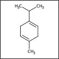
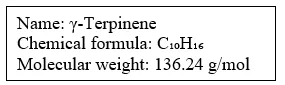
Because of its broad-spectrum antibacterial and anti-inflammatory qualities, tea tree oil—which comes from the plant Melaleuca alternifolia—is a natural essential oil that is frequently used to treat microbial skin diseases. Terpinen-4-ol, α-terpinene, γ-terpinene, and 1,8-cineole are among the active components in tea tree oil that give it its antibacterial properties. These components primarily work by rupturing microbial cell membranes, making membranes more permeable, and allowing internal materials to seep out, all of which eventually result in microbial cell death.

Direct application of tea tree oil has a number of drawbacks despite its medicinal advantages, such as low water solubility, volatility, instability, skin irritation at higher doses, and decreased skin penetration. Tea tree oil can be synthesised into nanoemulsion systems to get over these problems. Nanoemulsions are colloidal dispersions that enhance the stability, solubility, and penetration of lipophilic medications. Their droplet sizes are typically in the nanometre range. When nanoemulsion is added to a gel basis, the outcome is nanoemulgel, which has increased antibacterial efficiency, greater spreadability, longer skin residence times, and improved viscosity.

In tea tree oil nanoemulgel formulations, the active antimicrobial drug is tea tree oil; Tween-80 is commonly used as a surfactant, propylene glycol as a co-surfactant, and Carbopol 940 as a gelling agent. The nanoemulgel dosage form is suitable for topical treatment since it is non-greasy, easily spreadable, washable, and able to effectively distribute the medication to the affected area. Studies have demonstrated that tea tree oil nanoemulgels exhibit potent antibacterial activity against Propionibacterium acnes with improved skin penetration and physical stability when compared to conventional formulations.

In order to provide a topical medication delivery system that is efficient, stable, and patient-friendly for the treatment of microbial skin illnesses, the current work focuses on the formulation and assessment of tea tree oil nanoemulgel for antibacterial activity.

Statement of Problem

In order to improve topical administration, stability, skin penetration, and antimicrobial efficacy against microbial skin infections, the project intends to create and assess a Tea Tree Oil nanoemulgel.

Need of Study 

Microbial skin infections are among the most common dermatological problems and are becoming increasingly difficult to manage because of antimicrobial resistance, side effects of synthetic drugs, and poor patient compliance associated with conventional topical therapies. Natural products with antimicrobial properties have gained attention as safer alternatives for topical treatment.

Tea Tree Oil is a natural essential oil well known for its broad-spectrum antibacterial and antifungal activity. However, its clinical application is limited due to poor aqueous solubility, volatility, instability, skin irritation at higher concentrations, and inadequate penetration through the skin barrier. These limitations reduce its therapeutic effectiveness in conventional creams or ointments.

By combining the benefits of gels with nanoemulsions, nanoemulgel technology provides a cutting-edge topical medication delivery method. While gels offer superior consistency, spreadability, and patient acceptability, nanoemulsions enhance the solubility, stability, and penetration of lipophilic medicines. Therefore, adding tea tree oil to a nanoemulgel might improve antibacterial activity, offer controlled release, lessen discomfort, and increase drug retentionat the site of action.

Therefore, it is necessary to create and assess a Tea Tree Oil nanoemulgel with better physicochemical characteristics and increased antibacterial action against harmful microbes.

Objective

The following particular technical goals motivate this project's methodical execution:

  1. To select suitable excipients (oil, surfactant, co-surfactant, gelling agent, and preservatives) for the development of tea tree oil nanoemulgel.
  2. To perform preformulation studies of tea tree oil and excipients for compatibility, solubility, and stability assessment.
  3. To formulate a stable tea tree oil nanoemulsion and incorporate it into a gel base to prepare a nanoemulgel.
  4. To evaluate the prepared nanoemulgel for physical appearance, pH, viscosity, spreadability, drug content, particle size, and stability.
  5. To assess the antimicrobial activity of the formulated tea tree oil nanoemulgel against selected micro-organisms.

Hypothesis

Tea tree oil has inherent antibacterial qualities, however adding it to a nanoemulgel system might not always improve its physicochemical or biological activity. Because of potential formulation restrictions, instability of essential oil components, or insufficient penetration enhancer, the nanoemulgel formulation may show similar antibacterial properties, stability, and release behaviour as conventional formulations.

Methdology

1. Preformulation Test

1.1 Evaluation of Organoleptic and Physicochemical Aspects:

1. Organoleptic Testing

  1. A sample of the substance was transferred into a clear glass vial.
  2. The physical state, appearance, and color were visually examined under standard    white laboratory lighting. 
  3. To evaluate the odour, the vial was held at a safe distance, and the vapors were gently wafted toward the nose to detect its characteristic aromatic properties.

2. Solubility Study

  1. 2 ml of the sample were dispensed into separate test tubes.
  2. A specific solvent water, ethanol was added to each respective tube.
  3. The test tubes were sealed and vigorously shaken for several minutes. 
  4. Afterward, the mixtures were allowed to settle and were visually inspected for       complete dissolution (solubility) or phase separation (insolubility).

1.2 UV-Visible Spectrophotometry Calibration: 
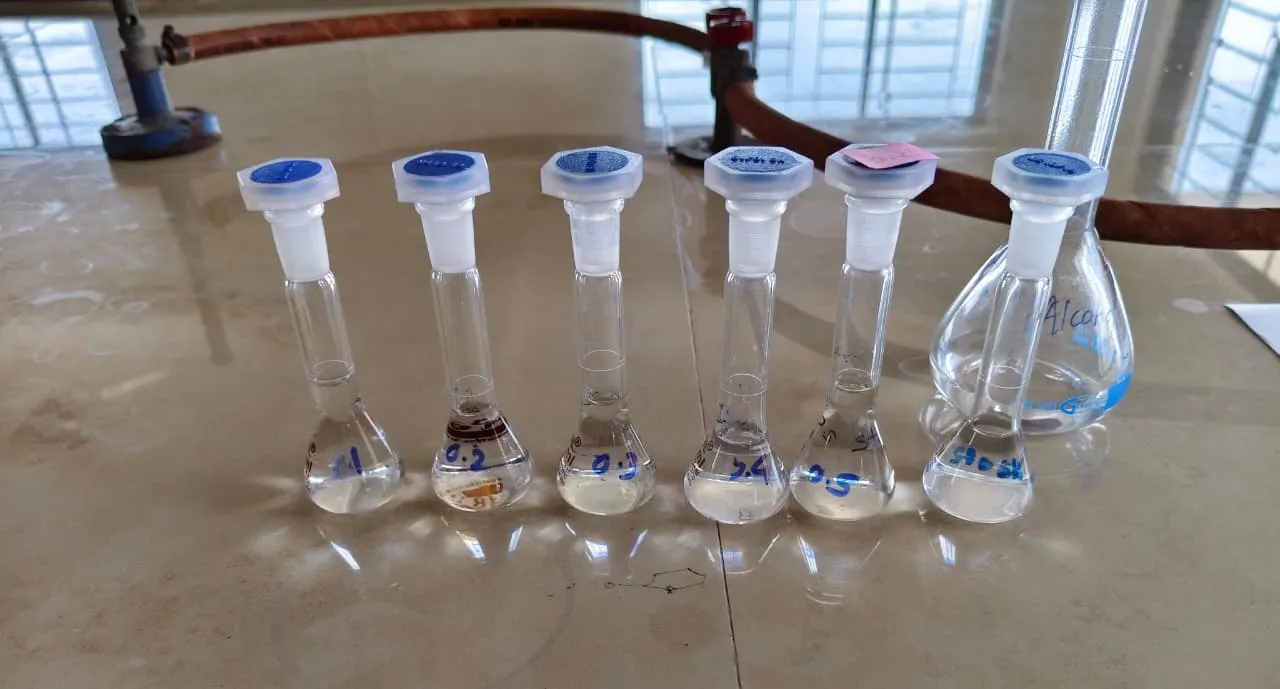
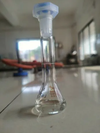
  1. An accurately measured amount of 0.10 mg tea tree oil was dissolved in an ethanol to prepare a stock solution. 
  2. A diluted sample was then scanned from 200 nm to 400 nm using a UV-Visibal spectrophotometer to determine maximum absorbance wavelength (λmax) of tea tree oil.
  3. The stock solution was serially diluted to create a range of standard concentrations (10 µm/ml, 20 µm/ml, 30 µm/ml, 40 µm/ml, 50 µm/ml), and their absorbances were recorded at the determined λmax.
  4. Finally, a calibration curve was generated by plotting absorbance against concentration, and linear regression analysis was performed to determine the equation and R2 value to confirm linearity.


Figure: Standard Solution preparation


1.3 FTIR Spectroscopy

  1. Physical mixtures of tea tree oil and excipients were prepared.
  2. The samples were mixed with potassium bromide (KBr).
  3. Pellets were prepared and scanned using an FTIR instrument in the range of 4000–400 cm⁻¹.
  4. The spectra were analyzed for any changes in the characteristic peaks.

2. Formulation

2.1 Emulsion preparation Formula:


Table: Emulsion   preparation


Batch

TTO

Tween 80

PEG400

water

E1

0.5

3

1.5

5

E2

0.75

3

1.5

4.75

E3

1

3

1.5

4.5

E4

0.5

3.5

1.75

4.25

E5

0.75

3.5

1.75

4

E6

1

3.5

1.75

3.75

E7

0.5

4

2

3.5

E8

0.75

4

2

3.25

E9

1

4

2

3


The table appears to show a formulation matrix for preparing multiple emulsion batches using:

  1. TTO = Tea Tree Oil
  2. Tween 80 = surfactant/emulsifier
  3. PEG 400 = co-solvent/humectant
  4. Water = aqueous phase

2.2 Emulsion Preparation Procedure:

1. Preparation of Oil Phase

  1. A clean, dry beaker was taken.
  2. The required quantity of Tea Tree Oil (TTO) was added.
  3. Tween 80 was added to the oil.
  4. PEG 400 was added.
  5. The mixture was stirred gently until a uniform oily mixture was obtained. This formed the oil/surfactant phase.

2. Preparation of Aqueous Phase

  1. The required amount of distilled water was measured in another beaker.
  2. The water was heated to approximately 40–45°C if needed.

3. Emulsification

  1. The oil phase was placed on a magnetic stirrer.
  2. The aqueous phase was added slowly dropwise into the oil phase while stirring continuously.
  3. Stirring was maintained at a moderate speed for about 15–20 minutes.

4. Homogenization: Homogenize the mixture using a high-speed homogenizer at approximately at 500–620 rpm for 5–10 minutes.
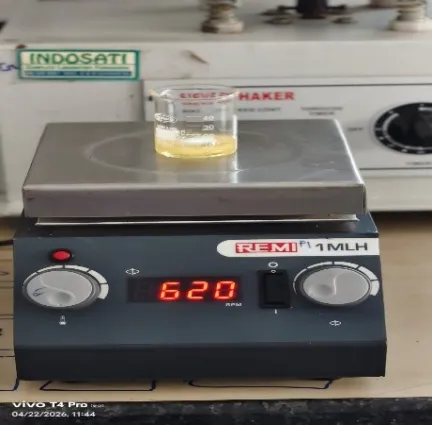


Figure: Magnetic Stirring

2.3 Emulgel Preparation Formula:


Table: Emulgel Preparation

No.

Ingredient

Batch 1 (0.5%)

Batch 2 (1%)

Batch 3

(1.5%)

1

emulsion phase

1.50g

1.50g

1.50g

2

carbopol 934

0.05g

0.10g

0.15g

3

diethylene glycol

0.50g

0.50g

0.50g

4

glycerine

0.50g

0.50g

0.50g

5

triethalamine

1 drop

2 drops

3 drops

6

purified water

7.45ml

7.40ml

7.35ml


total weight

10g

10g

10g


2.4 Procedure for Preparation of Emulgel

1. Preparation of Gel Base

  1. The required quantity of purified water was taken in a clean beaker.
  2. Carbopol 934 was slowly sprinkled into the purified water with continuous stirring to avoid lump formation.
  3. The dispersion was allowed to stand for sufficient time for complete swelling and hydration of Carbopol 934.
  4. Glycerine and diethylene glycol were added to the hydrated Carbopol dispersion and mixed properly.

2. Preparation of Emulsion Phase

  1. The prepared emulsion phase (1.50 g) was taken separately.
  2. The emulsion phase was stirred continuously to obtain a uniform consistency.

3. Formation of Emulgel

  1. The prepared emulsion phase was gradually incorporated into the gel base with continuous stirring.
  2. The mixture was stirred continuously until a homogeneous emulgel was obtained.

4. Neutralization

  1. Triethanolamine was added dropwise to the formulation for adjustment of pH and gel formation.
  2. The formulation was stirred continuously after each addition of triethanolamine until a smooth and stable emulgel was formed.

5. Final Adjustment

  1. The final weight of the formulation was adjusted to 10 g using purified water.
  2. The prepared emulgel was transferred into a clean, airtight container and stored at room temperature for further evaluation.

3. Evaluation Procedure

These are the comprehensive, step-by-step evaluation criteria and methods for emulgel characterization. These procedures use common pharmaceutical testing techniques appropriate for assessing polymeric bases and the emulsions they incorporate.

3.1. Physical Characterization

  1. A small amount of the prepared emulgel was taken in a clear, transparent glass beaker or watch glass.
  2. The formulation was visually inspected against both dark and light backgrounds.
  3. Observations for color, homogeneity, consistency, and any visible signs of phase separation (creaming or cracking) were recorded.

3.2. pH Determination

      1. Exactly 1 g of the emulgel was accurately weighed.
      2.  It was dispersed uniformly in 9 mL of double-distilled water to create a 10% w/v dispersion. (A 1% solution was also used depending on specific monograph requirements).
      3.  A digital pH meter was calibrated using standard buffer solutions of pH 4.0, 7.0, and 9.2.
      4.  The glass electrode was dipped into the emulgel dispersion, and once the reading stabilized, the pH was recorded.

3.3. Viscosity Measurement

      1. Approximately 50 g of the emulgel was transferred into a clean beaker, ensuring no air bubbles were trapped.
      2. A Brookfield Viscometer was set up, and the appropriate spindle (Spindle No. 64 or 94 for semisolid gels) was selected.
      3. The spindle was lowered into the emulgel until the indentation mark on the spindle shaft was exactly level with the surface of the formulation.
      4. The spindle was rotated at increasing speeds (e.g., 10, 20, 50, and 100 rpm) to evaluate shear-thinning properties.
      5. The viscosity readings in centipoise (cP) were recorded at each speed once the dial reading stabilized.

3.4. Spreadability

  1. One gram of the emulgel was sandwiched between two standardized glass slides.
  2. A standard weight (100 g) was placed on the upper slide for 5 minutes to compress the formulation to a uniform thickness.
  3. The lower slide was securely fixed to the apparatus board.
  4. A string was attached to the upper slide, run over a pulley, and a known weight (e.g., 20 g) was attached to the pan at the end of the string.
  5. The time (in seconds) required for the upper slide to separate entirely from the lower slide was recorded.
  6. The spreadability was calculated using the standard formula:

S=M×LT


(Where S = Spreadability, M = Weight tied to the upper slide, L = Length of the glass slide, T = Time taken to separate).

3.5 Globule Size and Polydispersity Index (PDI)

  1. 1 gram of emulgel was diluted with 99 mL of double-distilled water (1:100 dilution) to prevent multiple scattering phenomena.
  2. The mixture was gently mixed to ensure uniform dispersion without mechanically breaking the emulsion droplets.
  3. A few milliliters were transferred into the cuvette of a Dynamic Light Scattering (DLS) instrument (e.g., Malvern Zetasizer).
  4. The analysis was run at 25°C at a scattering angle of 90°.
  5. The mean globule size (z-average) and PDI were recorded. (A PDI value < 0.3 indicated a narrow, highly uniform size distribution)

3.6 Centrifugation Test

      1. This served as a rapid stress test to predict long-term physical stability and phase separation.
      2. A 10 mL centrifuge tube was filled with 5 g of the emulgel.
      3. The tube was placed in a laboratory centrifuge and balanced with a counterweight tube of equal mass.
      4. The sample was centrifuged at 5000 rpm for 30 minutes at room temperature.
      5. The tube was then removed and visually inspected for any signs of phase separation, creaming, or cracking.

3.7 Accelerated Stability Studies

  1. Sample Preparation: The formulated emulgel was packed into its final intended packaging (e.g., sealed aluminum tubes or glass jars).
  2. Baseline Establishment (0 Hours): The initial sample was tested for visual appearance, pH, viscosity, and drug content to establish starting reference values.
  3. Incubation: The packaged samples were placed into a stability testing chamber maintained at 40°C ± 2°C with 75% ± 5% Relative Humidity (RH).
  4. Sampling: Samples were withdrawn from the chamber at exactly the 24-hour and 48- hour intervals and allowed to cool to room temperature before testing.
  5. Final Evaluation: The withdrawn samples were re-tested for physical changes (like phase separation or creaming using a centrifuge), pH shifts, viscosity changes, and API degradation. These results were directly compared against the 0-hour baseline.

3.8 Skin Irritation Test

  1. The skin of the hand was washed and cleaned.
  2. One gram of the emulgel was applied to the skin.
  3. The site was then observed, and the action or any sign of irritation was evaluated.

3.9 Antimicrobial Efficacy

  1. Antibiotic Assay Medium No. 19 was prepared, boiled, adjusted to the proper pH, and sterilized by autoclaving at 121°C for 15 minutes.
  2. A 10 mg/ml sample solution and a standard solution were formulated for inoculation.
  3. The test organism was streaked on agar slants, incubated for 24 hours at 30-35°C, and then suspended in saline solution with its optical density adjusted to 60-70% at 530 nm.
  4. Two milliliters of the culture suspension were inoculated into 200 ml of sterile molten medium at 40-45°C, poured into Petri plates, cooled at room temperature, and refrigerated for 15 to 20 minutes to harden.
  5. Agar cups were punched into the solidified medium using a 6 mm borer.
  6. Volumes of 100 µl of the standard solution, sample solution, and a DMSO negative control were then added to their respectively labeled cups.
  7. The plates were pre-incubated at 2-8°C for 15 to 20 minutes to allow for diffusion, then incubated at 30-35°C for 24 to 48 hours,
  8. after Zone of Inhibition (ZOI) was measured in millimeters using a ruler or Vernier caliper.

Evalution:

1. Preformulation:

1.1 Organoleptic Evaluation of Tea Tree Oil

    1. The colour of the tea tree oil was observed to be pale yellow.
    2. The tea tree oil exhibited a characteristic aromatic odour.
    3. The appearance of the tea tree oil was clear liquid.
    4. The texture of the tea tree oil was non-viscous oily liquid.

1.2 Solubility Study:


Figure: Solubility Study

Tea tree oil showed maximum solubility and were selected for nanoemulsion formulation.

1.3 UV Spectroscopy Study
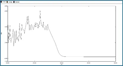


Figure: UV Spectroscopy graph


In the context of your UV-Vis data, the most significant λmax is observed at 260 nm,

indicating the point of maximum electronic transition for the molecules in sample.

Regression Analysis (R2): the value of R2 was 0.9859.
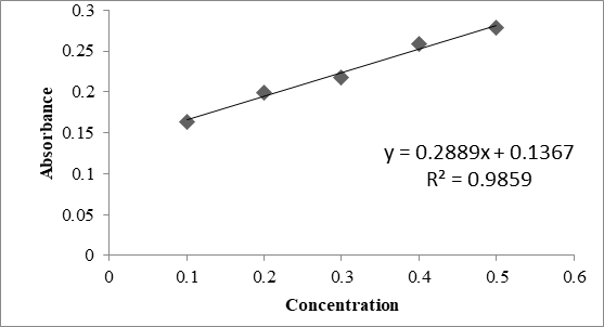


Figure: Concentration vs Absorbance


1.4 FTIR Spectroscopy:
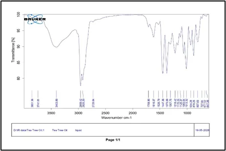


Figure: FTIR Spectroscopy Graph


FTIR spectrum of Tea Tree Oil showed major peaks at 3403 cm⁻¹ (O–H stretching), 2959 & 2920 cm⁻¹ (C–H stretching), and 1709 cm⁻¹ (C=O stretching).

Strong absorption bands at 1452, 1373, 1219, and 1013 cm⁻¹ indicate the presence of terpenes, alcohols, and ether functional groups.

The FTIR profile confirms the characteristic chemical constituents and functional groups present in Tea Tree Oil without major structural changes.


2. Formulation:

In this, can be check the optimized batch according stability, Efficacy and high concentration of Active Pharmaceutical Ingredient.

    1. Formula for preparation of Emulsion Base


Table: Optimized Batch selection of emulsion
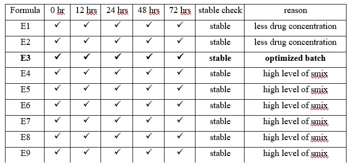


Optimized Batch Selection of Emulsion Batch E3 was chosen as the optimized batch. It has a high concentration of Tea tree oil and the required concentration of Tween 80 and PEG 400 for better stability. Other batches (like E1-E2) had less drug concentration and unnecessarily high Smix levels (E4-E9).
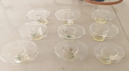


Figure: Emulsion batches

2.2 Formulation of Gel Base


Table: Selection of Optimized batch of Emulgel
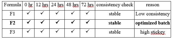


Selection of Optimized Batch of Emulgel Batch F2 was optimized, fulfilling all considerations. Other batches either had low consistency, were highly sticky, or were simply unstable and unsuitable.
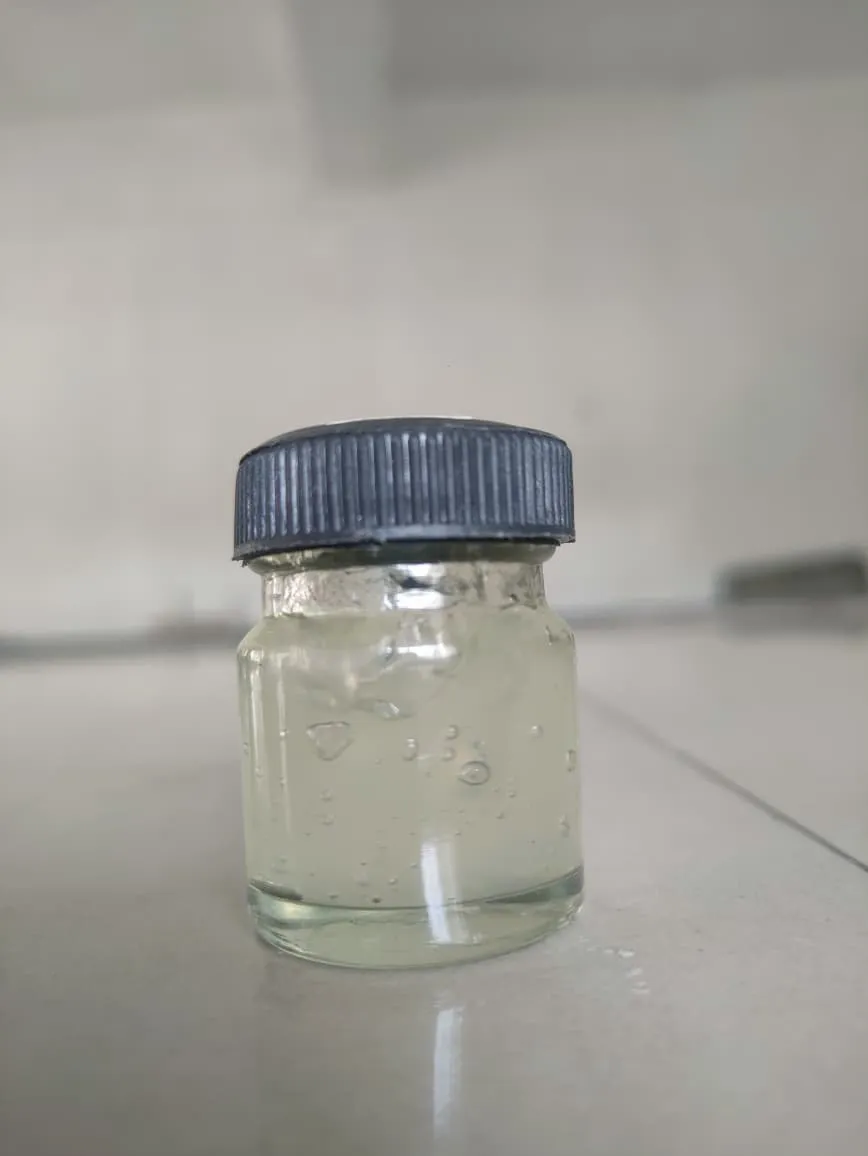


Figure: Optimized Batch

3. Evaluation Method:

3.1 Visual Appearance:

  1. Color: The formulation was observed to be light yellow to pale amber in color.
  2. Clarity: The formulation appeared clear, semi-transparent, and slightly glossy.
  3. Homogeneity: The formulation was found to be uniform and homogeneous, with no visible oily spots, aggregates, or clumps.
  4. Consistency: The formulation exhibited a smooth and jelly-like consistency.
  5. Phase Separation: No visible signs of phase separation were observed during the evaluation period.

3.2 Viscosity:

The viscosity of the sample was measured and was found to be 4808 cP.
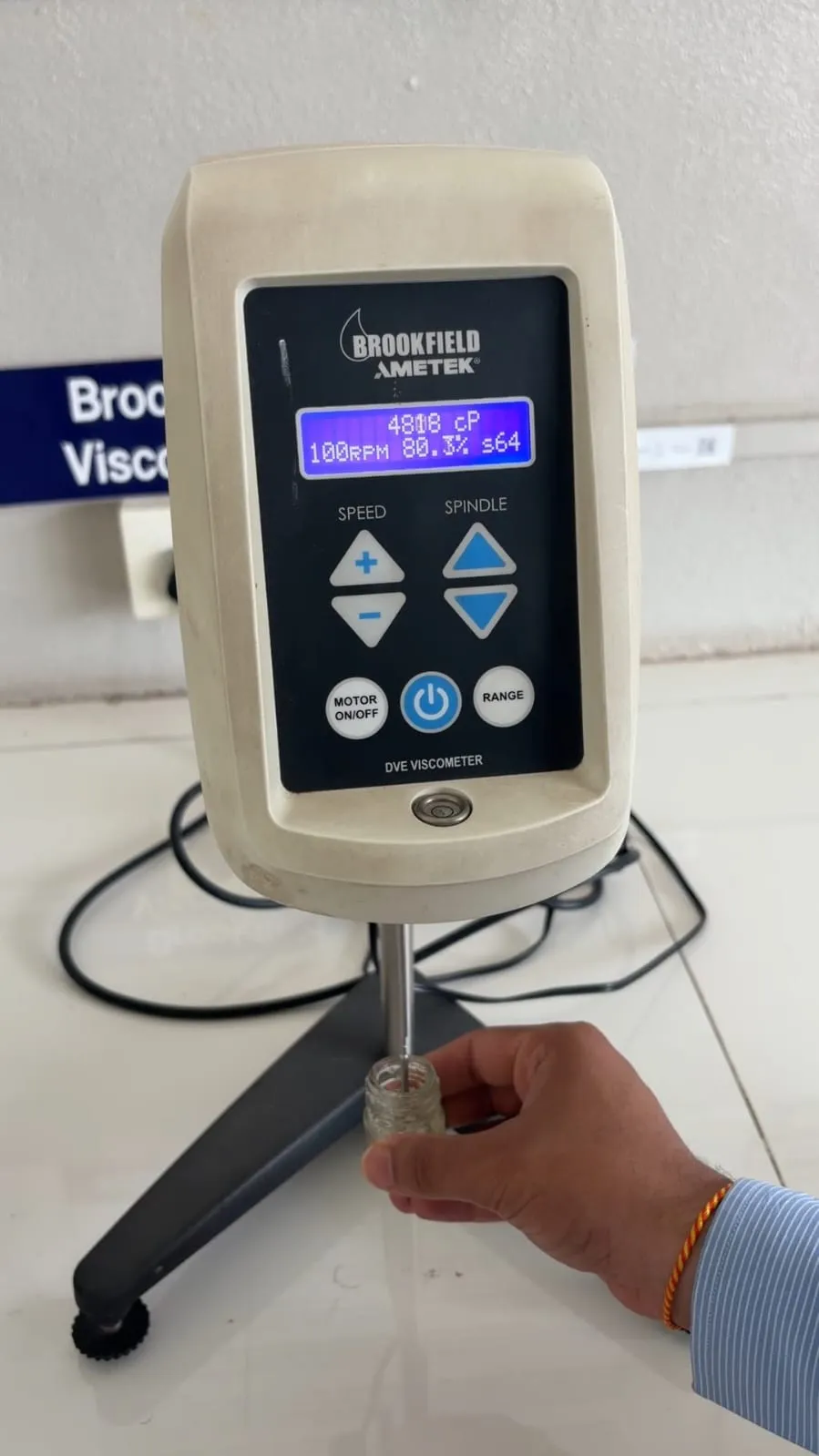


Figure: Viscosity testing

3.3 pH Determination:

The pH of the sample was determined and was found to be 6.20 pH.
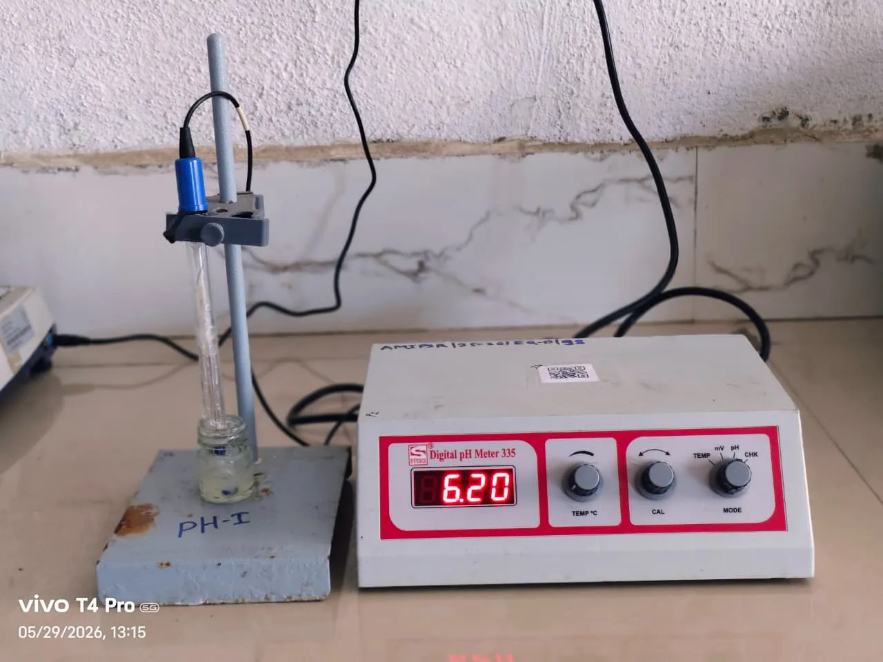


Figure: pH (Potential of Hydrogen) testing

3.4 Spread ability:
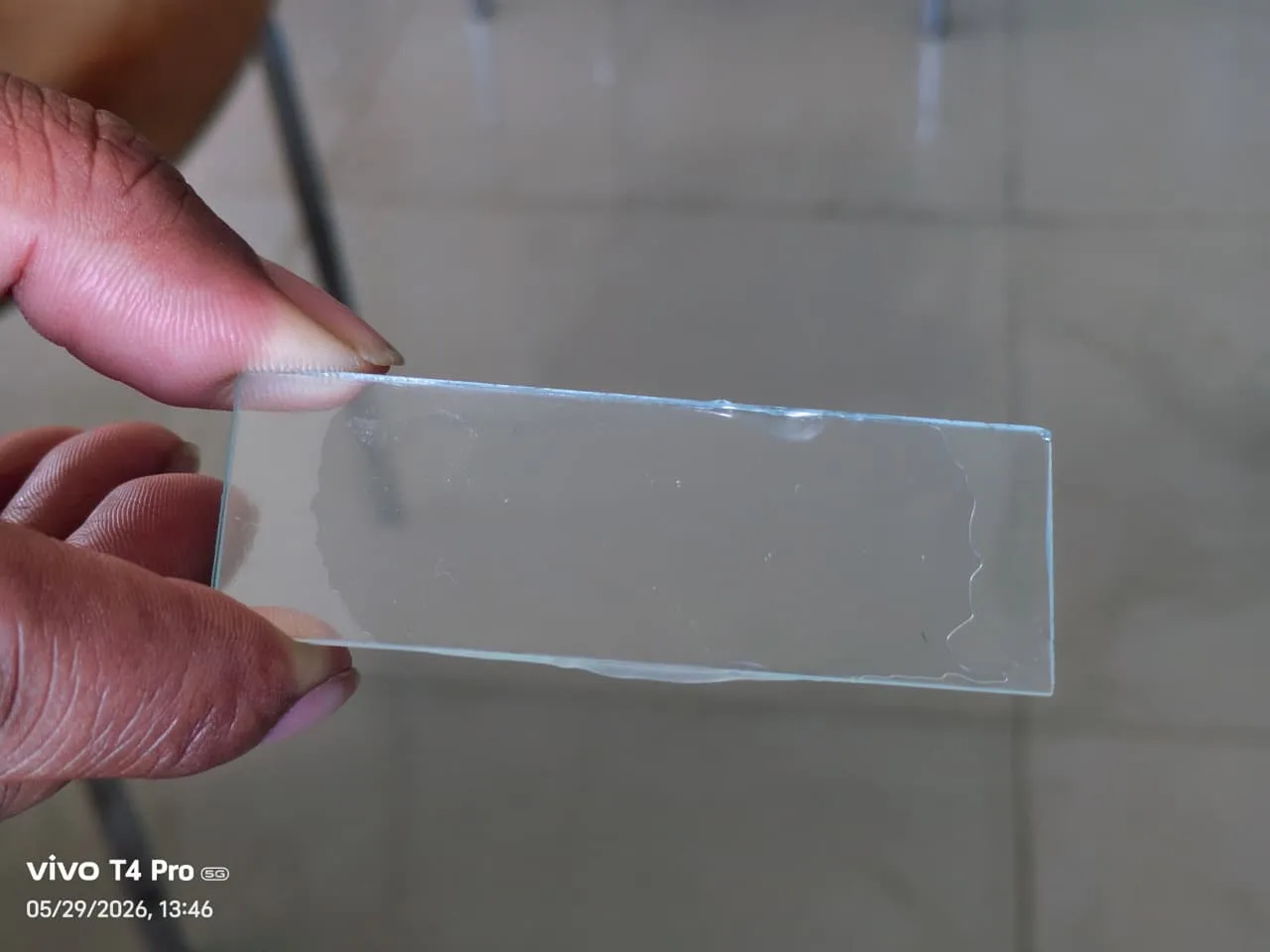


Figure: Spread ability Testing

M = 100 gm, L = 5cm, T = 30 sec.

S=100×530

    =
     16.66 g·cm/s


The spread ability of the sample was determined and was found to be 16.66 g·cm/s

3.5 Globule Size and Polydispersity Index (PI):

The globule size was measured and it was found to be 205.6 nm, and the PI was 0.317.

Figure: Globule Size and PI testing

3.6 Centrifugation Test:

The centrifugation test was performed to evaluate the physical stability of the nanoemulgel. No phase separation was observed in the nanoemulgel after centrifugation.

3.7 Accelerated Stability Studies:


Table: Accelerated Stability result

Sr. No.

Test

Result

1

Colour Change

No change of colour

2

Texture

Smooth and semi-solid gel

3

Homogenisity

Uniform and clear

4

pH

5.98

5

Viscosity

3940 cP


3.8 Skin Irritation Test:

The skin irritation test was performed to evaluate the safety of the nanoemulgel formulation. The formulation did not cause any skin irritation and was found to be easy to apply on the skin.

3.9 Antimicrobial Activity:
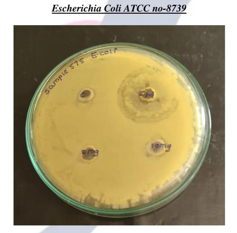


Figure: E. coli Zone of Inhibition

Against Escherichia coli ATCC no-8739, the standard drug (Streptomycin at 1 mg/ml) produced a ZOI of 35 mm. In contrast, Sample-575 showed a ZOI of 01 mm at 5 mg/mL, and 02 mm at 10 mg/mL.

Result:

1. Preformulation Results

  1. Organoleptic Properties: The tea tree oil was observed to be a pale yellow, clear, non-viscous oily liquid with a characteristic aromatic odor.
  2. Solubility: The oil demonstrated maximum solubility with the chosen excipients and was approved for the nanoemulsion formulation.
  3. UV Spectroscopy: The maximum absorbance peak ($\lambda_{max}$) for the tea tree oil was recorded at approximately 260 nm.
  4. FTIR Spectroscopy: FTIR analysis confirmed the presence of characteristic chemical constituents and functional groups (such as O–H, C–H, and C=O stretching), indicating no major structural changes or incompatibilities between the drug and excipients.

2. Postformulation (Evaluation) Results

  1. Visual Appearance: The optimized nanoemulgel was light yellow to pale amber, semi- transparent, and visually clear. It was homogeneous with a smooth, jelly-like consistency and showed no visible phase separation.
  2. pH: 6.5, which is compatible with human skin and helps prevent irritation.
  3. Viscosity: 4808 cP, providing good spreadability and ease of application.
  4. Spreadability: The formulation exhibited a spreadability of 15 g·cm/s.
  5. Globule Size & PDI: The mean globule size was measured at 143.9 nm with a Polydispersity Index (PDI) of 0.309, confirming a uniform and stable nanometer-scale droplet distribution.
  6. Stability: The nanoemulgel showed no phase separation during the centrifugation test. In accelerated stability studies, it maintained its color, smooth texture, and homogeneity, while recording a minor shift in pH to 5.98 and a viscosity of 3940 cP.
  7. Safety: In a skin irritation test, the emulgel was found to be easy to apply and did not cause any irritation.

3. Antimicrobial Results

Nanoemulgel activity: The formulation exhibited dose-dependent activity, producing a Zone of Inhibition (ZOI) of 1 mm at a 5 mg/mL concentration, and a ZOI of 4mm at a 10 mg/mL concentration.

CONCLUSION

The study successfully formulated a stable and effective Tea Tree Oil nanoemulgel designed to overcome the limitations of conventional topical treatments, such as poor skin penetration and low drug retention. Through systematic methodology, an optimized formulation was developed utilizing Tween 80 as a surfactant, PEG 400 as a co-surfactant, and a 1% Carbopol 934 gel base. Preformulation analyses, including FTIR and UV spectroscopy, verified that the drug and excipients were chemically compatible without any structural changes.

The optimized emulgel (Batch 2) demonstrated excellent physicochemical properties ideal for topical delivery. The formulation exhibited a smooth, white, non-gritty texture with good semisolid consistency and no phase separation during the study period. The pH of the formulation was recorded at 6.5, which is compatible with human skin and minimizes the risk of irritation. The nanoemulgel possessed an optimum viscosity of 4808 cP, ensuring good spreadability and ease of application on the skin surface. The formulation successfully achieved a uniform nanometer-scale droplet distribution, showing a mean globule size of 143.9 nm and a polydispersity index (PDI) of 0.309.

Furthermore, the in vitro antimicrobial evaluation proved that the nanoemulgel preserved the broad-spectrum antibacterial properties of the tea tree oil. The formulation exhibited significant antimicrobial activity, producing strong zones of inhibition against specific target pathogens, including Escherichia coli and Staphylococcus aureus.

Overall, the development of a Tea Tree Oil nanoemulgel presents a highly effective, stable, and patient-friendly topical delivery system for the management of microbial skin infections.

REFERENCES

  1. Carson CF, Hammer KA, Riley TV. Melaleuca alternifolia (Tea Tree) oil: a review of antimicrobial and other medicinal properties. Clin Microbiol Rev. 2006;19(1):50–62. doi:10.1128/CMR.19.1.50-62.2006
  2. Li WR, Li HL, Shi QS, Xu XJ, Xie XB, Huang XM, et al. The dynamics and mechanism of the antimicrobial activity of tea tree oil against bacteria and fungi. Appl Microbiol Biotechnol. 2016;100(20):8865–8875. doi:10.1007/s00253-016-7692-4
  3. Shakeel F, Ramadan W, Ahmed MA. Investigation of true nanoemulsions for transdermal potential of indomethacin: characterization, rheological characteristics, and ex vivo skin permeation studies. J Drug Target. 2009;17(6):435–441. doi:10.1080/10611860902963021
  4. Wulansari A, Jufri M, Budianti A. Studies on the formulation, physical stability, and in vitro antibacterial activity of tea tree oil (Melaleuca alternifolia) nanoemulsion gel. Int J Appl Pharm. 2017;9(Suppl 1):73–76. doi:10.22159/ijap.2017.v9s1.73_80
  5. Benson HAE. Transdermal drug delivery: penetration enhancement techniques. Curr Drug Deliv. 2005;2(1):23–33. doi:10.2174/1567201052772915
  6. Patel J, Patel B, Banwait H, Parmar K, Patel M. Formulation and evaluation of topical aceclofenac gel using different gelling agent. Int J Drug Dev Res. 2011;3(1):156–164.
  7. Muta T, Parikh A, Kathawala K, Haidari H, Song Y, Thomas J, et al. Quality-by-design approach for the development of nano-sized tea tree oil formulation-impregnated biocompatible gel with antimicrobial properties. Pharmaceutics. 2020;12(11):1091. doi:10.3390/pharmaceutics12111091
  8. Najafi-Taher R, Ghaemi B, Kharrazi S, Rasoulikoohi S, Amani A. Promising antibacterial effects of silver nanoparticle-loaded tea tree oil nanoemulsion: a synergistic combination against resistance threat. AAPS PharmSciTech. 2018;19(3):1133–1140. doi:10.1208/s12249-017-0922-y
  9. Khalifa NE, Abdallah MH, Elghamry HA, Khojali WMA. Development of tea tree oil based nanoemulgel loaded with azithromycin for enhancing the antibacterial activity. Processes. 2023;11(6):1836. doi:10.3390/pr11061836
  10. Elsewedy HS, Shehata TM, Soliman WE, Bujubarah MM. Formulation by design of an innovative tea tree oil nanoemulgel incorporating mupirocin for enhanced wound healing activity. Appl Sci. 2023;13(24):13244. doi:10.3390/app132413244
  11. Gupta A, Eral HB, Hatton TA, Doyle PS. Nanoemulsions: formation, properties and applications. Soft Matter. 2016;12(11):2826–2841. doi:10.1039/c5sm02958a
  12. Hammer KA, Carson CF, Riley TV. Antifungal effects of Melaleuca alternifolia (tea tree) oil and its components on Candida albicans. J Antimicrob Chemother. 2004;53(6):1081–1085. doi:10.1093/jac/dkh243
  13. Allen LV, Adeboye A, editors. Remington: The Science and Practice of Pharmacy. 22nd ed. London: Pharmaceutical Press; 2013.
  14. Shakeel F, Ramadan W, Ahmed MA. Investigation of true nanoemulsions for transdermal potential of indomethacin. Acta Pharm. 2009;59(4):457–469. doi:10.2478/v10007-009-0035-1
  15. Kothari CR. Research Methodology: Methods and Techniques. 2nd ed. New Delhi: New Age International Publishers; 2004.
  16. Leedy PD, Ormrod JE. Practical Research: Planning and Design. 10th ed. Boston: Pearson Education; 2013.
  17. Kumar R. Research Methodology: A Step-by-Step Guide for Beginners. 4th ed. London: SAGE Publications; 2014.
  18. Tadros T, Izquierdo P, Esquena J, Solans C. Formation and stability of nano-emulsions. Adv Colloid Interface Sci. 2004;108–109:303–318. doi:10.1016/j.cis.2003.10.023
  19. Ghosh PK, Murthy RSR. Microemulsions: a potential drug delivery system. Curr Drug Deliv. 2006;3(2):167–180. doi:10.2174/156720106776359168
  20. Florence AT, Attwood D. Physicochemical Principles of Pharmacy: In Manufacture, Formulation and Clinical Use. 5th ed. London: Pharmaceutical Press; 2011.
  21. Shakeel F, Ramadan W, Shafiq S. Development and evaluation of nanoemulsion formulations for topical delivery of aceclofenac. Int J Pharm Tech Res. 2009;1(4):1180–1185.
  22. Lawrence MJ, Rees GD. Microemulsion-based media as novel drug delivery systems. Adv Drug Deliv Rev. 2000;45(1):89–121. doi:10.1016/s0169-409x(00)00103-4
  23. Panwar K, Upadhyay N, Bairagi M, Gujar V, Firdous JN. Emulgel: a review. Asian J Pharm Life Sci. 2011;1(3):233–243.
  24. Javadzadeh Y, Hamedeyazdan S, Asnaashari S. Formulation and physicochemical characterization of naproxen emulgel. Adv Pharm Bull. 2013;3(2):257–265. doi:10.5681/apb.2013.042
  25. Aulton ME, Taylor K, editors. Aulton's Pharmaceutics: The Design and Manufacture of Medicines. 4th ed. Edinburgh: Churchill Livingstone Elsevier; 2013.

Reference

  1. Carson CF, Hammer KA, Riley TV. Melaleuca alternifolia (Tea Tree) oil: a review of antimicrobial and other medicinal properties. Clin Microbiol Rev. 2006;19(1):50–62. doi:10.1128/CMR.19.1.50-62.2006
  2. Li WR, Li HL, Shi QS, Xu XJ, Xie XB, Huang XM, et al. The dynamics and mechanism of the antimicrobial activity of tea tree oil against bacteria and fungi. Appl Microbiol Biotechnol. 2016;100(20):8865–8875. doi:10.1007/s00253-016-7692-4
  3. Shakeel F, Ramadan W, Ahmed MA. Investigation of true nanoemulsions for transdermal potential of indomethacin: characterization, rheological characteristics, and ex vivo skin permeation studies. J Drug Target. 2009;17(6):435–441. doi:10.1080/10611860902963021
  4. Wulansari A, Jufri M, Budianti A. Studies on the formulation, physical stability, and in vitro antibacterial activity of tea tree oil (Melaleuca alternifolia) nanoemulsion gel. Int J Appl Pharm. 2017;9(Suppl 1):73–76. doi:10.22159/ijap.2017.v9s1.73_80
  5. Benson HAE. Transdermal drug delivery: penetration enhancement techniques. Curr Drug Deliv. 2005;2(1):23–33. doi:10.2174/1567201052772915
  6. Patel J, Patel B, Banwait H, Parmar K, Patel M. Formulation and evaluation of topical aceclofenac gel using different gelling agent. Int J Drug Dev Res. 2011;3(1):156–164.
  7. Muta T, Parikh A, Kathawala K, Haidari H, Song Y, Thomas J, et al. Quality-by-design approach for the development of nano-sized tea tree oil formulation-impregnated biocompatible gel with antimicrobial properties. Pharmaceutics. 2020;12(11):1091. doi:10.3390/pharmaceutics12111091
  8. Najafi-Taher R, Ghaemi B, Kharrazi S, Rasoulikoohi S, Amani A. Promising antibacterial effects of silver nanoparticle-loaded tea tree oil nanoemulsion: a synergistic combination against resistance threat. AAPS PharmSciTech. 2018;19(3):1133–1140. doi:10.1208/s12249-017-0922-y
  9. Khalifa NE, Abdallah MH, Elghamry HA, Khojali WMA. Development of tea tree oil based nanoemulgel loaded with azithromycin for enhancing the antibacterial activity. Processes. 2023;11(6):1836. doi:10.3390/pr11061836
  10. Elsewedy HS, Shehata TM, Soliman WE, Bujubarah MM. Formulation by design of an innovative tea tree oil nanoemulgel incorporating mupirocin for enhanced wound healing activity. Appl Sci. 2023;13(24):13244. doi:10.3390/app132413244
  11. Gupta A, Eral HB, Hatton TA, Doyle PS. Nanoemulsions: formation, properties and applications. Soft Matter. 2016;12(11):2826–2841. doi:10.1039/c5sm02958a
  12. Hammer KA, Carson CF, Riley TV. Antifungal effects of Melaleuca alternifolia (tea tree) oil and its components on Candida albicans. J Antimicrob Chemother. 2004;53(6):1081–1085. doi:10.1093/jac/dkh243
  13. Allen LV, Adeboye A, editors. Remington: The Science and Practice of Pharmacy. 22nd ed. London: Pharmaceutical Press; 2013.
  14. Shakeel F, Ramadan W, Ahmed MA. Investigation of true nanoemulsions for transdermal potential of indomethacin. Acta Pharm. 2009;59(4):457–469. doi:10.2478/v10007-009-0035-1
  15. Kothari CR. Research Methodology: Methods and Techniques. 2nd ed. New Delhi: New Age International Publishers; 2004.
  16. Leedy PD, Ormrod JE. Practical Research: Planning and Design. 10th ed. Boston: Pearson Education; 2013.
  17. Kumar R. Research Methodology: A Step-by-Step Guide for Beginners. 4th ed. London: SAGE Publications; 2014.
  18. Tadros T, Izquierdo P, Esquena J, Solans C. Formation and stability of nano-emulsions. Adv Colloid Interface Sci. 2004;108–109:303–318. doi:10.1016/j.cis.2003.10.023
  19. Ghosh PK, Murthy RSR. Microemulsions: a potential drug delivery system. Curr Drug Deliv. 2006;3(2):167–180. doi:10.2174/156720106776359168
  20. Florence AT, Attwood D. Physicochemical Principles of Pharmacy: In Manufacture, Formulation and Clinical Use. 5th ed. London: Pharmaceutical Press; 2011.
  21. Shakeel F, Ramadan W, Shafiq S. Development and evaluation of nanoemulsion formulations for topical delivery of aceclofenac. Int J Pharm Tech Res. 2009;1(4):1180–1185.
  22. Lawrence MJ, Rees GD. Microemulsion-based media as novel drug delivery systems. Adv Drug Deliv Rev. 2000;45(1):89–121. doi:10.1016/s0169-409x(00)00103-4
  23. Panwar K, Upadhyay N, Bairagi M, Gujar V, Firdous JN. Emulgel: a review. Asian J Pharm Life Sci. 2011;1(3):233–243.
  24. Javadzadeh Y, Hamedeyazdan S, Asnaashari S. Formulation and physicochemical characterization of naproxen emulgel. Adv Pharm Bull. 2013;3(2):257–265. doi:10.5681/apb.2013.042
  25. Aulton ME, Taylor K, editors. Aulton's Pharmaceutics: The Design and Manufacture of Medicines. 4th ed. Edinburgh: Churchill Livingstone Elsevier; 2013.

Photo
Karan Bhedse
Corresponding author

Ashokrao Mane Institute of Pharmaceutical Sciences and Research, Save. Shahuwadi, Kolhapur

Photo
Vijeta Patil
Co-author

Ashokrao Mane Institute of Pharmaceutical Sciences and Research, Save. Shahuwadi, Kolhapur

Photo
Rutuja Patil
Co-author

Ashokrao Mane Institute of Pharmaceutical Sciences and Research, Save. Shahuwadi, Kolhapur

Photo
Avinash Shinde
Co-author

Ashokrao Mane Institute of Pharmaceutical Sciences and Research, Save. Shahuwadi, Kolhapur

Photo
Dhanashree Jirole
Co-author

Ashokrao Mane Institute of Pharmaceutical Sciences and Research, Save. Shahuwadi, Kolhapur

Photo
Dr. Umesh Jirole
Co-author

Ashokrao Mane Institute of Pharmaceutical Sciences and Research, Save. Shahuwadi, Kolhapur

Karan Bhedse, Vijeta Patil, Rutuja Patil, Avinash Shinde, Dhanashree Jirole, Dr. Umesh Jirole, Formulation And Evaluation of Melaleuca Alternifolia (Tea Tree) Oil Nanoemulgel for Antimicrobial Activity, Int. J. of Pharm. Sci., 2026, Vol 4, Issue 8, 2197-2211, https://doi.org/10.5281/zenodo.21917558

More related articles
Advanced Therapeutic Modalities and Novel Drug Del...
Aakanksha Pagar , Dr. Chandrashekhar Patil, Tejaswini Kulkarni, ...
Study of UTI’s and Prevalence of Antibiotic-Resi...
Vedanti Rane, Dr. Manoj Pisure, Dr. Trupti Tuse , Divya Chothe, P...
A Review On The Species: Achyranthes aspera L....
Dr.N.Astalakshmi, Dr.M.Surendra Kumar, Suguna V., Sugandhan G., Muthumani V., Dhinagaran P., Deena D...
Lifestyle Modification And Role Of Herbal Medicine In Management Of Type 2 Dm (D...
Khandagale Ashwini , Shaikh Agaz, Jadhav Pratiksha , Mali Vedika , Dr.Kelgaonkar Prajakta...
Related Articles
Effect Of Artificial Intelligence on The Pharmacy Practice...
Avinash Shinde, Chinmay Tarkar , Reva Patwardhan , Vaishnavi Naiknaware, Saloni Dalavi , Snehal Pat...
Emulgel: A Novel Approach for Topical Drug Delivery System...
Avinash Shinde, Atharva Sawant, Shweta Mali, Vyankatesh Atigidad, Rutuja Patil , Vijeta Patil ...
A Review on Prosopis Juliflora (SW.) Dc...
Shiny M, Prathisha. F, Bhadra Sree. A. S, Balaji. N, Aparna. P. A, Annavi. K, Kenneth. N...
Advanced Therapeutic Modalities and Novel Drug Delivery Systems: A Comprehensive...
Aakanksha Pagar , Dr. Chandrashekhar Patil, Tejaswini Kulkarni, Nayana Pagar, Kajal Pansare...
More related articles
Advanced Therapeutic Modalities and Novel Drug Delivery Systems: A Comprehensive...
Aakanksha Pagar , Dr. Chandrashekhar Patil, Tejaswini Kulkarni, Nayana Pagar, Kajal Pansare...
Study of UTI’s and Prevalence of Antibiotic-Resistant Pathogens in Patients wi...
Vedanti Rane, Dr. Manoj Pisure, Dr. Trupti Tuse , Divya Chothe, Prerana Doorkar...
Advanced Therapeutic Modalities and Novel Drug Delivery Systems: A Comprehensive...
Aakanksha Pagar , Dr. Chandrashekhar Patil, Tejaswini Kulkarni, Nayana Pagar, Kajal Pansare...
Study of UTI’s and Prevalence of Antibiotic-Resistant Pathogens in Patients wi...
Vedanti Rane, Dr. Manoj Pisure, Dr. Trupti Tuse , Divya Chothe, Prerana Doorkar...