

We use cookies to ensure our website works properly and to personalise your experience. Cookies policy
PRES‘S College of Pharmacy (For Women) Chincholi, Nashik Maharashtra, India 422103
Cancer remains a significant global healthcare challenge due to the difficulty in effectively targeting and eliminating cancer cells while minimizing damage to healthy cells. Despite a variety of available drugs, biological barriers such as renal and hepatic clearance complicate drug delivery. Advances in nanomedicine enable the development of drug-loaded nanoparticles designed to bypass these barriers, thus enhancing treatment efficacy and reducing side effects. Notable achievements in this field include the clinical approval of first-generation nanomedicines, such as Doxil® and Onivyde®, which illustrate the progress made in improving therapeutic outcomes for cancer patients. This review elaborates on the key biological obstacles to effective drug delivery and provides a summary of various nanoparticles utilized in cancer therapies. It also highlights the successful clinical trials for Onivyde, approved by the FDA in 2015, serving as a case example of nanomedicine’s potential. Future developments in nanomedicine must focus on more precise targeting of cancerous tissues while navigating challenges like biomarker identification, synthesis scalability, and consistency in characterization. Collaborative efforts among academia, industry, and regulatory bodies are essential to address these hurdles and move closer to the goal of eradicating cancer.
Cancer is still one of the most deadly and aggressive diseases. It is the second most common cause of death in the United States [1], accounts for millions of deaths worldwide [2], and is predicted to increase dramatically over the next 20 years due to a number of risk factors, including but not limited to environmental pollution, changes in the population, and an increase in the prevalence of certain lifestyle choices [3].Using lipid-based, polymeric, and inorganic nanoparticles to target cancer cells by improving drug solubility and circulation, improving tumour penetration, and controlling the release of the drug, multifunctional nanomedicine has seen a significant expansion of research in oncology over the past 30 years . This ensures a more precise and safe delivery of treatments to tumour sites [4,5].
1. Physico‑chemical properties
The nanomaterials used in cancer research can be altered in terms of dimensions, form, and surface properties for personalization to address particular tumours. Size is crucial. For passage thru the circulation and subsequent delivery of the nanocarriers to the tumor tissue. Even tho smaller Nanoparticles are more likely to gather in the leaky blood vessels of tumors compared to those that are bigger, they can penetrate healthy tissue as well. Conversely Larger nanoparticles, however, are more difficult to extravasate and as a result, their bloodstream distribution is extremely variable [6]. Optimizing the size of nanoparticles could Enhance targeted absorption into tumor tissue. The form Of the nanocarriers could affect fluid dynamics, so Impact uptake. At the moment, spherical nano Carriers seem to be more prevalent than that of the Nonspherical variety as a result of difficulties in synthesis and Testing.[7]
2. Solubility, degradation and clearance
The charge of nanocarriers may also affect their stability and distribution in the blood. Positively charged Nanoparticles were previously shown to most effectively Target tumor vessels, but a switch to a neutral charge after Extravasation allowed quicker diffusion of the nanoparticles to the tumor tissue [8]. The surface of the nanocarriers can also be modified with ligands that may prolong Blood circulation and promote specific types of endocytosis and cellular uptake into tumor tissue. The reticuloendothelial system (RES) recognizes Hydrophobic materials as foreign and eliminates them From the bloodstream, taking them up in the liver or The spleen. Foreign materials coated with opsonin proteins are more easily recognized by monocytes and macrophages [10]. Opsonization of hydrophobic molecules Can reduce their ability to reach the tumor tissue and Trigger inflammation following the secretion of cytokines From the phagocytic cells [9,10]
3. Targeting
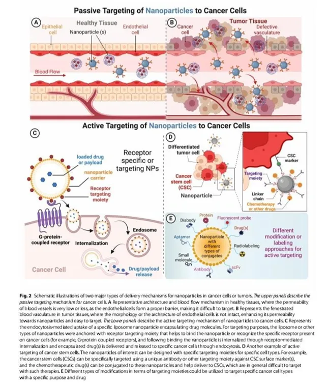
The attachment of ligands to the nanocarriers’ surface is used in active targeting. These ligands are highly specific to receptors and other cancer-specific targets, like glycans, [11]that are overexpressed on the surface of tumor cells. By conjugating these ligands, non-specific nanocarrier uptake to tissues other than tumor tissue may be prevented. Transferrin, folic acid, enzymes, modified antibodies, and macromolecules like proteins and carbohydrates are examples of such ligands. In order to prolong the time that nanoparticles circulate in the bloodstream, the density of these ligands should be optimized to prevent recognition by the RES and interaction with serum proteins[5,12].
4. Drug Release
Several pertinent studies in the field of stimuli-responsive nanomedicine should be taken into consideration in addition to the results previously described in our paper. The ability of stimuli-responsive nanomedicines to react to particular physical or biological triggers and subsequently regulate drug release profiles in terms of space, time, and dosage constitutes a novel strategy for improving drug delivery efficiency [13].
5. Nanoparticle Types:
5.1. Protein-Drug Conjugated Nanoparticles:
These consist of proteins directly linked to drug molecules. The connection is usually biodegradable once inside the cell, which can lead to early drug release due to breakdown by proteases and redox agents in the blood. Recent advancements have introduced systems with stable linkers that remain intact until reaching the target site, allowing for precise and controlled drug delivery, thereby reducing treatment toxicity[14]. Because protein-drug conjugated nanoparticles are usually very small (10 nm), they can be delivered to the target tumor site more easily and have a long half-life in vivo[15].
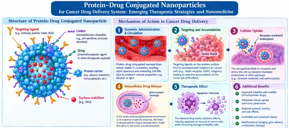
Fig 5.1 Protein drug conjugated Nanoparticles
5.2. Liposomal Nanoparticles
Sonication, extrusion, reverse-phase evaporation, and solvent injection are common laboratory techniques used to produce liposomal nanoparticles[16]. Thermosensitive liposomes can release encapsulated drugs at specific temperatures. These liposomes enable targeted drug delivery to areas exposed to energy sources like radio frequencies, microwaves, and high-intensity ultrasound[17]. High-frequency ultrasound has been successfully used in clinical applications to improve drug delivery[18].
While hydrophobic and amphiphilic drugs easily penetrate the lipid bilayer, potentially causing toxicity, hydrophilic drugs do not. Liposomes can address this by either fusing with the cell membrane or lysing in harsh environments like peroxisomes, releasing the drug. A polymeric shell can contain hydrophobic drugs, though it may hinder fusion with target cells, requiring additional release mechanisms. Polymeric coatings also enhance circulation time, bioavailability, targeting efficiency, and alter surface charge[14].Polymeric nanoparticles, made from synthetic polymers, allow customization of key properties like molecular weight, hydrophobicity, and biodegradability. Their synthesis is well researched, with effective drug encapsulation methods such as emulsification, electrospray, and nanoprecipitation. These nanoparticles, typically composed of dense matrices with known degradation curves, make drug release easier to control compared to other systems[14].Polymeric nanoparticles face issues with limited shape and broad size distribution. Typically spherical, they can vary widely in size during synthesis. New methods, like Particle Replication in Nonwetting Templates (PRINT), are being explored to address these issues. PRINT allows for uniform nanoparticles, enabling customization of size, shape, and properties, including drug uptake pathways and aesthetic qualities[19].
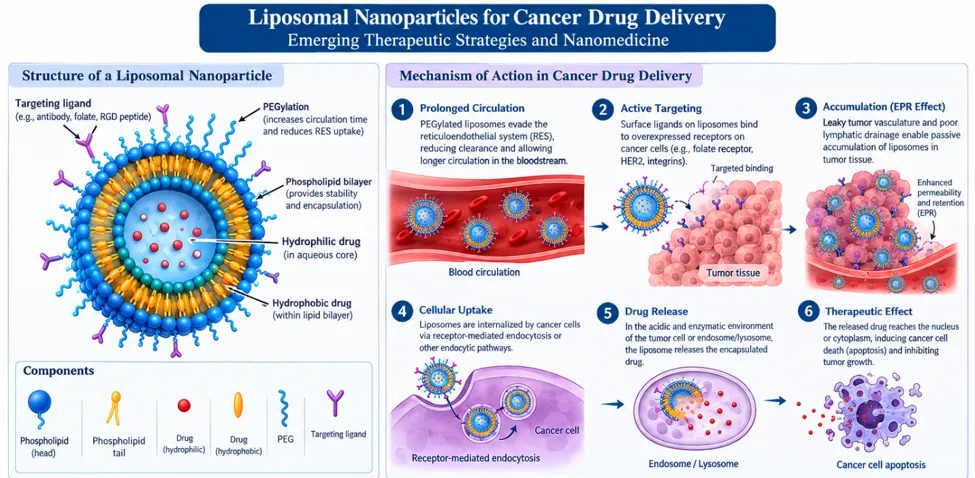
Fig 5.2 Liposomal Nanoparticles
5.3. Dendrimeric Nanoparticles
Dendrimers, spherical macromolecules with numerous branches from a central point, make up dendrimeric nanoparticles. These are produced layer by layer, integrating the initial core before forming branches. Using specific initiator cores allows manipulation of size and branching, reducing nanoparticle polydispersity. Carefully planning cores and branches specifies molecular weight, size, branch density, flexibility, and water solubility[14]
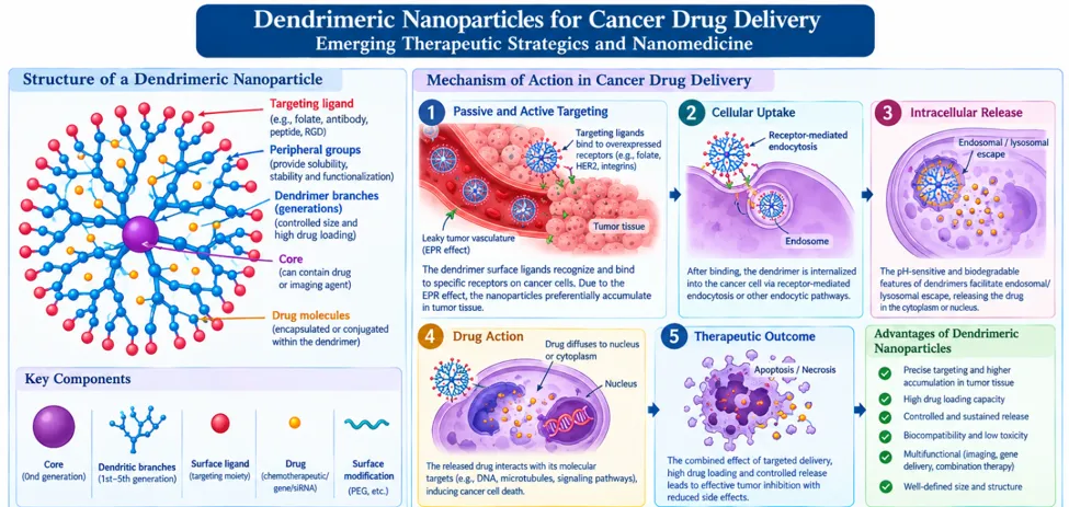
Fig 5.3 Dendrimeric Nanoparticles
5.4. Hydrogel
Hydrogels are three-dimensional networks of cross-linked, water-soluble polymers capable of holding large amounts of fluid. While most synthetic hydrogels aren’t biodegradable, adding enzymatic, hydrolytic, and stimuli-responsive components can create biodegradable nanoparticles under certain conditions. Hydrogels’ unique fluid retention resembles biological tissues, reducing tension and enhancing biocompatibility. [20] The hydrogel’s porosity can be adjusted to control drug loading and release rates by modifying the cross-linking in the matrix. Hydrogels have a positive surface charge, enhancing interaction with negatively charged cell membranes and increasing drug uptake. However, they may bind to negatively charged serum proteins, reducing circulation time[14].
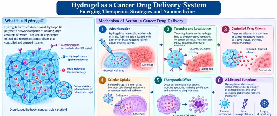
Fig 5.4 Hydrogel
5.5. Other Nanoparticle platforms
Gold is a well-known inorganic metallic nanoparticle, extensively used for detection and direct cancer treatment, with or without drug loading. Its strong optical absorbance aids detection, while photothermal properties make it suitable for anticancer therapy. Complex gold structures enhance drug release efficiency, with drug molecules conjugated to surfaces or hollow interiors for better encapsulation. Thermoresponsive polymers on the surface allow controlled drug release by expanding or contracting with temperature changes. This, combined with gold’s photothermal properties, enables targeted drug delivery. For instance, lasers can heat gold nanoparticles near tumors, optimizing drug loading and reducing toxicity[14].Carbon nanotubes have been studied for cancer treatment. They can bind to various biological materials and enter cells through endocytosis. Single-walled carbon nanotubes (SWCNTs) form stable suspensions in physiological buffers, making them suitable for biological use. They bind to molecules via cleavable disulfide bonds, allowing drug release in the presence of certain enzymes. Due to their optical properties, carbon nanotubes show promise in treatments, similar to gold, using photothermal and photodynamic therapy to damage tumor cells[21].Silver nanoparticles are another potential cancer treatment. Though their exact mechanism is unclear, they react with the acidic environment in cancer cells to generate reactive oxygen species, causing damage and apoptosis. Silver also inhibits vascular endothelial growth factor (VEGF), providing anti-angiogenic effects[22]. A study showed that silver nanoparticles in nanofibers have antitumor effects, with higher silver content increasing cancer cell death. These nanofibers also inhibited bacterial growth. Toxicity concerns can be managed by using silver cores with biocompatible shells that degrade in specific environments, allowing drug conjugation[23].
Prospective View
Prospects and Challenges of Nanomedicine in Tumor Therapeutics
Despite nanotechnology’s promise in tumor treatment with its unique molecular properties for targeted, less toxic therapies, several obstacles must be overcome. A major challenge is the biological barriers that prevent nanomedicine from effectively accumulating and penetrating tumors, from the bloodstream to tissues and cells[24].Endothelial cells lining blood vessels are closely linked to major drug delivery obstacles at the blood level. Conditions like cancer or inflammation often cause poor blood flow, hindering drug delivery. Additionally, the permeability of tumor vasculature and its reliance on vascular endothelial growth factor (VEGF) can significantly impede drug delivery[25].Barriers at the tissue level include inadequate vascularization, leading to hypoxic regions that impair drug effectiveness and delivery. Increased interstitial fluid pressures in the tumor microenvironment (TME) can hinder drug penetration. Additionally, heterogeneity in tissue characteristics, such as cell types, genetic mutations, or vasculature, complicates drug delivery and efficacy[24,26].At the cellular level, the main challenge is that most drug delivery methods are passive. They depend on diffusion and concentration gradients to move drugs from high-concentration areas (blood) to low-concentration areas (tumor interstitium), which limits the drug amount reaching tumor cells[24].
Active transport mechanisms use cellular energy like ATP or GTP to enable selective molecular transport against concentration gradients. While these barriers are significant, they can be overcome. Strategies such as vascular promotion therapy, vessel normalization, and iRGD-based drug delivery are being developed. However, advancing these strategies to clinical use requires further research.
Concerns about nanomedicine’s toxicity and safety, including effects on organs and reproductive systems, are crucial for long-term use. Refining nanocarriers’ drug loading and release control is also necessary for optimal effectiveness. Challenges like complex synthesis, high costs, limited biocompatibility, and nanomaterial stability hinder broader clinical application. Additionally, the lack of suitable animal models that mimic human tumors complicates translating preclinical findings into clinical trials. Overcoming these obstacles through innovation and extensive research is critical to unlocking nanotechnology’s potential in tumor therapeutics and providing personalized cancer treatments.
Directions for Novel Nanomedicine Development in Tumor Treatment.
Nanomedicine shows great promise in early cancer diagnosis and treatment but is still in early development and faces limitations that must be resolved before market approval. Key areas for advancement in tumor therapy include:
Reducing Nanomaterial Toxicity: Establishing safety evaluation standards is urgent.
Improving Delivery Strategies: Develop precise nanodrug delivery methods to enhance efficiency by targeting specific tumor types and overcoming biological barriers.
Ensuring Stability and Controlled Release: This avoids systemic toxicity and enhances drug delivery efficacy.
Simplifying Nano-Design: Simplification aids clinical translation by addressing complexity challenges. Innovative manufacturing technologies, such as microfluidic technology and non-infiltrating template microprinting, can support large-scale nanomedicine production. Establishing in vivo and ex vivo evaluation models, including human-derived tumor xenograft models and tumor-like organs or microarrays, is vital for comprehensive evaluation and better correlation with experimental outcomes.
6. Nanotechnology in Cancer
Nano-oncology, or nanotechnology in oncology, refers to the use of nanoscale pharmaceuticals as anticancer treatments[27]. Over the past 20 years, this new strategy has demonstrated encouraging results in cancer detection[28], treatment[29], and prevention, making it a state-of-the-art method in oncology. Let’s now explore the different uses of nanoparticles in oncology, beginning with liposomes[30].
6.1. Liposomes
Liposomes are nano-therapeutic platforms derived from natural or synthetic phospholipids like cholesterol, making them highly commercially viable. One of the most commercially successful nano-therapeutic platforms is liposomes, which are made from synthetic or natural phospholipids like cholesterol[31]. They improve the solubility and stability of therapeutic agents by encasing both fat-soluble and water-soluble medications. Their biocompatibility and biodegradability make them ideal for directed therapy, reducing the systemic toxicity linked to conventional chemotherapy[32].Furthermore, targeting molecules can be added to liposomes to precisely deliver cytotoxic agents to tumor sites[33]. Longer circulation times, better pharmacokinetic profiles, and passive targeting of tumors via the enhanced permeability and retention (EPR) effect are some advantages of liposomes. They also reduce systemic toxicity by improving drug solubility and providing a slow, sustained release. Fulton et al. Examined their use in overcoming pharmacokinetic constraints and enhancing the therapeutic efficacy of chemotherapeutic drugs[34] Nevertheless, liposomes still have potential toxicity and lack precise targeting and disposal, despite their effectiveness in treating various cancers. These issues have impeded their clinical translation, suggesting that more targeted research is needed to address these obstacles[35].
Examples
Medication: doxorubicin PEGylated liposome as the carrier.
Applications include multiple myeloma, ovarian cancer, and Kaposi sarcoma associated with AIDS.
Mechanism:
PEGylated liposome → increased blood flow → tumor growth → doxorubicin uptake/release via liposomes → DNA intercalation → topoisomerase II inhibition → DNA damage → apoptosis
Doxorubicin inhibits DNA/RNA synthesis by intercalating between DNA base pairs and interfering with topoisomerase II. Additionally, it has the potential to produce reactive oxygen species, which can harm cells.
Medication: Daunorubicin Use: Kaposi sarcoma associated with AIDS.
Method: DaunoXome → tumor growth → release of daunorubicin → DNA intercalation → suppression of topoisomerase activity and DNA synthesis → DNA damage → apoptosis
Mechanism:
Additionally, daunorubicin can produce free radicals, which can lead to oxidative damage to membrane lipids, proteins, and DNA.
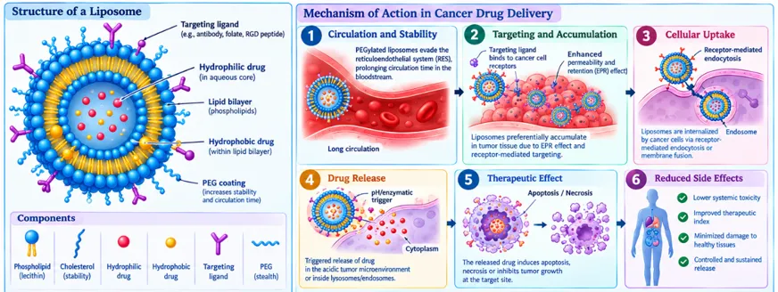
Fig 6.1 Liposomes
6.2. Extracellular vesicles
Because of their distinct biological makeup and effective cellular absorption, extracellular vesicles (EVs) present exceptional potential for drug delivery. Their surfaces can be altered to specifically target tumor regions[36]. Because of their innate capacity to pass through the blood-brain barrier, they are especially attractive for treating brain cancer[37]. EVs can also carry a variety of bioactive compounds that are helpful for ongoing disease evaluation and early cancer detection[38]. They are great options for immunotherapy and personalized medicine due to their natural origin and capacity for cell communication[39].
Examples
1. Exosome doxorubicin
Mechanism
Exosome-doxorubicin DoxorubicinDNA damage and intracellular release of doxorubicin after EV-mediated absorption in breast, lung, and other malignancies. Following intracellular release, doxorubicin can damage DNA and trigger apoptotic pathways by entering the nucleus, intercalating with DNA, and inhibiting topoisomerase II. Thus, doxorubicin-loaded EV → cancer-cell uptake → intracellular drug release → nuclear localization → DNA damage/topoisomerase-II inhibition → death can be used to illustrate the mechanism.
2. Exosome paclitaxel
Mechanism:
Exosome-paclitaxel Paclitaxel is delivered to tumor cells, stabilizes microtubules, and inhibits cell proliferation in breast, ovarian, and other malignancies.
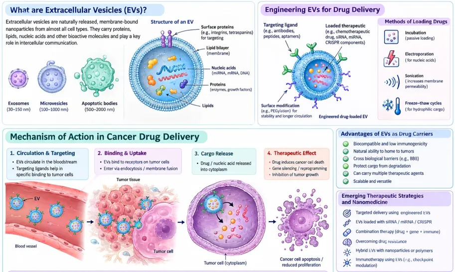
Fig 6.2 Extracellular Vesicles
6.3. Nano emulsion
Medication is delivered to tumor sites using nanoemulsions, which are nanoscale emulsions. They can be made with oils that are generally recognized as safe (GRAS), reducing adverse effects[40,41].Their tiny droplet size increases the solubility and effectiveness of hydrophobic agents, improving results and lowering dosages[42]. Additionally, nanoemulsions can be customized to precisely target particular tumor cells, ensuring accurate therapeutic agent administration[43]. Multi-drug-resistant (MDR) tumors are a major barrier to cancer chemotherapy and a significant cause of cancer-related mortality. Several anticancer drugs encapsulated in nanoemulsions have been studied for their potential to overcome MDR. Ganta et al. Developed a novel theranostic nanomedicine using a clinically relevant GRAS-grade nanoemulsion to bypass MDR processes. Tested on ovarian cancer cell lines, MRI tracked tumor accumulation, and the study showed that the nanoemulsion outperformed docetaxel alone by increasing cytotoxicity and overcoming taxane resistance through receptor-mediated uptake[44].
Examples:
Use: Ovarian, breast, and other malignancies
Paclitaxel has a low solubility in water and is quite hydrophobic. Its dispersion can be improved and delivery made easier by adding it to a nanoemulsion.
Mechanism:
Paclitaxel-loaded nanoemulsion → absorption by cancer cells → release of paclitaxel → binding to β-tubulin → stabilization of microtubules → suppression of microtubule depolymerization → mitotic arrest → apoptotic pathway activation → death of cancer cells.
Therefore, whereas paclitaxel generates the pharmacological anticancer action, the nanoemulsion mainly enhances transport and bioavailability.
Curcumin is a medication used in experiments to treat pancreatic, colon, breast, and other malignancies.
Curcumin has a limited bioavailability and poor water solubility. Its apparent solubility and interaction with biological membranes can both be enhanced via nanoemulsification.
Mechanism
Curcumin nanoemulsion → higher cellular absorption → intracellular curcumin release → regulation of survival signaling pathways like PI3K/Akt and NF-κB → decreased proliferation and increased apoptosis → reduction of tumor-cell development.
Rather than being proven clinical cancer medications, curcumin nanoemulsions are mostly studied in preclinical cancer research.
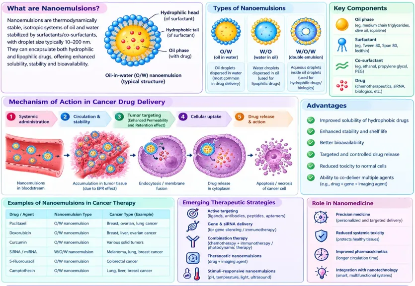
Fig 6.3 Nanoemulsions
6.4. Inorganic Nanoparticles
Inorganic nanoparticles, such as gold and silver nanoparticles, carbon nanotubes, carbon quantum dots, metallic oxide fragments, and mesoporous silica nanoparticles, are widely used in medical applications due to their precise targeting, favorable pharmacokinetics, and diagnostic functions. Their unique optical and magnetic properties, ease of functionalization, and inherent stability make them highly valuable[45]. Similar to this, drugs can be encapsulated in mesoporous silica nanoparticles and released in response to particular stimuli, improving targeted drug delivery and lowering systemic toxicity[46]. Furthermore, Pugazhendhi et al. Offered a thorough analysis of the function of inorganic nanoparticles in pharmacotherapy, examining methods for creating efficient drug delivery systems and contrasting the advantages and disadvantages of conventional chemotherapy versus nanotherapy for enhancing cancer treatment[47].
Examples
Gold nanoparticles loaded with doxorubicin and AuNPs functionalized with ligands or antibodies are two examples.
Mechanism
Targeting compounds and anticancer medications can be used to functionalize gold nanoparticles. Receptor-mediated absorption can raise the concentration of intracellular nanoparticles once they have entered cancer cells. After that, the medication is released and has its typical cytotoxic effect.
Additionally, AuNPs can function as photothermal therapeutic agents. Gold nanostructures absorb near-infrared (NIR) light, which results in localized heat. The ensuing rise in temperature has the potential to harm cellular membranes and proteins, interfere with mitochondria, and trigger apoptosis.
Mechanism;
AuNPs + targeting ligand → attachment to cancer cells → endocytosis → release of medication
either AuNP → NIR irradiation → light absorption → heat production → damage to proteins and membranes → apoptosis → tumor destruction
Mechanism
Drug: injected into silica pores; nanoparticle arrives at the tumor; cellular absorption; acidic endosomal/lysosomal environment; stimulus-responsive drug release; intracellular drug activity; apoptosis.
For instance, after internalization, MSNs laden with doxorubicin may release doxorubicin. After intercalating into DNA, doxorubicin inhibits topoisomerase II, causing DNA damage and apoptosis.
Additionally, "gatekeeper" molecules that stay closed under normal circumstances but open in response to low pH, enzymes, light, or other stimuli can be designed into MSNs to provide regulated drug release.
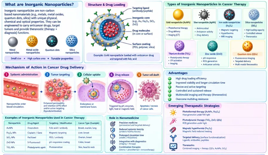
Fig 6.4 Inorganic Nanoparticles
6.5. Solid lipid Nanoparticles
Solid lipid nanoparticles (SLNs) offer flexibility for various administration routes, including intravenous, oral, local, and topical, by using solid lipids in place of liquid lipids in a colloidal drug delivery system[48]. SLNs provide controlled drug release, systemic toxicity is decreased and dosing frequency is minimized[49]. They can also be designed to deliver drugs to particular cancer cells, improving the accuracy of drug delivery[50].
The intrinsic and acquired resistance of cisplatin, a commonly used chemotherapeutic medication, as well as its serious side effects, present a noteworthy challenge. Raut et al. Showed that by increasing cisplatin’s effectiveness and lowering its toxicity, SLN encapsulation successfully resolves these problems[51]
Examples
Medication: Methotrexate
Cancer: Leukemia and solid tumors were examined.
SLNs loaded with methotrexate have been studied as a way to enhance drug delivery and regulate release.
Mechanism
Methotrexate-SLN → cellular absorption → methotrexate release → dihydrofolate reductase (DHFR) inhibition → decreased tetrahydrofolate availability → defective nucleotide synthesis → DNA replication inhibition → cancer-cell growth inhibition/apoptosis.
Medication: 5-Fluorouracil (5-FU)
Skin, colon, and other types of cancer, to enhance localization and offer controlled release, 5-FU can be added to lipid nanoparticle systems.
Mechanism
5-FU-SLN → drug release → production of active metabolites → thymidylate synthase inhibition and disruption of RNA/DNA synthesis → reduced proliferation of cancer cells → cell death.
SLNs have also been studied for topical and localized distribution, where the carrier may affect drug retention and penetration.
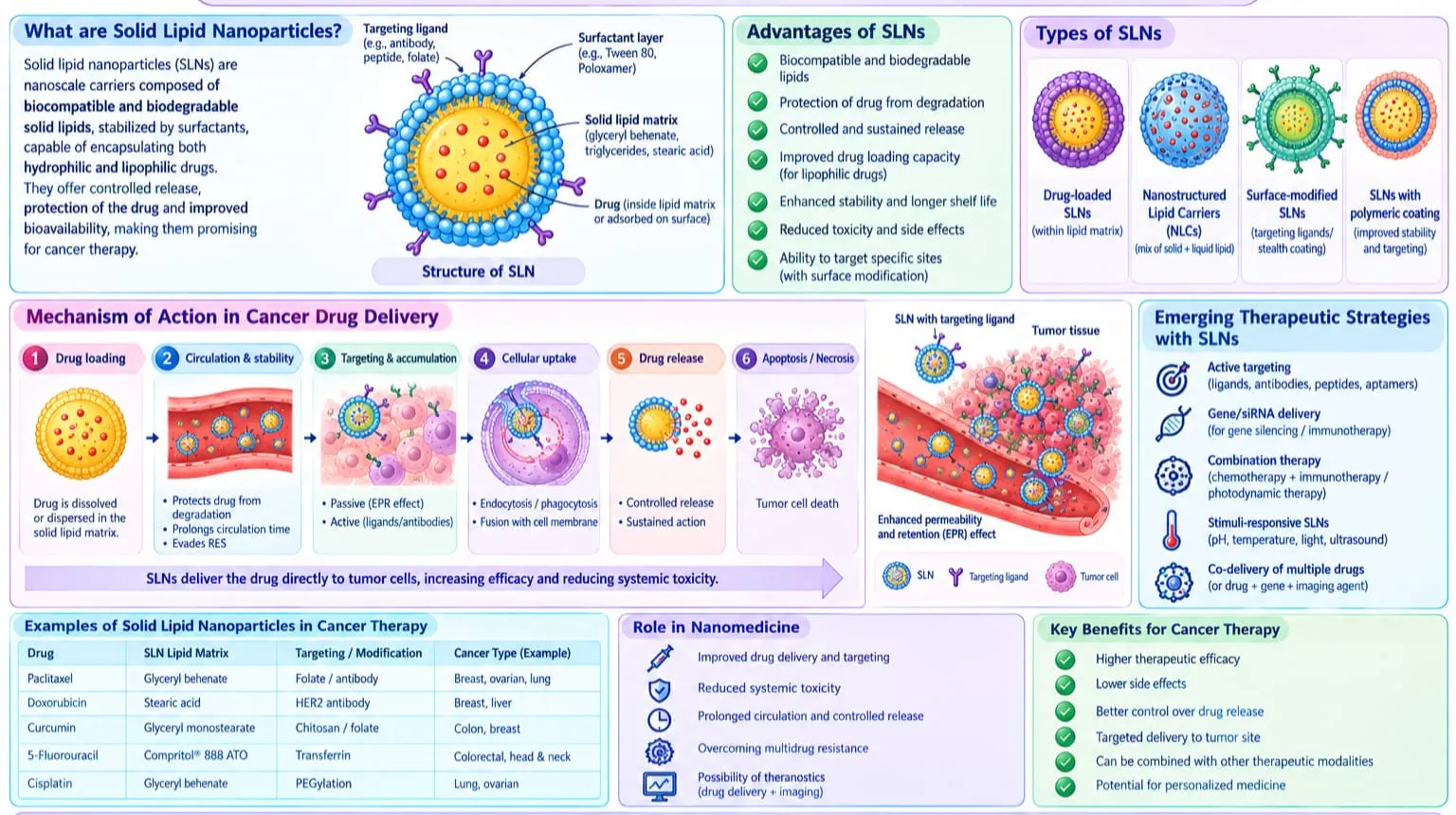
Fig 6.5 Solid Lipid Nanoparticles
6.6. Polymeric Nanoparticles
Polymeric nanoparticles are becoming more and more popular in cancer treatment because of their capacity to enhance patient care by releasing therapeutic agents precisely and under control. By maintaining ideal drug concentrations at tumor sites, these nanoparticles can improve the effectiveness of treatment[52].Their biocompatibility and capacity to encapsulate both hydrophilic and hydrophobic medications make them ideal for a variety of anticancer treatments[53].Furthermore, precise drug delivery to cancer cells can be accomplished by functionalizing polymeric nanoparticles with targeting ligands[54].The therapeutic potential of polymeric nanoparticles can be further increased by combining them with other materials. For example, they can be created by combining organic substances like lipids, proteins, and phospholipids with inorganic elements like carbon nanotubes, polymers, silica, metal oxide nanoparticles, and graphene. Compared to conventional drug formulations, this hybrid approach offers several advantages. In their review of the creation of polymer-hybrid nanoparticles for biomedical applications, Ferreira Soares et al. Emphasized state-of-the-art synthesis methods and their applications in both in vitro and in vivo settings[55].
Examples
For instance, folic acid-functionalized PLGA nanoparticles containing paclitaxel or doxorubicin. The folate receptor is overexpressed in certain cancer cells. In order to promote receptor-mediated uptake, folic acid can be affixed to the surface of the nanoparticle.
Mechanism
Folic acid-PNP leads to folate receptor binding, receptor-mediated endocytosis, intracellular nanoparticle trafficking, drug release, anticancer activity, and apoptosis.
Instead than depending only on passive tumor accumulation, this is an example of active targeting.
Medication: Cisplatin, PLGA and other polymeric systems
Cancer: Testicular, ovarian, lung, and other cancers
The use of polymeric nanoparticles as cisplatin and similar platinum agent carriers has been studied.
Mechanism
Cisplatin nanoparticle → cellular absorption → regulated cisplatin release → aquation/activation → platinum-DNA adduct formation → DNA cross-linking → transcription/replication inhibition → DNA damage → apoptosis
Modifying distribution and possibly increasing tumor exposure while lowering nonspecific toxicity is the aim of nanoparticle administration.
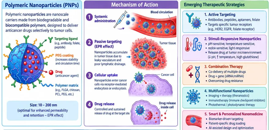
Fig 6.6 Polymeric Nanoparticles
6.7. Quantum Dots
The remarkable optical and electronic characteristics of quantum dots (QDs), semiconductor nanoparticles that range in size from two to ten nanometers, are essential for improving cancer diagnosis and treatment. Highly sensitive imaging methods made possible by QDs make it easier to detect and track the growth of tumors early on[56].Furthermore, QDs can be used as carriers to precisely deliver therapeutics to cancer sites, greatly reducing the impact on nearby healthy tissues[57].In photodynamic therapy, QDs selectively kill cancer cells by producing reactive oxygen species when activated by light[58].
Examples
Carbon quantum dots are carbon-based fluorescent nanoparticles that have garnered a lot of attention because, in comparison to some heavy-metal-containing QDs, they may have better biocompatibility and desirable optical properties, depending on their surface chemistry and formulation.
Targeting compounds or anticancer medications can be used to functionalize them.
Mechanism
CQD-drug → intracellular drug release → drug-specific molecular action → apoptosis → cancer-cell uptake.
Additionally, some CQDs have photochemical characteristics that can be used to produce reactive oxygen species when exposed to light.
To deliver siRNA into cancer cells, QDs can be mixed with polymers or other carriers.
Mechanism
The QD-siRNA combination leads to tumor cell uptake, intracellular siRNA release, RISC activation, complementary target mRNA destruction, reduced expression of oncogenic proteins, reduction of proliferation and survival, and ultimately cancer cell death.
Cellular uptake and distribution can be monitored using QDs' fluorescence characteristics.
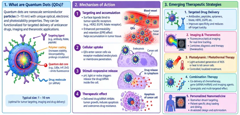
Fig 6.7 Quantum Dots
Cancer Nanomedicine
By combining pharmaceuticals with nanoscale technology, cancer nanomedicine uses nanotechnology to detect, track, and treat cancer. By maintaining targeted treatment dosages, these developments can be incorporated into biological machinery, nanobiosensors, and medical equipment, greatly improving therapeutic outcomes in complex and deadly diseases[59].Additionally, a number of additional nanomedicine formulations that demonstrate the variety in composition, mechanisms of action, and therapeutic targets have been approved. Hensify (NBTXR3)[60].Drug delivery to cancer sites can be significantly improved by ligand-coated liposomes and nanoparticles. For example, doxorubicin-loaded liposomes and a blood clotting factor XIII analog increased drug accumulation at tumor sites by 40 times[61].The focus of recent developments has been on nanoparticles that can transport both therapeutic and diagnostic agents. These nanomedicines are designed to overcome the shortcomings of traditional cancer therapies. Theranostic liposomes coated with RGD-TPGS (arginine-glycine-aspartic acid peptide with D-α-tocopheryl polyethylene glycol 1000 succinate) and loaded with docetaxel and QDs are a prime example. A significant decrease in the production of reactive oxygen species without causing edema or brain damage indicates that these liposomes are much more effective than conventional treatments[62].Furthermore, formulations of nanoparticles such as Abraxane[63].
Promising role of Nanomedicine in tumor Therapies
Nanomaterials’ exceptional qualities and potential have propelled them to the forefront of tumor therapeutics research. Numerous nanomaterials are showing promise in revolutionizing cancer treatment, such as liposomes, solid lipid nanoparticles (SLNs), polymeric micelles (PMs), dendrimers, mesoporous silica nanoparticles (MSNs), quantum dots (QDs), carbon nanotubes (CNTs), and metallic/magnetic NPs. These intriguing nanomaterials offer better penetration into tumor tissues, enhanced stability, controlled release, and targeted drug delivery. They also support personalized medicine techniques, photothermal therapy, and multimodal imaging. As a result, nanomedicine presents an exciting opportunity to advance the development of tumor therapeutics and solve the challenges posed by traditional cancer treatments. The specific functions of nanomaterials in various aspects of cancer treatment, such as surgery, chemotherapy, immunotherapy, radiotherapy, and more, will be covered in the sections that follow, highlighting their important role in improving patient outcomes and transforming cancer care.
Surgery
Nanomaterials provide a useful tool for effective and quick hemostasis, improving safety during surgical procedures, in contrast to conventional surgery, which frequently involves removing diseased tissues and possibly damaging nearby healthy ones[64].
Carbon nanotubes and chitosan derivatives were successfully combined to create a nanocomposite porous gel that showed remarkable hemostatic performance on deep traumatic bleeding. Furthermore, surgical efficacy is seriously hampered by microscopic tumors, which are frequently invisible to the unaided eye. However, sentinel lymph nodes can be mapped, residual tumor cells and micrometastases can be efficiently labeled, and tumor margins and surrounding critical structures can be illuminated with the help of nanotechnology. This offers useful guidance for tumor removal and significantly increases detection resolution[65]. created CLIO-Cy5.5, a dual-purpose NP that can be used as a near-infrared fluorescence optical probe and an MRI contrast agent. It has shown promise in identifying the boundaries of orthotopic malignancies in hosts. In addition to these advantages, NPs have been used to increase the effectiveness of cryosurgery by controlling the freezing range, improving the distribution of freezing temperatures, and directing the production of ice balls, all of which lessen the possibility of injury to nearby healthy tissues [66].
Chemotherapy:
Although conventional chemotherapy is frequently used, it can cause discomfort for patients because it is not tumor-specific and can harm healthy tissue. Additionally, because of its quick elimination from the body, it requires frequent administration and might lead to drug resistance, which lowers patient survival rates [67]. Nonetheless, NPs can be designed to specifically bind to cancer cells and accumulate at the tumor location, producing a very effective, low-toxicity therapeutic strategy that can get beyond tumor drug resistance. NPs can accomplish targeted drug delivery and localized drug release by taking use of TME properties as pH, redox gradients, enzyme activity, hypoxia, or particular cell surface receptors [68]. To address the shortcomings of non-specific chemotherapeutic medicines, for example, lactic acid-modified mesoporous silica nanoparticles have shown selective binding to desialylated glycoprotein receptors on hepatocellular carcinoma cells. In order to maximize treatment success, nanocarriers may also carry several medicines at once, regardless of their characteristics. As an example, Dai et al. [67]. developed a method for co-delivering vincristine and paclitaxel to brain tumors. Therapeutic results are frequently improved by these integrated approaches, which include vincristine's complex processes (such as apoptosis induction, anti-angiogenesis, and TME regulation).
Immunotherapy
In spite of the capabilities of cancer immunotherapies, the response rate is limited, with around 10% of patients showing considerable progress. The fusion of nanotechnology and immunotherapy could yield groundbreaking methods to address this challenge. NPs have proven their potential to boost tumor immunotherapy by enhancing the performance of tumor vaccines and modifying the tumor microenvironment to elicit anti-tumor immune responses. As an illustration, nano vaccines utilizing nanoparticles allow for the concurrent administration of antigens and adjuvants, thereby mitigating the shortcomings of standard cancer vaccinations. Importantly, the introduction of nanoparticles to antigen-presenting cells (APCs) has augmented antigen intake and triggered APC activation. Additionally, innovative nanoparticle-based treatments, which employ metals such as iron, zinc, and copper, have exhibited potential in the realm of tumor immunotherapy. As an illustration, iron nanoparticles can activate apoptosis in neoplastic cells through the Fenton process.
For instance, iron nanoparticles can stimulate a strong adaptive immune response by inducing death in tumor cells via Fenton reactions. It has been discovered that zinc nanoparticles cause immunogenic cell death, which stimulates the immune system.
Copper nanoparticles have been used as adjuvants in cancer vaccines to enhance antigen presentation and dendritic cell maturation. Additionally, by co-delivering PD-1/PD-L1 inhibitors with immune stimulants or chemotherapeutic medications, nanomedicine can serve as a platform for combination therapy, providing a synergistic strategy to address tumor immune evasion. Thus, combining immunotherapy and nanomedicine could provide novel and efficient ways to boost the body's immune response against malignancies.
Radiotherapy
High-energy X-rays are used in radiotherapy, a common clinical cancer treatment, to kill rapidly proliferating cancer cells and stop tumor development [69]. However, the efficiency of the treatment is compromised since solid tumors are two to three times more radioresistant than normal tissues due to their hypoxic nature caused by an inadequate oxygen supply. Moreover, weak radiation is delivered to tumor cells that are far from the radiation site [70].
In order to increase tumor oxidation, Liu et al. created tantalum oxide nanoparticles (NPs) with adsorbed perfluorocarbon. The NPs concentrated radiation energy on the tumor, while the adsorbed perfluorocarbon continually produced oxygen. NPs have potential as radiosensitizers as well. Swanner et al., for instance [71]. showed how silver nanoparticles (AgNPs) significantly cytotoxically and radiosensitively affected triple-negative breast cancer cells both in vivo and in vitro. It has been demonstrated that AgNPs cause more DNA damage [72].
Photodynamic and Photothermal Therapy
Two cutting-edge light-based cancer treatment approaches are photodynamic therapy (PDT) and photothermal therapy (PTT). PDT uses light-activated photosensitizers (PSs) to create harmful reactive oxygen species (ROS) that destroy tumor cells [73]. For oxygen-enhanced PDT, Liang et al. created a TME-responsive AuNC@MnO2(AM) nanoplatform that successfully suppresses tumor growth and spread in metastatic breast cancer. In a similar vein, a TME-responsive nanoplatform has shown promise in treating PDT tumor hypoxia [74]. PTT, on the other hand, works by taking advantage of tumor cells' high sensitivity to heat and turning light energy into heat, which results in intratumoral damage [75]. This method makes use of a range of near-infrared (NIR) nanomaterials, such as carbon nanotubes and gold nanorods [76]. Heat shock proteins (HSPs) cause thermal tolerance in tumor cells, which is a drawback of PTT and calls for the creation of more effective NIR-absorbing materials [77]. Both fields have seen exciting developments. For example, a porphyrin-based micelle has shown promise for improved chemosensitivity under NIR irradiation and dual-drug delivery [78].
Gene therapy:
Gene therapy uses techniques like gene overexpression and RNA interference (RNAi) to modify or correct abnormal genes [79]. Genetic material can be delivered to cells using a variety of viral and non-viral vectors. However, because of their extreme toxicity and immunogenicity, many viral gene carriers are restricted [80]. For gene delivery, safer substitutes include nanocarriers including gold nanoparticles (GNs), polymer NPs, and lipid NPs. These NPs improve medication distribution, carry nucleic acid molecules effectively, and protect them from deterioration.
For instance, in prostate cancer models, systemic administration of PTEN mRNA using polymer–lipid hybrid NPs slowed tumor growth [81]. Similarly, overexpressed oncogenes can be silenced by RNA interference. Because of its low cytotoxicity and high gene transfection effectiveness, Wu et al. [82] proposed PEI-PEG as a viable non-viral carrier for the treatment of gastric cancer. By creating a dual receptor-mediated siRNA delivery system for efficient, low-toxicity siRNA administration, Ju et al. [83] successfully stopped tumor growth and achieved targeted gene silencing.
Diagnosis and Imaging
Nanotechnology opens up new possibilities for biomedical imaging by using NPs like QDs, liposomes, and AuNPs to create multifunctional, tumor-specific devices. By combining with optical, magnetic, and acoustic imaging technologies, these NPs improve multimodal imaging and offer thorough diagnostic data. Because of their special optical characteristics and capacity to increase the specificity of cancer detection, QDs stand out as excellent optical imaging nanoprobes. The selection of successful targeted therapy is guided by the detection of liposome accumulation within tumor tissues. Because of their tiny size and biocompatibility, AuNPs are excellent contrast agents that allow for the monitoring of tumor cell proliferation under X-ray irradiation and the detection of early-stage malignancies. Additionally, NPs encapsulated with iodine, a CT contrast agent, minimize iodine cytotoxicity while producing better imaging results.
Nanoparticle-enhanced CT imaging and SPECT/CT show great promise for early cancer diagnosis, with encouraging results in melanoma and breast cancer models [84].
CONCLUSION
Nanomedicine has deep learning can significantly aid clinicans in cancer diagnosis and research by selecting the best nanomedicine.
Our review includes various Nanoparticles types showing the potential and application through nanomedicine.
Drug delivery has advanced significantly with the introduction of nanomedicines. The list of possible uses for nanoparticles is constantly expanding, and the options for their design and function are so diverse that the drug delivery system can be customized to best fit the chosen medication.
A more promising approach to treating drug resistance in cancer is made possible by the rapid advancement of nanotechnology and drug delivery systems.
The future of nanomedicine in cancer treatments is bright, despite obstacles like improving biocompatibility, controlling drug release, streamlining nano-design, and creating appropriate evaluation models.
By offering controlled platforms for drug protection, pharmacokinetic modulation, targeting, prolonged release, combination therapy, and theranostics, nanomedicine has revolutionized cancer drug-delivery research. While polymeric nanoparticles, SLNs, nanoemulsions, extracellular vesicles, inorganic nanoparticles, dendrimers, and quantum dots present unique prospects, liposomes have shown the best clinical maturity. Tumor heterogeneity, fluctuating EPR, biological barriers, protein-corona effects, immunological clearance, restricted tumor penetration, manufacturing complexity, and regulatory constraints are still significant challenges. Therefore, nanocarrier engineering should be combined with tumor biology, biomarker-driven patient selection, scalable manufacturing, stringent safety testing, and clinically significant endpoints in the next generation of cancer nanomedicine. Instead of focusing on complexity for its own purpose, a successful platform will offer a repeatable and observable improvement in patient outcomes.
Emerging future strategies
New Approaches for the Future Rather than relying solely on more complex nanoparticles, future cancer nanomedicine is anticipated to prioritize biologically informed and clinically measurable targeting. Research is now being conducted on biomimetic carriers, cell-membrane-coated nanoparticles, exosome–liposome hybrids, biodegradable inorganic materials, and programmable nucleic acid delivery systems. In addition to identifying patient or tumor characteristics linked to response, artificial intelligence and machine learning may aid in the optimization of formulation variables. Compared to traditional monolayer cultures, organ-on-a-chip and three-dimensional tumor models might offer better predictive testing. The therapeutic spectrum may be further expanded by combining nanomedicine with targeted therapy, immunotherapy, radiation, and gene editing. A therapeutically beneficial increase in the therapeutic index, rather than just better nanoparticle characterisation, should be the primary translational goal. In addition to mechanistic discoveries, future research should provide pharmacokinetics,biodistribution, toxicity, manufacturing repeatability, and therapeutically meaningful efficacy.
REFERENCES


Pranali Shinde, Sheetal Gosavi, Nisha Avhad, Sakshi Khedkar, Review on Cancer Drug Delivery Systems Emerging Therapeutic Strategies and Nanomedicine, Int. J. of Pharm. Sci., 2026, Vol 4, Issue 10, 1563-1586. https://doi.org/10.5281/zenodo.23330789
 10.5281/zenodo.23330789