

We use cookies to ensure our website works properly and to personalise your experience. Cookies policy
Ashokrao Mane Institute of Pharmacy, Ambap, Maharashtra, India.
Wound infections and delayed tissue repair remain significant challenges in healthcare management [1,3]. Herbal formulations are widely preferred due to their reduced side effects and better patient compliance [6]. Ocimum sanctum (Tulsi) possesses antimicrobial, antioxidant, anti-inflammatory, and wound healing activities.[4,5]Tulsi extract contains important phytoconstituents such as eugenol, ursolic acid, and flavonoids responsible for therapeutic activity. [7]Nanogel drug delivery systems improve topical retention, penetration, and controlled release of active constituents. [9] Nanogels prepared using suitable polymers and stabilizers show improved physicochemical stability.[53]Evaluation parameters including pH, viscosity, spreadability, homogeneity, and drug content are essential for topical nanogel characterization.[11]Antimicrobial studies against wound-infecting microorganisms are commonly performed using standard microbiological techniques [10]. In-vitro wound healing models help determine the tissue repair potential of herbal formulations.[5]Herbal nanogels have shown promising effectiveness in enhancing wound contraction and reducing microbial growth.[7,8] Tulsi-based nanogel formulations may serve as economical and effective alternatives to conventional wound healing therapies.[14]
The History of Ayurveda:
Origins and Early Development
Ayurveda, one of the world's oldest holistic healing systems, originated in India more than 5,000 years ago. [1] Its roots can be traced back to the Vedic period, with the earliest references found in the Rig Veda, a sacred text composed around 1500 BCE. [1] The term "Ayurveda" is derived from two Sanskrit words: "Ayur" meaning life, and "Veda," meaning knowledge or science, collectively translating to "the science of life".[1] Classical Texts.
The core principles of Ayurveda are detailed in several classical texts:
Philosophical Foundations
Ayurveda is deeply intertwined with Indian philosophy, particularly the Sankhya school of thought, which posits that the universe is composed of two fundamental entities: Purusha (consciousness) and Prakriti (matter). It also incorporates the concept of the five elements (Panchamahabhutas) - earth, water, fire, air, and ether - which form the basis of all matter and life.[7]
These elements combine to form three primary life forces or doshas: Vata (air and ether), Pitta (fire and water), and Kapha (earth and water). [7] The balance of these doshas within the body determines a person's health and constitution.[7]
Spread and Influence
Ayurveda flourished in ancient India and influenced medical practices in neighboring regions. It significantly impacted traditional medicine systems in countries such as China, Tibet, Sri Lanka, and Thailand. [8]The spread of Buddhism from India facilitated the dissemination of Ayurvedic knowledge across Asia. [8]
Decline and Revival
With the advent of Islamic rule in India and later British colonialism, Ayurveda experienced a period of decline. [9] The introduction of Western medicine marginalized traditional practices. However, in the late19th and early 20th centuries, there was a resurgence of interest in Ayurveda, spurred by the nationalist movement and a renewed appreciation for indigenous knowledge systems.[9]
Modern Era
In contemporary times, Ayurveda has gained global recognition. It is integrated into the healthcare systems of countries like India and Sri Lanka, where it is practiced alongside modern medicine. [10] Ayurvedic principles are also applied in various fields such as diet, lifestyle, and holistic wellness. [10] Institutions dedicated to Ayurvedic education and research continue to proliferate, ensuring the growth and adaptation of this ancient science to meet modern health challenges.[10]
Reason for using Nanogel formulation
Introduction to Nanogel Drug Delivery Systems
Nanogel drug delivery systems are advanced nanosized hydrogel formulations widely used in modern pharmaceutical and biomedical applications for controlled and targeted drug delivery. [1] Nanogels are three-dimensional cross-linked polymeric networks capable of absorbing large amounts of water or biological fluids while maintaining their structural integrity. [1] The particle size of nanogels generally ranges from 1 to 1000 nm, which enhances penetration of drugs through biological membranes and improves therapeutic effectiveness. [1] Nanogels combine the advantages of nanoparticles and hydrogels, resulting in improved drug loading capacity, stability, bioavailability, and controlled drug release. [2] Nanogel systems possess several desirable properties such as high-water content, flexibility, biocompatibility, biodegradability, and non-irritant nature. These properties make them highly suitable for topical and transdermal drug delivery applications.
Nanogels can encapsulate both hydrophilic and hydrophobic drugs.[4] and protect them from degradation caused by environmental conditions or enzymatic activity. Due to their nanosize structure, nanogels improve retention of drugs at the site of application and provide sustained release for prolonged therapeutic action. Nanogel formulations are easy to apply, non-greasy, and provide better patient compliance compared to conventional dosage forms. They are extensively used in wound healing, antimicrobial therapy, anti-inflammatory treatment, ophthalmic delivery, cosmetic preparations, and cancer therapy. In wound healing applications, nanogels maintain a moist environment at the wound site, enhance tissue regeneration, reduce microbial growth, and accelerate healing process. Herbal extract-loaded nanogels have gained significant attention because they combine the therapeutic benefits of medicinal plants with advanced nanotechnology-based drug delivery systems. Therefore, nanogel drug delivery systems represent a promising and innovative approach for development of effective and safe pharmaceutical formulations. [4]
Factors Affecting Nanogel System
Type of Polymer
Nature and concentration of polymer affect viscosity, swelling, and drug release.
Particle Size
Smaller particle size improves penetration and drug delivery efficiency.
Cross-linking Density
Higher cross-linking affects swelling behavior and drug release rate.
pH of Formulation pH influences stability, swelling, and compatibility with skin.
Drug Solubility
Solubility of drug affects entrapment efficiency and release pattern.
Temperature
Temperature may affect viscosity, stability, and gel structure.
Surfactant Concentration
Surfactants influence particle formation and stability of nanogel.
Viscosity.
Proper viscosity is necessary for spreadability and retention at application site.
Method of Preparation
Preparation technique affects particle size and uniformity.
Storage Conditions
Light, humidity, and temperature affect stability of nanogel formulation.
Swelling Capacity
Swelling behavior influences drug release and penetration.
Drug-Polymer Interaction
Interaction between drug and polymer affects entrapment efficiency and therapeutic activity
LITERATURE REVIEW
AIM
OBJECTIVES
PLANT PROFILE
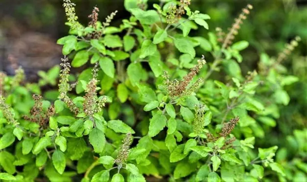
(Figure.1)
Introduction
Tulsi, scientifically known as Ocimum sanctum Linn. or Ocimum tenuiflorum, is one of the most important medicinal plants used in Ayurveda and traditional medicine systems. [21] It belongs to family Lamiaceae and is commonly known as Holy Basil. [21] Tulsi is considered a sacred plant in India and is worshipped in many households because of its medicinal and spiritual importance. [21] The plant possesses significant antimicrobial, antioxidant, antiinflammatory, analgesic, antipyretic, immunomodulatory, antidiabetic, and wound healing activities. Due to its wide range of pharmacological properties, Tulsi is extensively used in pharmaceutical, cosmetic, nutraceutical, and herbal formulations. [21]
2. Taxonomical Classification[22]
Category Classification
Kingdom - Plantae
Subkingdom - Tracheobionta
Division - Magnoliophyta
Class - Magnoliopsida
Subclass - Asteridae
Order - Lamiales
Family - Lamiaceae
Genus - Ocimum
Species - sanctum
3. Synonyms [23]
4. Vernacular Names [24]
Language
English - Holy Basil
Hindi - Tulsi
Marathi - Tulas
Sanskrit - Tulasi
Tamil - Thulasi
Telugu - Tulasi
Kannada - Tulasi
Malayalam - Tulasi
5. Biological Source
Tulsi consists of fresh and dried leaves and flowering tops of Ocimum sanctum Linn. [25] belonging to family Lamiaceae. [25]
6. Geographical Distribution
Tulsi is widely distributed throughout India and cultivated in tropical and subtropical regions of Asia, Africa, and America. [26] It grows well in warm climatic conditions with moderate rainfall. [26] The plant is commonly cultivated in household gardens, herbal farms, and medicinal plant cultivation areas. [26]
7. Cultivation and Collection
Tulsi is propagated mainly by seeds.
Seeds are sown during spring season. [27]
The plant requires fertile and well-drained soil. [27]
Adequate sunlight and moderate watering are necessary for proper growth. [27]
Leaves are collected before flowering stage for maximum phytoconstituent content. [27]
Shade drying is preferred to preserve volatile oils and active constituents.[27]
8. Macroscopic Characteristics
Upper and lower epidermis with stomata. [29]
Presence of multicellular covering trichomes. [29]
Glandular trichomes containing volatile oils. [29]
Collenchymatous cells beneath epidermis. [29]
Vascular bundles in midrib region. [29]
Oil glands distributed throughout leaf surface.[29]
Plant
Tulsi is an erect, aromatic, branched herb or undershrub growing up to 30–60 cm in height. [30]
Stem
Quadrangular in shape
Hairy surface
Green or purplish color
Aromatic odor
Leaves
Opposite arrangement
Simple and ovate shape
Serrated margins
Acute apex
Petiolate leaves
Characteristic aromatic smell
Green or purple coloration
Flowers
Small purple or white flowers
Arranged in elongated racemes
Bilabiate corolla
Fruits
Small nutlets containing seeds
Seeds
Reddish-black in color
Mucilaginous when soaked in water [28]
9. Microscopic Characteristics
Microscopic examination of Tulsi leaves shows:
Upper and lower epidermis with stomata. [29]
Presence of multicellular covering trichomes. [29]
Glandular trichomes containing volatile oils.
Collenchymatous cells beneath epidermis.
Vascular bundles in midrib region. [29]
Oil glands distributed throughout leaf surface.[29]
10. Powder Characteristics[30]
Powdered Tulsi leaves show:
11. Chemical Constituents
Tulsi contains several biologically active phytoconstituents responsible for its therapeutic activities
Volatile Oil Constituents
Eugenol -Antimicrobial, analgesic, anti-inflammatory, antiseptic activity
Ursolic acid- Wound healing, antioxidant, anti-inflammatory, anticancer activity
Rosmarinic acid - ntioxidant, antimicrobial, anti-inflammatory activity
Linalool - Antimicrobial, calming and stress-relieving activity
Carvacrol - Antibacterial, antifungal, preservative activity
Caryophyllene - Anti-inflammatory and analgesic activity
Flavonoids - Antioxidant, free radical scavenging, tissue protective activity
Tannins - Astringent, antimicrobial, wound healing activity
Saponins - Immune boosting, antimicrobial, healing activity
Phenolic compounds - Antioxidant and antimicrobial activity
Essential oils - Antiseptic, antimicrobial, preservative activity
Orientin - Antioxidant and radioprotective activity
Vicenin - Antioxidant and anti-inflammatory activity
Oleanolic acid - Hepatoprotective and anti-inflammatory activity
Methyl eugenol -Antimicrobial and aromatic activity
Glycosides - Therapeutic and metabolic activity
Alkaloids - Pharmacological and antimicrobial activity
Caffeic acid -Antioxidant and anti-inflammatory activity [31,32]
13. Pharmacological Activities
13.1 Antimicrobial Activity
Tulsi exhibits broad-spectrum antimicrobial activity against bacteria, fungi, and viruses. [33] Essential oils and eugenol disrupt microbial cell membranes and inhibit growth of pathogens responsible for wound infections. [33]
13.2 Anti-inflammatory Activity
Tulsi inhibits inflammatory mediators such as prostaglandins and cyclooxygenase enzymes, thereby reducing swelling and inflammation. [34]
13.3 Antioxidant Activity
Flavonoids and phenolic compounds present in Tulsi neutralize free radicals and reduce oxidative stress. [35]Antioxidant activity protects tissues from cellular damage. [35]
13.4 Wound Healing Activity
Tulsi accelerates wound contraction, collagen synthesis, angiogenesis, and tissue regeneration. [36] Antimicrobial and antioxidant activities further support wound healing process. [36]
13.5 Analgesic Activity
Tulsi helps reduce pain and discomfort by inhibiting inflammatory pathways and pain mediators. [37]
13.6 Antipyretic Activity
Tulsi reduces fever by acting on temperature-regulating centers and improving immune response. [38]
13.7 Immunomodulatory Activity
Tulsi stimulates immune cells and enhances body defense mechanisms against infections and diseases. [39]
13.8 Antidiabetic Activity
Tulsi helps regulate blood glucose levels and improves carbohydrate metabolism. [40]
13.9 Adaptogenic Activity
Tulsi acts as an adaptogen and helps the body cope with physical and mental stress. [41]
14. Therapeutic Uses [42]
15. Uses in Pharmaceutical Formulations [43]
16. Advantages of Tulsi in Nanogel Formulation [44]
MATERIALS AND INSTRUMENTS
Materials Used
(Table.1)
|
Sr. No. |
Material/ Chemical |
Quantity |
Category/ Use |
|
1 |
Tulsi (Ocimum sanctum) aqueous extract |
6gm |
Plant material |
|
2 |
Carbopol 940 |
0.72gm |
Gelling agent |
|
3 |
Tween 80 |
7.2gm |
Surfactant |
|
4 |
Pure honey |
3gm |
Co-solvent |
|
5 |
Olive oil |
3gm |
Oil phase |
|
6 |
Distilled water |
Sufficient quantity |
Vehicle |
Instrument
(Table.2)
|
Sr. No. |
Instrument |
Use |
|
1 |
Soxhlet apparatus |
Extraction of Tulsi leaves |
|
2 |
Magnetic stirrer |
Continuous stirring and mixing |
|
3 |
Digital weighing balance |
Accurate weighing of materials |
|
4 |
Brookfield viscometer |
Measurement of viscosity |
|
5 |
Beaker |
Preparation of formulation |
|
6 |
Measuring cylinder |
Measurement of liquids |
|
7 |
Funnel |
Filtration |
|
8 |
Glass rod |
Stirring |
|
9 |
Centrifuge |
Separation of particles |
|
10 |
Micropipette |
Transfer of small liquid volumes |
EXPERIMENTAL METHODOLOGY
1.Collection of Plant Material
Fresh leaves of Ocimum sanctum were collected from local area and the collected leaves were washed thoroughly with distilled water to remove dirt and foreign matter. [25]
2. Drying and Powdering
The cleaned leaves were shade dried at room temperature for 7–10 days to preserve active phytoconstituents.[25] Dried leaves were powdered using mechanical grinder and passed through sieve to obtain uniform powder.[25]
3. Preparation of Tulsi Extract
Soxhlet Extraction Method
About 50 g of dried Tulsi leaf powder was packed in Soxhlet apparatus. [25] Ethanol (500 mL) was used as extraction solvent. [25] Extraction was carried out for 6–8 hours until complete extraction occurred. [25] The extract obtained was filtered using filter paper. [25] Filtrate was concentrated using water bath to obtain semisolid extract. [25] The extract was stored in airtight container for further use.
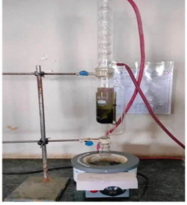
(Figure.2)
5. Procedure for Preparation of Nanogel
Step 1: Preparation of Gel Base
Required quantity of Carbopol 940 was dispersed slowly in distilled water with continuous stirring using magnetic stirrer. [26] The dispersion was allowed to hydrate properly for 1–2 hours.
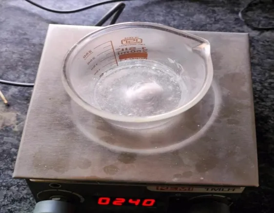
(Figure.3)
Step 2: Preparation of Extract Solution
Tulsi extract was dissolved separately in small quantity of ethanol or distilled water. [26]
Tween 80 was added to improve dispersion and solubility.
Step 3: Incorporation of Extract
The extract solution was added slowly into hydrated Carbopol gel base under continuous stirring. [26]
Methyl paraben was added as preservative.
Step 4: pH Adjustment
Triethanolamine was added dropwise to adjust pH and obtain gel consistency. [26]
Stirring was continued until homogeneous nanogel was formed. [26]
Step 5: Storage
Prepared nanogel was transferred into airtight containers and stored at room temperature for further evaluation.[26]
Evaluation of Nanogel Formulation
Appearance – Smooth, clear/translucent, homogeneous gel with no visible lumps, grittiness, or phase separation. The formulation should possess a uniform texture indicating proper mixing and stability of all ingredients. Absence of air bubbles and particulate matter confirms good formulation quality and aesthetic acceptability.
Color and Odor – Light green to brownish-green in color with a characteristic pleasant odor of Tulsi. The natural color indicates the presence of phytoconstituents in the extract, while the pleasant herbal odor enhances patient acceptability. Any significant change in color or odor during storage may indicate instability or degradation of active constituents.
pH Determination – The pH of the nanogel should be maintained within the range of 5.5–7.0, which is compatible with the normal skin pH and minimizes the risk of irritation or sensitization. [29] Stable pH indicates good formulation stability. [26]
Viscosity – The nanogel should exhibit moderate viscosity in the range of 5000–20000 cps, ensuring easy application, good consistency, and adequate retention on the skin surface. Viscosity is determined using a Brookfield viscometer at controlled temperature and rpm. Proper viscosity improves spreadability and drug release characteristics.
Spreadability – The formulation should possess good spreadability, generally in the range of 5–7 cm within 1 minute under standard applied weight, indicating ease of application on the skin with minimal friction. Good spreadability ensures uniform distribution of the gel over the affected area and improves patient compliance.
Homogeneity – The prepared nanogel should show excellent homogeneity without the presence of aggregates or coarse particles. Uniform distribution of drug and excipients throughout the formulation ensures consistent therapeutic effect and appearance.
Extrudability – The gel should exhibit satisfactory extrudability from collapsible tubes or containers with slight pressure. Good extrudability ensures convenient handling and accurate dose application by the user.
Drug Content Uniformity – The formulation should contain uniform distribution of the active constituents throughout the gel matrix. Drug content analysis helps to confirm formulation accuracy and reproducibility.
Stability Study – The prepared nanogel should remain physically and chemically stable during storage conditions without significant changes in color, odor, pH, viscosity, or phase separation. Stability studies are generally carried out at room temperature and accelerated conditions for a specified period.
Skin Irritation Test – The formulation should be non-irritant and safe for topical application. The absence of redness, itching, edema, or inflammation after application indicates good skin compatibility of the Tulsi nanogel formulation.
Antimicrobial activity - Agar Well Diffusion Method. Nutrient agar plates were prepared and inoculated with selected microorganisms.[30] Wells were made in agar plates using sterile borer.[30] Nanogel formulation was introduced into wells.[30]Plates were incubated at 37°C for 24 hours. [30]Zone of inhibition was measured to determine antimicrobial activity. [30]
Test Organisms
• Staphylococcus aureus
RESULT
Observation Table
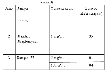
The antibacterial study against S. aureus demonstrates that the standard drug Streptomycin (1 mg/mL) exhibited a strong inhibitory effect with a zone of inhibition of 35 mm, confirming its high efficacy. In comparison, the Sample PP showed very weak antibacterial activity, with zones of inhibition of only 1 mm at 5 mg/mL and 4 mm at 10 mg/mL. Although there is a slight increase in activity with higher concentration, the effect remains minimal.
Overall, it can be concluded that Sample PP possesses negligible antibacterial activity against S. aureus and is significantly less effective than the standard antibiotic.
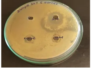
(Figure.4)
CONCLUSION
The present study was carried out to evaluate the antibacterial activity of Sample–PP against Staphylococcus aureus using the agar well diffusion method. [46] The antimicrobial potential of the sample was compared with the standard antibiotic Streptomycin. [46] The results obtained from the study clearly indicate a significant difference between the activity of the standard drug and the test sample. [46] Streptomycin, used as the standard reference drug at a concentration of 1 mg/mL, showed a prominent zone of inhibition of 35 mm against S. aureus. This large inhibition zone confirms the strong antibacterial efficacy of the standard antibiotic and validates the suitability of the experimental procedure and test conditions used in the study. In contrast, sample–PP exhibited only very weak antibacterial activity. [46] At a concentration of 5 mg/mL, the sample produced a zone of inhibition of only 1 mm, while at 10 mg/mL the inhibition zone slightly increased to 4 mm. The increase in inhibition zone with increase in concentration suggests that the antibacterial effect of the sample is concentration dependent. However, even at the higher concentration, the inhibitory effect remained extremely low when compared with the standard drug. The negligible antibacterial activity of Sample–PP may be due to the low presence or absence of potent antimicrobial phytoconstituents capable of inhibiting the growth of Staphylococcus aureus. It may also indicate poor diffusion of active constituents through the agar medium or insufficient concentration of bioactive compounds in the sample.
From the overall findings, it can be concluded that Sample–PP possesses minimal antibacterial activity against Staphylococcus aureus under the experimental conditions employed in this study. The sample was significantly less effective than Streptomycin and therefore cannot be considered a strong antibacterial agent against the tested microorganism. Further studies involving purification, isolation of active constituents, higher concentrations, or combination with other antimicrobial agents may be required to improve its antibacterial potential.
REFERENCES


Pankaj Patil, V. A. Mahajan, Formulation and Evaluation of Tulsi Extract Nanogel for Wound Healing, Int. J. of Pharm. Sci., 2026, Vol 4, Issue 8, 2660-2674. https://doi.org/10.5281/zenodo.21972584
 10.5281/zenodo.21972584