

We use cookies to ensure our website works properly and to personalise your experience. Cookies policy
1,2Assistant Professor, Department of Pharmaceutics JBVP’S Vidya Niketan Collage of Pharmacy, Lakhewadi, Tal- Indapur Dist- Pune (413103), Maharashtra, India.
3,4,5,6,7Students JBVP’S Vidya Niketan Collage of Pharmacy, Lakhewadi, Tal-Indapur, Dist- Pune (413103), Maharashtra, India.
Eye is one of the organs that is well protected; therefore giving medicine to the eye is difficult because of several natural barriers. These barriers include the change of tears blinking, tear production and drainage from the nose to the eye all of which take away most of the medicine that is put in the eye with drops (usually less than five percent) and the medicine stays only a few minutes. Because of this only a small amount of the medicine reaches the place in the eye is taken up by the eye tissue and the amount that actually works is low. This low working amount means that many doses are needed. A better way than eye drop is to use in-situ forming gels. In-situ forming gels stay liquid when they are applied and then they change into a gel after they meet a trigger such as a change in pH, temperature or salt level. When in-situ forming gels are formed in the eye in-situ forming gels stays there longer than drops do so the medicine stays inside the part of the eye for a longer time the amount that works increases the dose needed goes down and the eye gets better faster. This review looks at how in-situ ocular gels are made what natural barriers exist, how the gel is made and which polymers and helper substances are used. The review also talks about how the gel's made how its quality is checked how mathematical models help predict how the medicine leaves the gel and how these gels are useful for many types of medicine. The review also shows the bad sides of in-situ gels how they are regulated today and where new research could go. The review gives examples of gels that use polymers like Carbopol hydroxypropyl methylcellulose, gellan gum, sodium alginate, xanthan gum, ethylcellulose, chitosan and poloxamer. These polymers are used to make gels that respond to pH, salt or temperature. They aim to treat eye infections caused by bacteria and fungi. The medicines that are put in these gels include drugs such as ciprofloxacin, ofloxacin,
Topical ocular delivery is preferred for its -invasive nature; however the formulations are quickly cleared from the eye by blinking, lacrimal dilution and nasolacrimal drainage which limits precorneal residence time, drug absorption and bioavailability. Sustained release ocular systems offer an opportunity to prolong the contact time on the ocular surface and reduce the dosing frequency to enhance therapeutic efficacy and patient compliance. (1,2) Sustained-release ocular dosage forms are designed to prolong residence time and provide a state of drug concentration for an extended period on the eye surface by counteracting the short retention time on the cornea. (2)
Advanced ocular dosage forms such as inserts, polymeric films, nanoparticles and liposomes can be considered as novel approaches to the clearance and low bioavailability of conventional-eye drops. In-situ gelling systems are nanosuspension, microemulsions and in-situ gelling systems that have been developed to overcome the aforementioned of a gel undergoing a sol to gel transition on the ocular surface in response to physiological triggers because they combine the convenience of instilling a low viscosity liquid with the extended residence time such as pH, temperature or ionic composition of tears. (3,4)
In activated transparent gels are instilled as clear liquids that gel on the ocular surface avoiding blurred vision while prolonging precorneal residence. They provide bioadhesion, tolerability and sustained release over traditional eye drops and are used in the treatment of glaucoma dry eye Sjögren’s syndrome, age related macular degeneration (AMD) and trachoma. Low acyl gellan gum is preferred for its excellent ion triggered gelation and good mechanical and rheological- properties. (5)
Interest in this class of materials has grown exponentially over the two decades following the broader development of stimulus-responsive polymers. This literature review aims to highlight the trends and milestones in the field starting from the pH-triggered systems for antibiotic delivery and expanding to temperature ion or enzyme sensitive polymers that were used to carry antiglaucoma, antimicrobial and protein therapeutics. (7,8,9)
Natural and synthetic polymers are widely studied for developing in gelling drug delivery systems. The polymers such as pectin, gellan gum, chitosan, alginic acid, guar gum, carbopol, xyloglucan, xanthan gum, hydroxypropyl methylcellulose and poloxamers can be used for preparing ocular, transdermal, buccal, injectable, rectal and vaginal dosage forms. By virtue of their response to stimuli these polymers offer opportunities to improve drug absorption, prolong release increase bioavailability reduce dosing frequency and enhance patient adherence. (5)
For applications natural polymers such as gellan gum, sodium alginate, chitosan and xanthan gum contain functional groups that can increase the solution viscosity, mucoadhesive property and induce gel formation, which prolongs the precorneal residence time and improves the drug retention on the ocular surface with minimal impact on the ease of administration (4)
Fluoroquinolone antibiotics such as moxifloxacin and besifloxacin have gained tremendous interest in ocular application due to their broad antibacterial activity. Moxifloxacin and besifloxacin are mainly used in the treatment of conjunctivitis and keratitis. The conventional eye drop formulations of these drugs showed enhancement of residence time on the ocular surface; hence an alternative approach was needed to develop prolonged drug release formulations. In-situ gelling systems can be used in combination with these antibiotics to increase their retention on the surface. (3,4)
Nanotechnology has also been incorporated into this system of drug delivery. Nanoparticles can provide retention, better cellular interaction and controlled release of drugs which would result in better therapeutic efficacy. The combination of nanoparticles with an ions pH or temperature gel can be used as a multifunctional drug delivery system with enhanced targeting ability. The system not helps in achieving prolonged drug release but also increases the bioavailability due to increased retention at the target site. This would reduce the dosing frequency. Improve patient compliance. (3)
Conventional eye drops and suspensions suffer from ocular bioavailability due to rapid removal by tears, nasolacrimal drainage or short residence time of the formulations on the ocular surface. These drawbacks can be overcome by using in-situ gels due to their ability to undergo sol-gel transition on the surface in response to the change in pH, temperature and ions. The gel formed provides contact time of the formulation with the cornea which ultimately enhances drug absorption reduces dosing frequency and improves patient compliance. (6)
The development of an in-situ gel requires assessment of physicochemical properties such as appearance, pH, viscosity, gelation capacity, drug content, rheological properties and in vitro drug release to confirm the stability and efficacy of the formulation after administration (7)
Ophthalmic in-situ gel is an attractive dosage form for improving drug bioavailability and patient compliance by extending drug residence time on the ocular surface after instillation due to the sol-gel transition that occurs after administration, which prolongs residence time reduces loss of drug from the eye and provides an extended drug release rate. (7)
The conventional ocular dosage forms have reduced residence time on the eye due to tearing, nasolacrimal drainage and blinking. These factors limit the drug’s contact time with the tissues resulting in reduced drug absorption and necessitating frequent dosing or higher doses to obtain the desired therapeutic effect. (8)
Over the three decades research in the area of controlled release has been dominated by polymeric systems, among which in-situ gels have attracted considerable attention due to their potential clinical applications. Upon instillation these types of gels are primarily solutions that undergo a sol–gel transition in response to a change in pH, temperature or a solvent and enable controlled delivery of therapeutics with improved convenience over conventional injectable and oral routes particularly for biologics. A wide variety of natural and synthetic polymers have been explored since the 1970 s to generate these stimuli-responsive smart materials for drug delivery applications (10)
Conventional and novel ocular delivery systems face many challenges including corneal permeability, anatomical barrier, stability of the drug and formulation bioavailability of the therapeutic and tolerance of the formulation. Current and future development of these systems remains a priority if ocular therapeutics are to become more efficient and long lasting. Advanced systems such as in gels, nanoparticles, liposomes, nano-suspension, microemulsions and ocular inserts represent potential opportunities to improve drug availability and efficacy with prolonged residence time on the cornea. (11)
This review focuses on an overview of the development of in-situ gels, which includes gelation mechanism, types of stimulus-responsive gelling system, polymers and excipients, formulation strategies, physicochemical characterizations and evaluations therapeutic applications based on drug category comparison with other ocular dosage forms, formulation stability, regulatory aspects, advantages, limitations and recent developments with a special focus on the use of these systems in prolonging drug residence time and enhancing drug bioavailability and patient compliance with conventional ocular dosage forms. In short in-situ ocular gels improve retention by changing from liquid to viscoelastic systems in response to chemical and physical stimuli. (12)
Gels are semi two-phase systems consisting of a dispersed phase and a continuous phase. The dispersed phase is interconnected by a network structure that binds a majority phase forming a colloidal dispersion of inorganic or organic particles in a liquid medium. The binding sites are achieved by covalent bonds or other physical interactions such as hydrogen bonding and hydrophobic forces. USP describes gels as mixtures of divided substances in which the inorganic substance is suspended in a liquid solution, in which the organic matter is dissolved, swollen and entangled to form a colloidal dispersion. The rigidity of the gels depends on the nature of the dispersed substance and the type of bonds, which range from primary valence bonds to hydrogen bonds and Van der Waals forces that are broken down at the slightest heating (13)
In gels are low-viscosity solutions that form gels when triggered by physiological pH, temperature or ionic strength changes. In-situ gelling formulations prolong the precorneal contact time and residence period of drugs compared to eye drops resulting in improved ocular drug delivery. (17)
2. Anatomy of the Human Eye
The eye is an organ that lets you see light and make pictures of the world. The eye is almost round because air pushes it. The front part is a layer called the cornea. The eye also has built‑in guards: eyelashes, eyelids, blinking and tears. Eyelashes stop dust and other stuff from entering the eye. Blinking spreads tears over the cornea. Tears clean the eye and kill germs so the eye stays healthy. Those same guards keep medicines from reaching the eye surface. That makes the eye a tough place, for drug delivery. Generally the eye is usually split into two parts: the part and the back part. The front part holds layers focuses light lets light in circulates fluid in the front chamber and helps the eye adjust focus. The back part keeps the eye shape gives food to eye tissues spots light and sends nerve messages to the brain. (7)
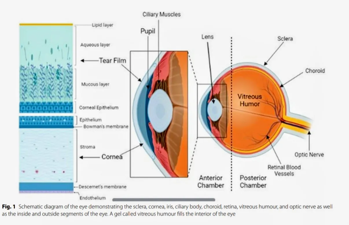
Figure 1: Structure of Eye (1)
2.1 Major structure of the eye (7)
| Eye segment | Structure | Primary role or significance |
|---|---|---|
| Anterior segment | Cornea | The front part of the eye is usually clear and shaped like a curve. The front part of the eye lets light come into the eye. The front part of the eye is what gives the eye its ability to focus. A thin layer of mucus that covers the part of the eye that's visible and also covers the inside of the eyelids. It works to keep the eye safe and to make it smooth and wet. |
| Anterior segment | Conjunctivitis | A thin layer of mucus that covers the part of the eye that's visible and also covers the inside of the eyelids. It works to keep the eye safe and to make it smooth and wet. |
| Anterior segment | Iris | The pigmented structure that controls the amount of light entering the eye. |
| Anterior segment | Pupil | The central opening of the iris is the doorway that lets light enter the eye. |
| Anterior segment | Ciliary body Structure | The lens changes its shape when you focus on things and makes a fluid called humor. |
| Eye segment | Structure | Primary role or significance |
| Anterior segment | Chamber of the anterior eye | The fluid filled space in between the cornea and the iris. |
| Anterior segment | Aqueous humor | The fluid that nourishes the cornea the lens and also maintains the pressure, inside the eye is called aqueous humor. |
| Anterior segment | Lens | A clear bendable part that helps to focus light on the retina. |
| Anterior segment | Trabecular meshwork | A type of tissue that lets the fluid from the eye flow out and keeps the pressure, inside the eye under control. |
| Posterior segment | Vitreous humor | A clear, gel like substance that helps to maintain the shape of the eyeball. |
| Posterior segment | Sclerae | The tough, white outer coat that provides protection and support for the eye. |
| Posterior segment | Retinal | A light sensitive membrane that contains specialized cells that convert light to electrical nerve impulses. |
| Posterior segment | Choroid coat | A vascular layer of tissue in between the retina and the sclera. It supplies oxygen and nutrients to the part of the retina that it is in. It is absolutely necessary for the health and function of the retina. In the absence of it the outer retina will not survive. It is a part of the eye that plays a role in vision. This layer is known as the choroid. |
| Posterior segment | Maculae | A part of the retina that gives you straight vision. |
| Posterior segment | Optic nerve | A group of nerve wires that sends messages from the eyes to the brain. |
Corneal Permeability is a factor that determines how much drug finally reaches the aqueous humour. The corneal epithelium is very tough for drugs and this mainly decides the speed and amount of transcorneal absorption. (9)
Beneath the epithelium lies the hydrophilic stroma. Its organised collagen framework limits how highly lipophilic compounds can spread. The conjunctiva by contrast is larger and more permeable. However its blood vessels and lymphatic channels can carry drug away into circulation, which reduces ocular bioavailability. (9)
Each of the eye structures has its own role. The conjunctiva. Lubricates. The sclera gives support. The choroid nourishes the retina and absorbs excess light. The retina converts light into signals. The vitreous humour. Supports the space between the lens and the retina. (8)
2.2 Barriers to Ocular Drug Delivery
Conjunctival Cul‑de‑Sac Capacity
The conjunctival cul‑de‑sac lies between the bulbar conjunctival. Its acts as a reservoir that keeps foreign material out and helps shape the upper eyelid crease. Its maximum volume is 30 µL. However after blinking the effective capacity falls to 70–80 % and it can fall even more during allergic reactions or swelling. This small volume dilutes medications, which lowers drug concentration and therapeutic effectiveness. (1)
Lacrimal Fluid and Drug Loss
Tear turnover and solution drainage are precorneal barriers that keep drugs out. Continuous tear fluid refreshment keeps the eye hydrated. Removes foreign agents but it also washes away administered medications. Protein binding and enzyme metabolism further block drug absorption. This means that formulation strategies must increase residence time to allow the drug to act. (1)
Corneal Barriers
The cornea protects the eye and guides light to the retina. It also creates several permeability challenges. The epithelial layer stops molecules and hydrophilic drugs. The hydrophilic stroma blocks medication from penetrating. The endothelium keeps the cornea clear. Only lets hydrophilic drugs and macromolecules enter the aqueous humour. Transcorneal drug penetration depends on ionization state, molecular mass, charge and hydrophobicity. (1)
Blood‑Ocular Barriers
The blood aqueous barrier (BAB) and blood retinal barrier (BRB) stop chemicals from reaching intraocular tissues. The anterior BAB blocks intraocular drugs but let small hydrophobic compounds pass. These compounds are cleared quickly than larger hydrophilic molecules. The posterior BRB, made of retinal pigment epithelium and vascular endothelium protects the retina from toxins and controls water and plasma movement. (1)
3. Mechanisms of Sol‑to‑Gel Transition
3.1 Swelling
Swelling based systems absorb water and swell. The swelling can create a structure or matrix where the drug is delivered. Sometimes the water makes organised phases. Myverol 18‑99 a glycerol mono-oleate is an example that forms crystalline structures in water to keep the drug released slowly. (14)
3.2 Diffusion
Diffusion based in‑situ systems form when the solvent leaves a polymer formulation and mixes with the surrounding tissue fluid. As the solvent moves the polymer becomes more concentrated until it precipitates separates or solidifies into a matrix. This depot holds the drug. Releases it slowly. The speed and type of matrix formation depend on miscibility, polymer concentration, tissue‑fluid composition and the drug’s properties. (14)
3.3 PH‑Triggered Gelation
Many in‑situ gelling eye formulations use change to form a gel. The systems are given as acidic solutions (pH 4.0–6.0) and turn from a liquid to a gel when they meet tear fluid at physiological pH (~7.4). Carbopol® (polyacrylic acid) is the polymer in these formulations. When the pH rises the carboxylic acid groups lose a proton creating repulsion. This makes the polymer chains expand and cross‑link into a gel network. (4)
3.4 Ion‑Activated Gelation
Another class of polymers reacts to the cations in fluid such as sodium, potassium, calcium and magnesium not to pH. Gellan gum, in its deacetylated form and sodium alginate are the most studied. Divalent cations connect polymer chains forming three‑dimensional junction zones that lock the material into a stable gel. These formulations can keep fluoroquinolone release going for times. (2,15)
3.5 Temperature‑Responsive Gelation
Thermosensitive systems use an approach they are liquid at room temperature, roughly 20–25 °C and they become firm when they reach the warmer corneal surface, roughly 34–35 °C. Poloxamers, sold commercially as Pluronic® F‑127 and F‑68 are the choice: above their lower critical solution temperature the polymer micelles aggregate and entangle with one another to form the gel. (3,16)
3.6 Dual and Combination Trigger Systems
Dual stimulus systems demonstrate shorter gelling times and better resistance to dilution from tear secretion compared to single‑trigger formulations. Dual‑stimulus systems use mechanisms, which reduces the total polymer concentration required and improves ocular retention while prolonging drug release. However this performance benefit comes with increased formulation complexity and demanding manufacturing processes as precise adjustments, in both pH and temperature are necessary to achieve optimal gelation characteristics. Dual stimulus systems also minimize irritation risk by allowing individual polymer concentrations while maintaining acceptable gelation temperature and gel strength. (9)
4. Ophthalmic Gels
4.1 Gel eye drops
Gel eye drops are thick formulations that stay gel like after being placed into the eye. They are mainly used as a replacement for tears to help treat dry eye conditions. The thicker texture helps them stay on the surface of the eye longer and slows down the loss of tears through drainage. However the high thickness comes with some problems. These include vision sticking to eyelashes eye irritation, extra tear production and difficulty in giving the correct amount of medicine.
Most eye gel formulations use polymers to make them thicker and to help keep them on the eye longer. Common ingredients include cellulose-based materials like carboxymethylcellulose and hydroxypropyl methylcellulose. Carbomer and hyaluronic acid are also used to stabilize the film and help manage dry eye symptoms. (16)
4.2 In-Situ Gels
In-situ gels starts off as a liquid when put into the eye. Once they are in place they change into a gel-like material. This helps them stay on the eye longer and stop from draining fast. This change happens because certain polymers react to the body’s conditions like temperature, pH or salt levels. When the gel forms it sticks to the eye’s surface. Stays there longer. This improves drug release increases how much medicine gets absorbed and makes the treatment more effective.
Compared to gels in-situ gels are easier to use. Since they start as a liquid they are easier to measure and put into the eye. They are also simpler to make. The amount of polymer used controls how quickly the gel forms, how long it stays and how fast the medicine is released. Because these gels can be used often patients are more likely to stick with their treatment. During development important things like clarity, sterility, comfort how fast the gel forms and safety must be carefully considered. (16)
5. Formulation components and excipients
Besides the gelling polymers many other ingredients can be used when making a working in-situ gel. These include:
5.1 pH triggered System
pH triggered in-situ gels use polymers that have groups that can gain or lose charge based on the level. These are called polyelectrolytes. Anionic polymers like Carbopol® (acid) swell up quickly when the pH goes up. Cationic polymers on the hand shrink at higher pH levels. Because polyacrylic acid solutions are naturally acidic and often too strong for use in the eye a mix of acid and HPMC is used. This creates a solution that gets thicker at pH 4 and turns into a gel at the eye pH of 7.4. Another method is to blend (polymethacrylic acid) with glycol to get a gel that forms based on pH changes. (10)
5.2 Viscosity Enhancers and Gel-Strength
Cellulose based polymers like hydroxypropyl methylcellulose (HPMC) hydroxyethylcellulose (HEC) and methylcellulose (MC) are often used in eye treatments to increase thickness. These are not the gelling agents but they help make the solution thicker. Since they attract water adding them increases the viscosity of the eye drop. This helps the solution stay longer on the eye slows down drainage and keeps the medicine in place even before the gel forms. Adjusting the amount of these polymers can change how long the medicine stays and how well it works. (4)
5.3 Thermally triggered System
Thermally triggered in-situ gels use polymers that change with temperature. These are split into three types: thermosensitive (LCST) positively thermosensitive (UCST) and thermally reversible polymers. PNIPAAm and Pluronics (poloxamers) are liquids when cold but turn into gels when warmed up. This happens because the tiny structures inside them change shape when the temperature rises. ProLastins are another type a protein based system that forms a gel within minutes after injection. This gives a release of medicine that can last weeks or even months. (10)
5.4 Preservatives and Antioxidants
Products that are used more than once often need preservatives to stop bacteria from growing. The most common is benzalkonium chloride.. Too much of this can damage the eye surface. So there is a move toward using harmful preservatives like polyquaternium-1 or making single use packages. Some medicines break down when exposed to air so antioxidants such, as sodium metabisulfite are added to keep them stable. (12,14)
6. Advantages of In-Situ Gels
In gels provide controlled and sustained release of the drug.
In gels make drug administration easier.
In gels can be administered to unconscious patients.
In gels increase patient compliance and comfort by minimizing dose frequency and reducing drug toxicity.
In gels increase the bioavailability of the drug.
In gels that use natural polymers offer biocompatibility and biodegradation.
Natural polymers in gels have inherent biocompatibility, biodegradability and biologically recognized groups that support cellular activities.
Synthetic polymers in in gels are usually well defined.
These structures can be modified to provide degradability and functionality.
In gels can also be engineered to be bioadhesive helping target drug delivery through mucus membranes for non-invasive administration.
In gels have a stealth characteristic in vivo; their hydrophilicity helps the delivery device circulate longer by evading the immune system.
In gels reduce host immune response and decrease phagocytic activities (7).
7. Importance of in gelling system
In gels help achieve controlled and sustained release of the drug through their special sol-gel transition.
In gels help reduce the frequency of drug administration in the body.
In gels mean a low dose of the drug is needed, preventing drug accumulation and side effects.
In gels increase the bioavailability of the drug and gel formation keeps the drug in the body longer.
In gels reduce drug wastage.
A liquid dosage form that can sustain drug release and stay in contact with the cornea for a time is ideal.
In gels reduce systemic absorption of drug drained through the nasolacrimal duct, which may lessen undesirable side effects (7).
8. Disadvantages of in gel system
In gels require a high level of fluids.
The sol form of the drug is more vulnerable to degradation.
There is a chance of stability problems because of degradation.
After placing the drug eating and drinking can be limited for a hours.
The amount and uniformity of drug loading into hydrogels can be limited, for hydrophobic drugs.
Only drugs with dose requirements can be used.
The lower mechanical strength of in gels may cause premature dissolution or the hydrogel to flow away from the target site (7).
9. Evaluation Parameters
9.1 Physical Parameters
1) Clarity test
Clarity is checked visually against both white backgrounds. (12)
2) PH
The pH of each formulation is measured with a pH meter (Equip-Tronics). The meter is first calibrated using buffer solutions at pH 4.0 7.0 and 9.2. (4)
3) Gelling capacity
A drop of formulation is placed in a vial with 2 mL of simulated tear fluid. Gel formation is seen by eye and the time to gel is noted. (4)
9.2 Rheological Studies
Viscosity of in-situ gels was measured with a Brookfield viscometer (LVDVI-Prime, Middleborough, MA, USA) at 34 ± 1 °C over speeds from 0.5 to 100 rpm.
Each rheological test used a controlled ramp increasing and decreasing speed every 6 seconds at each setting.
To mimic conditions after eye drops are used gel viscosity was also measured after mixing the formulation with tear fluid (STF) at a 25:7 ratio reproducing the typical dilution by lacrimal fluid. (5)
9.3 In Vitro Gelation Assessment
The gelation behavior of formulations was tested in polypropylene vials with tear fluid (STF) kept at 34 ± 0.5 °C in a water bath.
Each test used 100 µL of the preparation, which was transferred into a vial then 2 mL of STF was added slowly with a micropipette.
Gelling capacity was judged by eye noting the time to form a gel and how long the gel stayed before dissolving. (5)
9.4 Antimicrobial Efficacy Studies
Antibacterial activity is usually measured with the cup-plate agar diffusion test.
Ofloxacin standards and test samples diluted in buffer at pH 6.0 are placed in wells cut into sterile nutrient agar that has been inoculated with Pseudomonas aeruginosa and Staphylococcus aureus.
After 2 hours of diffusion plates are incubated at 37 , °C for 24 hours. The result is judged by looking at the zones of inhibition compared to the control.
All steps, except incubation are done under aseptic laminar-airflow conditions. Samples are run in triplicate with both negative controls. (2)
9.5 Ocular Irritation Studies
Ocular tolerability is typically assessed with the Draize method on six albino rabbits that weigh between 2 and 3 kilograms. Sterile formulations are instilled daily for a period of 21 days and the eyes are examined repeatedly for redness, swelling, watering and other visible reactions. The level of irritation is then graded using Draize criteria that cover corneal, iris and conjunctival response. (2)
9.6 Sterility Testing
Sterility assessment requires incubating the formulation for a minimum of 14 days in two culture media. Fluid thioglycollate medium is maintained at temperatures between 30 and 35 degrees Celsius to detect growth, including both aerobic and anaerobic organisms. Soybean casein digest medium is incubated at temperatures between 20 and 25 degrees Celsius to identify contamination and aerobic bacteria. If no microbial growth is observed after the 14-day incubation period the formulation is confirmed to meet sterility requirements. (14)
9.7 Viscosity
Viscosity is measured before. After gelation to identify compositions that exhibit suitable in-situ gelling behaviour. This is typically performed with a Brookfield viscometer operating at 12 revolutions per minute and using spindle number 61. Pre-gelation readings indicate how easily the formulation can be instilled, while post gelation readings reveal how well the formulation can resist drainage and extend residence. This comparison is routinely used when optimising in-situ gels. (3)
9.8 Gelation Study
Gelation behaviour can be studied using purpose-built Teflon® cells. Each cell contains a reservoir of 3 millilitres and a transparent 250-microlitre plastic cup at the base to hold the formulation as it gels. (15)
9.9 Gel Strength
Gel strength assessment is performed with a rheometer or a texture analyser. The formulation is first converted from a sol to a gel state in a beaker following the preparation method. During testing the beaker containing the gel is raised at a controlled rate causing an analytical probe to slowly penetrate downward through the gel matrix. The instrument records load variations as a function of probe immersion depth below the gel surface and the transition from gel resistance to space provides quantitative gel strength measurements. (14)
10. Therapeutic Applications by Drug Class
In-situ gel technology has been applied across a span of ophthalmic therapeutic categories. The choice of gelling mechanism is often tailored to the properties of the drug and to the dosing-frequency needs of the therapy in question.
10.1 Antimicrobials
Fluoroquinolone antibiotics such as ofloxacin, ciprofloxacin and besifloxacin are the extensively studied actives in this area. This focus is largely because bacterial keratitis and conjunctivitis both require local concentrations that remain well above the minimum inhibitory concentration. Ion-activated gellan gum systems loaded with ciprofloxacin hydrochloride have demonstrated release profiles that are well suited to reducing dosing frequency from hourly regimens to only a few applications per day. PH-triggered ofloxacin gels have shown benefits extending precorneal residence compared with the equivalent conventional solution. (2,4,15)
10.2 Anti-Inflammatory and Antiglaucoma Agents
Sustained delivery is equally valuable for conditions such as glaucoma, where poor adherence to multiple-times-daily drop regimens is a well-recognised driver of treatment failure. Thermosensitive poloxamer gels have been investigated as vehicles for agents with the goal of reducing dosing to once or twice daily while maintaining therapeutic drug levels between administrations. (3,16)
10.3 Antiviral and Nanoparticle-Loaded Systems
Recents work has combined in-situ gel bases with nanocarriers, polymeric nanoparticles, liposomes and micelles to extend the approach to poorly soluble or rapidly degraded actives. Embedding nanoparticles, within a gelling matrix appears to add a layer of controlled release: the nanoparticle shell slows drug diffusion out of the particle itself while the surrounding gel matrix slows diffusion out of the precorneal pocket as a whole. (3,9)
11. Comparison with Other Advanced Ocular Delivery Systems
In-situ gels are one of approaches developed to prolong ocular residence time and it is worth comparing their position against the alternatives. Ocular inserts provide the residence times of any topical formulation but are frequently reported as uncomfortable or challenging to self-administer for patients reducing real world compliance. Polymeric films provide dosing but encounter similar insertion challenges. Liposomes and nanoparticle suspensions offer improved penetration, for lipophilic actives but do not generally prolong precorneal residence unless using a mucoadhesive or gelling carrier. (3,11,18)
12. Stability and Regulatory Considerations
Regulatory authorities consider in-situ gels to be liquid dosage forms for the purpose of manufacture and packaging assessment. Require extensive characterisation of the gelled state as this reflects the in-vivo condition following administration. Submission dossiers should include profiling in simulated tear fluid, mucoadhesive property assessment and extended in-vitro release studies to demonstrate sustained drug delivery beyond the initial gelling matrix. Beyond ophthalmic requirements (sterility testing, endotoxin limits and preservative-efficacy validation) in-situ gel formulations must demonstrate reliable gelation under physiological conditions and maintain appropriate mechanical properties throughout the intended dosing period. Stability-indicating methods should demonstrate that critical quality attributes including gel strength, viscosity (5-1500 cps for sol state; 50-50000 cps for gel state) drug content, pH, clarity and release kinetics are maintained within specification across the proposed shelf-life under recommended storage conditions (40 ±2°C/75 ±5% RH according to ICH guidelines). Stability testing at predetermined intervals evaluate these parameters to establish re-test periods or shelf-life and provide evidence of quality maintenance over time against factors including temperature, humidity and light exposure. (12,13)
12.1 Packaging Considerations
As gelling polymers can be sensitive multi-dose dropper bottles are typically evaluated for their effect on viscosity after repeated dispensing and single dose preservative free presentations are increasingly favoured where formulation cost allows, both to avoid preservative related irritation and simplify the stability package by eliminating term preservative efficacy testing as a requirement. (12,14)
13. Future Perspectives
Next generation in-situ ophthalmic gels are expanding beyond pH, temperature and ionic-strength triggers to include enzyme and light responsive (UV radiation) mechanisms that can detect disease specific biomarkers rather than general physiological triggers. Dual and multi-stimuli systems (pH/enzyme, thermo/pH combinations) are under development to achieve greater spatial and temporal control over gelation and drug release kinetics in response to pathological microenvironmental changes. (3,8)
Ophthalmic in-situ gels integrate nanocarrier gel hybrids for biologics and peptide delivery incorporating stimuli-responsive mechanism including enzyme triggered and light activated systems that enable disease-specific targeting and precise spatiotemporal drug release beyond conventional pH and temperature triggers. (9,11)
RESULTS AND DISCUSSION
Across the literature reviewed here ophthalmic in-situ gel systems consistently emerge as an approach to enhance topical ocular drug delivery. Conventional formulations are rapidly cleared from the surface via tear turnover blinking, lacrimation and nasolacrimal drainage and this clearance is the principal reason for limited precorneal residence, limited drug absorption and low ocular bioavailability. In-situ gels circumvent this by undergoing a sol-to-gel transition upon administration as a result of conditions such as ocular pH, temperature or tear ions.
The literature also highlights a range of natural and synthetic polymers capable of forming responsive ophthalmic gel systems Carbopol, gellan gum, sodium alginate, hydroxypropyl methylcellulose, xanthan gum, ethylcellulose, chitosan and poloxamers are among them. Each contribute to viscosity, mucoadhesion, gel strength and prolonged retention in their way and the resulting gel layer generally remains at the site of application for a significantly longer period than a rapidly cleared liquid formulation would aiding in controlled drug release.
A formulations performance correlates strongly with its rheological characteristics. Appearance, clarity, pH, gelling capacity, viscosity, rheological behaviour, drug content, vitro release, sterility, isotonicity, stability, texture, mucoadhesive strength and ocular tolerability are all evaluation parameters. Viscosity measurement before and after gelation prove to be particularly useful as the formulation requires fluidity for administration but thickens sufficiently to meet ocular conditions.
In-vitro drug release testing helps indicate if the gel matrix is capable of sustaining release over time.
Diffusion-based studies combined with release models particularly the Higuchi and Korsmeyer-Peppas equations allow characterisation of that release behaviour and determination of the dominant mechanism at play. Formulations pairing ion-activated gellan gum with a viscositymodifying cellulose derivative have reported drug release ranging from approximately 8 to 12 hours without eliciting irritation in ocular safety models. Antimicrobial testing has been utilised for antibiotic-loaded formulations while sterility and ocular irritation testing remain crucial for ensuring that a formulation is safe and suitable for ophthalmic use.
Accurate polymer concentration is crucial to ensure comfort and safety as too much polymer can lead to patient discomfort or blurred vision while too little can fail to sustain the structure against the continual wash of lacrimation. Across the drug classes reviewed in Section 10 a trend emerges wherein pHtriggered and ion-activated systems dominate the literature most likely due to the acute nature of infection benefitting from a rapid and reliable gelling response while thermosensitive poloxamer systems are more common in chronic disease applications such as glaucoma, wherein formulation elegance and patient comfort in long term use are prioritised over gelling speed.
Overall the findings reviewed here support the assertion that ophthalmic in-situ gels can significantly increase retention reduce rapid drug loss encourage sustained release and enhance drug availability at the ocular surface. Formulation optimisation remains a consideration as drug loading tear fluid variability, stability, gelation behaviour, comfort and ocular safety can all impact final performance. Combining polymers with advanced carriers such as nanoparticles and increasingly with dual trigger gelation strategies are promising approaches to further enhance drug retention, corneal permeation and therapeutic performance.
CONCLUSION
Ophthalmic drug delivery poses a challenge primarily because the eye is so adept at doing exactly what it is evolved to do: clear away any foreign entities, including the drug formulations intended to assist it. Tear turnover, blinking, lacrimal drainage, corneal barriers and blood-ocular barriers all contribute to reducing precorneal residence and limiting bioavailability. In-situ ophthalmic gels provide a route through several of these obstacles as they are applied as easily instilled liquids and only gel once they meet the physiological conditions of the eye. That sol-to-gel change can increase residence and aid in reduced drug loss and controlled release.
How well these systems perform depends on the polymers chosen and their combination along with formulation characteristics including pH, viscosity, gelation, rheology, drug release, sterility, isotonicity, stability and ocular tolerability. PH, ion and temperature-responsive systems using Carbopol, gellan gum, sodium alginate, hydroxypropyl methylcellulose, xanthan gum, ethylcellulose and poloxamers have all shown promise for use with the additional benefit of reducing dosing frequency and enhancing convenience of treatment adherence.
However significant challenges remain drug loading, tear fluid variability, formulation stability, occasional discomfort or temporary visual disturbance and the need to guarantee sterility and ocular safety. Future work should focus on optimising polymer combinations enhancing drug retention and corneal permeation and incorporating technologies such as nanoparticles and dualtrigger gelation systems. Overall ophthalmic in-situ gels appear to be an promising platform one with good potential to increase the duration of drug action raise ocular bioavailability reduce dosing frequency and enhance topical ophthalmic therapy.
REFERENCES







Anant More, Ajay More, Nikhil Shinde, Dnyaneshwari Shende, Harshada Shinde, Anuradha Salunke, Abhishek Salunkhe, In Situ Ophthalmic Gel for Treatment of Eye Disease, Int. J. of Pharm. Sci., 2026, Vol 4, Issue 11, 1679-1691. https://doi.org/10.5281/zenodo.24143607
 10.5281/zenodo.24143607