

We use cookies to ensure our website works properly and to personalise your experience. Cookies policy
1,2,3,4,5School of Pharmaceutical Sciences, Jiwaji University, Gwalior, Madhya Pradesh, India
6PhD scholar ITM University.
Background: Diabetic retinopathy (DR) is a microvascular and neurodegenerative complication of diabetes and remains a leading cause of preventable visual impairment in working-age adults. Because the number of people living with diabetes continues to grow, the absolute burden of DR is expected to rise for decades.Objective: This review summarises the molecular and cellular mechanisms that drive DR, maps them onto established and emerging pharmacological targets, and appraises the clinical evidence for current drug-based management.Findings: Chronic hyperglycaemia activates several interconnected injury pathways (polyol flux, advanced glycation, protein kinase C activation, oxidative stress and low-grade inflammation) that converge on endothelial dysfunction, pericyte loss, breakdown of the blood–retinal barrier and, in advanced disease, VEGF-driven neovascularisation. Systemic control of glucose, blood pressure and lipids reduces the risk and slows progression. Fenofibrate has now shown benefit in a dedicated randomised trial. Intravitreal anti-VEGF agents, including the bispecific antibody faricimab, and corticosteroid implants are the mainstay for vision-threatening disease, while several oral agents targeting individual pathways have not translated into clinical practice.Conclusion: Effective pharmacotherapy of DR requires combining systemic risk-factor control with targeted intraocular therapy. Less invasive, longer-acting and neuroprotective strategies are the main unmet needs.
Diabetic retinopathy is among the commonest microvascular complications of diabetes mellitus. A large meta-analysis estimated that roughly one in five people with diabetes worldwide has some degree of retinopathy, corresponding to about 103 million adults in 2020, and projected that this figure could reach approximately 160 million by 2045 [1]. An earlier pooled analysis of population-based studies reported that around a third of people with diabetes had retinopathy and about one in ten had a vision-threatening form, with longer diabetes duration, poorer glycaemic control and hypertension emerging as the dominant risk factors [2
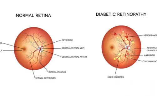
Fig 01: Position Of retinopathy in diabetes. Source: Carolina Eye care (2021)
Clinically, DR is divided into non-proliferative (NPDR) and proliferative (PDR) stages, and diabetic macular oedema (DME) can complicate either stage and is the commonest cause of central vision loss in diabetes [3]. Disease that begins silently can progress to haemorrhage, retinal ischaemia, tractional detachment and neovascular glaucoma. Because early lesions are asymptomatic, pharmacological strategies operate at two levels: preventing or delaying onset and progression, and rescuing vision once sight-threatening complications have appeared.
This article reviews (i) the pathophysiological mechanisms of DR, (ii) the drug targets these mechanisms expose, and (iii) the clinical evidence for the pharmacological approaches currently used or under investigation.
2. Pathophysiology
2.1 Metabolic pathways activated by hyperglycaemia
Sustained hyperglycaemia overloads glucose metabolism in retinal cells and diverts substrate into several injurious routes. These include increased flux through the polyol (aldose reductase) pathway, enhanced formation of advanced glycation end-products (AGEs), activation of protein kinase C (PKC) isoforms and increased flux through the hexosamine pathway. A unifying proposal is that excess mitochondrial superoxide production, arising from glucose-driven electron transport, inhibits glyceraldehyde-3-phosphate dehydrogenase and thereby pushes glycolytic intermediates into each of these branches [4]. This concept is important pharmacologically, since it explains why blocking one branch alone has generally produced modest clinical effects.
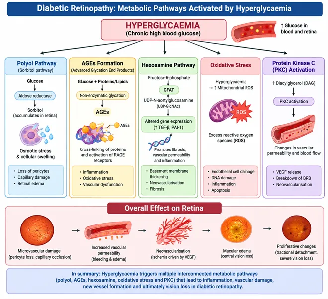
Fig:02 Metabolic pathway by hyperglycaemia
Source : Adapted and redrawn from published literature on hyperglycaemia-induced metabolic pathways in diabetic retinopathy
2.2 Oxidative stress
The retina has a high oxygen consumption and a lipid-rich composition, making it especially vulnerable to reactive oxygen species. In diabetes, superoxide generation rises while antioxidant defences fall, and damage to mitochondrial DNA and proteins creates a self-reinforcing cycle of further oxidant production. Oxidative stress also acts as an upstream signal for inflammatory and angiogenic responses, and it has been implicated in the accelerated death of retinal capillary cells [5].
2.3 Inflammation and leukostasis
DR is increasingly viewed as a low-grade inflammatory disease. Diabetic retinas show elevated cytokines and chemokines, activation of microglia and Müller cells, upregulated adhesion molecules and increased adherence of leukocytes to the capillary wall. Leukostasis can occlude capillaries and injure endothelium directly, contributing to vascular non-perfusion and barrier failure [6]. This inflammatory component provides the rationale for corticosteroid therapy and for interest in more selective anti-inflammatory targets.
2.4 Hypoxia, VEGF and angiogenesis
As capillaries drop out, areas of the retina become ischaemic and upregulate hypoxia-responsive genes. Vascular endothelial growth factor (VEGF) is central to this response. Seminal work measured markedly elevated VEGF concentrations in the ocular fluid of patients with active proliferative retinopathy, and levels fell after successful laser treatment, linking VEGF to neovascular activity [7]. VEGF increases vascular permeability by loosening endothelial junctions and stimulates the growth of fragile new vessels that bleed and promote fibrovascular traction. Other mediators, notably angiopoietin-2 acting through the Tie2 receptor, destabilise vessels and sensitise them to VEGF, which is the biological basis for dual-pathway inhibition.
2.5 Neurodegeneration and the neurovascular unit
Neuronal changes precede or accompany overt vascular lesions. Apoptosis of retinal neurons, including ganglion cells, has been demonstrated early in both experimental and human diabetes [8]. The neurovascular unit (neurons, glia, pericytes and endothelial cells) therefore appears to fail as a whole, and loss of neuronal and glial support may weaken vascular control. This shifts the therapeutic question from vascular protection alone towards neuroprotection, although no neuroprotective drug has yet reached routine use.
2.6 Blood–retinal barrier breakdown and macular oedema
Characteristic vascular lesions include basement-membrane thickening, pericyte loss, acellular capillaries and microaneurysms. Loss of pericytes and disruption of endothelial tight junctions compromise the inner blood–retinal barrier, allowing plasma constituents to leak into the retinal tissue and produce oedema. When leakage involves the centre of the macula, central vision falls [9]. Reviews of the field emphasise that vascular, neural, glial and immune abnormalities interact rather than act in isolation [10,11]
3. Pharmacological Targets
The mechanisms above translate into a set of tractable targets. Table 1 links each target to representative drug classes and their status.
Table 1. Pathophysiological targets and corresponding pharmacological approaches
|
S.No |
Target / Mechanism |
Drug Class / Agent |
Status |
|
1 |
Hyperglycaemia |
Insulin oral and injectable glucose- lowering drug |
Established (Risk reduction ) |
|
2 |
Systemic hypertension renin angiotensin system |
Anti hypertensives ACE inhibitors, ARBs (e.g., candesartan) |
Established for BP Control retinal- specification benefit modest |
|
3 |
Dulipidaemia PPAR- α Signalling |
Fenofibrate |
Emerging, supported by randomised trials |
|
4 |
VEGF-A |
Ranibzumab, aflibercept, bevacizumab (offlabel) |
Established first-line for centre- involvingDME and PDR |
|
5 |
VEGF_A plus angiopoietin-2 |
Faricimab |
Approved for DME dual-pathway approch |
|
6 |
Intraocular inflammation |
Various inhibitors and antioxidants |
Largely unscessful clinically |
|
7 |
PKC- β |
Ruboxistaurin |
Investigational not approved |
|
8 |
Adose reductase, AGES,oxidative stress |
Various inhibitors and antioxidants |
Largely unsuccessful Clinically |
|
9 |
Angiopoietin-Tie2, neurodegeneration |
Tie2 activators neuroprotective agents |
Experimental |
4. Therapeutic Approaches
4.1 Systemic pharmacological control
Glycaemic control. The Diabetes Control and Complications Trial showed that intensive insulin therapy substantially lowered the development of retinopathy in people with type 1 diabetes without baseline retinopathy and slowed progression in those with early disease [12]. In type 2 diabetes, the UK Prospective Diabetes Study demonstrated that intensive glucose control reduced microvascular complications overall [13]. Effects are durable but must be balanced against a well-recognised early worsening that can follow very rapid glucose lowering.
Blood pressure. In the UKPDS blood pressure study, tighter control reduced the risk of retinopathy progression and deterioration in visual acuity [14]. The ACCORD Eye study, however, found no added retinal benefit from intensive systolic pressure targets beyond standard care [15]. Renin–angiotensin blockade was tested specifically in the DIRECT programme: candesartan showed a trend towards reduced incidence of retinopathy in type 1 diabetes (25% versus 31%, borderline significance) and no effect on progression in those with established retinopathy [16], while in type 2 diabetes it increased the likelihood of regression without meeting the primary progression endpoint [17]. Overall, ARBs should be chosen for their systemic indications, with any retinal benefit regarded as a bonus rather than a primary reason for prescribing.
Lipid-modifying therapy and fenofibrate. In the FIELD study, fenofibrate reduced the need for first laser treatment in type 2 diabetes [18], and in ACCORD Eye, adding fenofibrate to simvastatin slowed retinopathy progression compared with simvastatin alone [15]. The LENS trial, the first large randomised study designed specifically for this question, enrolled 1,151 adults with early non-referable retinopathy or maculopathy. Over a median of four years the composite of progression to referable disease or treatment occurred in 22.7% of those on fenofibrate versus 29.2% on placebo (hazard ratio 0.73), with no effect on visual acuity, and estimated glomerular filtration rate was lower in the fenofibrate group, so renal function needs monitoring [19]. Fenofibrate is a PPAR-α agonist, and its retinal effects are thought to involve actions beyond lipid lowering, although the mechanism is not settled.
Incretin-based therapy. The SUSTAIN-6 cardiovascular outcome trial reported more retinopathy complications with semaglutide than with placebo in high-risk type 2 diabetes, probably related to rapid early glycaemic improvement in people with existing retinopathy [20]. This does not argue against these drugs but supports baseline retinal assessment and follow-up in patients starting potent glucose-lowering treatment.
4.2 Intravitreal anti-VEGF therapy
Anti-VEGF agents are the cornerstone of treatment for centre-involving DME and are also used for PDR. In DME, the DRCR Retina Network head-to-head comparison found all three agents (aflibercept, bevacizumab, ranibizumab) improved vision, with aflibercept giving greater gains at one year when baseline visual acuity was worse (approximately 20/50 or below) [21]. Registration studies of ranibizumab (RISE and RIDE) and aflibercept (VISTA and VIVID) established durable visual and anatomical benefit compared with sham or laser [22,23].
In PDR, the DRCR Protocol S trial showed that ranibizumab was non-inferior to panretinal photocoagulation for visual acuity at two years, with less peripheral visual field loss and a lower rate of vitrectomy, though anti-VEGF therapy demands reliable follow-up because benefit is lost if injections lapse [24].
Anti-VEGF has also been studied earlier in the disease course. In PANORAMA, aflibercept improved retinopathy severity in moderately severe to severe NPDR without DME, although in the second year fixed dosing appeared necessary to sustain the anatomical benefit [25]. The Protocol W trial found that preventive aflibercept reduced the development of vision-threatening complications over two years [26]. However, at four years the cumulative probability of PDR or centre-involving DME with vision loss was 33.9% with aflibercept and 56.9% with sham, yet visual acuity outcomes were not different (mean change −2.7 versus −2.4 letters) [27]. The investigators concluded that a preventive strategy may not be generally warranted for NPDR without centre-involving DME, and that regular monitoring with treatment on development of complications is a reasonable approach. The choice between early and deferred treatment therefore has to weigh anatomical benefit against injection burden, cost and procedural risk.
4.3 Dual-pathway inhibition: faricimab
Faricimab is a bispecific antibody that neutralises both VEGF-A and angiopoietin-2. In the two phase 3 trials YOSEMITE and RHINE, it achieved visual gains non-inferior to aflibercept in DME while permitting dosing intervals of up to 16 weeks in a substantial proportion of patients [28]. Its significance is conceptual as well as practical: it shows that targeting a second pathway can extend durability, which is valuable given the treatment burden that anti-VEGF regimens impose.
4.4 Corticosteroids
Corticosteroids suppress inflammatory mediators, reduce VEGF expression and stabilise the blood–retinal barrier. Sustained-release intravitreal implants are used mainly in eyes with an inadequate response to anti-VEGF, in pseudophakic eyes, or where injection frequency is a problem. The dexamethasone implant improved vision in DME over three years in the MEAD trial [29], and the fluocinolone acetonide insert offered long-term benefit in the FAME studies [30]. Both are limited by cataract progression and elevation of intraocular pressure, which need routine monitoring.
4.5 Agents that failed or remain investigational
Several mechanism-based oral drugs illustrate the difficulty of translating pathway biology into clinical benefit. Ruboxistaurin, an oral inhibitor of PKC-β, reduced sustained moderate visual loss in a phase 3 study of patients with non-proliferative retinopathy [31], yet it did not become an approved therapy. Aldose reductase inhibitors and antioxidant strategies have likewise not shown convincing clinical benefit. Current experimental directions include Tie2 activation, longer-acting or sustained-release anti-VEGF delivery systems, gene therapy delivering anti-angiogenic proteins, and neuroprotective agents. These approaches remain outside routine practice, and claims about them should be viewed as preliminary until supported by phase 3 evidence.
5. Challenges and Limitations of Current Pharmacotherapy
Treatment burden. Frequent injections and visits strain patients and health systems, and adherence lapses can erase gains.
Non-responders. A meaningful proportion of eyes respond incompletely to anti-VEGF monotherapy, pointing to non-VEGF drivers such as inflammation.
Invasive delivery. Intravitreal injection carries small but real risks of endophthalmitis and pressure spikes, and steroid implants add cataract and glaucoma risk.
Late intervention. Most drugs act on established sight-threatening disease, while treatment that prevents early progression, such as fenofibrate, produces only modest absolute effects.
Access and cost. Availability of screening and of high-cost biologics differs widely between and within countries, so the burden of avoidable blindness falls unequally.
Neurodegeneration unaddressed. No approved drug protects retinal neurons.
FUTURE PERSPECTIVES
Priorities include oral or topical agents that could act earlier and reduce reliance on injections, longer-lasting delivery platforms, combination or multi-target regimens building on the dual-pathway concept, and neuroprotective drugs. Biomarker-guided, individualised therapy, using imaging and systemic markers to identify patients likely to benefit from early treatment, could address the modest population-level benefit of preventive strategies seen in trials such as Protocol W. Repurposing established drugs such as fenofibrate is attractive because of low cost and wide availability, provided that safety, including renal monitoring, is respected.
CONCLUSION
Diabetic retinopathy arises from interacting metabolic, oxidative, inflammatory, vascular and neurodegenerative processes. This complexity explains why pharmacological management is layered: systemic control of glucose, blood pressure and lipids to reduce risk; anti-VEGF agents, including dual-pathway inhibition, as the mainstay for vision-threatening disease; and corticosteroid implants as an alternative when inflammation predominates or anti-VEGF response is inadequate. Evidence from Protocol W and LENS reminds us that anatomical improvement does not always mean better vision, and that low-cost systemic drugs may still add value. Future progress depends on less invasive, longer-acting and neuroprotective therapies delivered within equitable screening programmes.
REFERENCES




Gajendra Jayant, Peeyush Dixit, Omkar Rajput, Satyam Singh Jayant, Manoj Sharma,Pooja Gourav, Role of Pharmacology in Diabetic Retinopathy: A Review of Pathophysiology, Pharmacological Targets and Therapeutic Approaches, Int. J. of Pharm. Sci., 2026, Vol 4, Issue 10, 1206-1213, https://doi.org/10.5281/zenodo.23237807
 10.5281/zenodo.23237807