

We use cookies to ensure our website works properly and to personalise your experience. Cookies policy
Department of Pharmaceutical Chemistry, Sarojini Naidu Vanita Pharmacy Maha Vidyalaya, Tarnaka, Secunderabad, Telangana, India – 500017
Phytoconstituents such as polyphenols exhibit significant therapeutic potential; however, their clinical application is often limited by poor aqueous solubility, low membrane permeability, extensive first-pass metabolism, and inadequate bioavailability. To overcome these challenges, various nanocarrier-based delivery systems—including liposomes, nanoemulsions, and polymeric nanoparticles—have been explored. Among these, phytosome technology has emerged as a promising and superior approach for phytoconstituent delivery. Phytosomes are formed through the molecular complexation of plant bio actives with phospholipids via hydrogen bonding, resulting in lipid-compatible structures that closely mimic biological membranes. This unique architecture enhances drug stability, absorption, and systemic availability, particularly for polyphenolic compounds such as ellagic acid and resveratrol. Comparative evaluation reveals that, while liposomes offer higher encapsulation of hydrophilic compounds and polymeric nanoparticles enable sustained release, these systems are associated with limitations including drug leakage, burst release, potential toxicity, and complex manufacturing processes. Nanoemulsions, although physically stable and easy to prepare, lack vesicular organization and targeted delivery capability. Overall, phytosomes provide an optimal balance of biocompatibility, formulation simplicity, and enhanced bioavailability, making them a clinically relevant and efficient delivery platform for phytoconstituents in oral, topical, and systemic applications.Phytoconstituents such as polyphenols exhibit significant therapeutic potential; however, their clinical application is often limited by poor aqueous solubility, low membrane permeability, extensive first-pass metabolism, and inadequate bioavailability. To overcome these challenges, various nanocarrier-based delivery systems—including liposomes, nanoemulsions, and polymeric nanoparticles—have been explored. Among these, phytosome technology has emerged as a promising and superior approach for phytoconstituent delivery. Phytosomes are formed through the molecular complexation of plant bio actives with phospholipids via hydrogen bonding, resulting in lipid-compatible structures that closely mimic biological membranes. This unique architecture enhances drug stability, absorption, and systemic availability, particularly for polyphenolic compounds such as ellagic acid and resveratrol. Comparative evaluation reveals that, while liposomes offer higher encapsulation of hydrophilic compounds and polymeric nanoparticles enable sustained release, these systems are associated with limitations including drug leakage, burst release, potential toxicity, and complex manufacturing processes. Nanoemulsions, although physically stable and easy to prepare, lack vesicular organization and targeted delivery capability. Overall, phytosomes provide an optimal balance of biocompatibility, formulation simplicity, and enhanced bioavailability, making them a clinically relevant and efficient delivery platform for phytoconstituents in oral, topical, and systemic applications.
Ellagic acid (EA), a naturally occurring polyphenolic compound, has garnered significant attention in recent decades for its diverse pharmacological properties and potential therapeutic applications. Primarily derived from hydrolysable tannins known as ellagitannins found in various fruits, nuts, and plants, EA exhibits potent antioxidant, anti-inflammatory, anticancer, and other bioactive effects. Despite its promising health benefits, including protection against oxidative stress, inflammation, and chronic diseases such as cancer and metabolic disorders, the clinical utility of conventional EA is hindered by challenges related to bioavailability. These limitations have spurred research into advanced delivery systems to enhance its efficacy and therapeutic potential [1].
Ellagic acid (EA) is a naturally occurring polyphenolic compound widely distributed in flowering plants and is predominantly present as hydrolysable tannins known as ellagitannins, which release free EA upon acid- or base-catalysed hydrolysis. EA represents an important dietary polyphenol owing to its widespread occurrence in edible and medicinal plant species. Dietary intake of EA is mainly derived from fruits and berries, where total EA and ellagitannin contents typically range from 100 to 1500 mg/kg dry weight. Pomegranate (Punica granatum) is among the richest sources of EA, with exceptionally high concentrations reported in the peel and mesocarp. Berries such as raspberries, strawberries, blackberries, cloudberries, and muscadine grapes also contain substantial amounts of EA, particularly concentrated in the fruit skin.¹ Kakadu plum (Terminalia ferdinandiana) has been identified as one of the richest edible sources of EA, with levels reaching up to 140 g/kg dry weight. Camu-camu (Myrciaria dubia) fruit flour also represents a significant source of EA, containing more than 5600 mg/kg dry weight. In addition to fruits, EA is present in nuts and seeds such as walnuts, pecans, and almonds. Agro-forestry and wood-processing by-products serve as important industrial sources of EA and ellagitannins, particularly in tannin-rich tree species. Significant levels of EA have been reported in the bark and wood of Eucalyptus species, sweet chestnut (Castanea sativa), and oak (Quercus spp.). EA is highly abundant in medicinal plants, and is commonly used as a chemotaxonomic marker, particularly in the Rosaceae family. Geraniaceae family and other tannin-rich species EA is produced by the hydrolysis of compounds like geraniin [1].
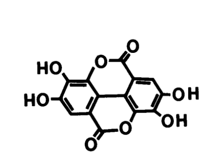
Fig. 1. Chemical structure of ellagic acid (2,3,7,8-tetrahydroxy-chromeno[5,4,3-cde] chromene-5,10-dione; molecular formula: C??H?O?).
Ellagic acid (EA) is a naturally occurring polyphenolic compound that demonstrates a broad spectrum of pharmacological activities, supported by extensive in vitro, in vivo, and some clinical studies. These effects are largely attributed to its potent antioxidant and anti-inflammatory properties, with therapeutic potential in diverse pathological conditions, including cancer, metabolic disorders, neurodegenerative diseases, inflammatory conditions, infections, cardiovascular diseases, and dermatological disorders [2].
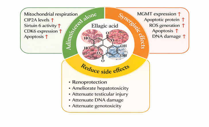
Fig. 2. Mechanistic overview of ellagic acid–mediated therapeutic and protective effects
Among its most intensively investigated actions are the anticancer and chemo preventive effects against various malignancies, including prostate, colon, breast, skin, ovarian, brain, oesophageal, lung, and liver cancers. EA exhibits antiproliferative, antimutagenic, and pro-apoptotic effects, inhibiting cancer cell growth through multiple mechanisms such as inhibition of procarcinogen activation, scavenging of reactive carcinogenic species, prevention of DNA damage, cell cycle arrest, angiogenesis inhibition, and suppression of metastasis. It induces mitochondrial-dependent apoptosis via modulation of the Bax/Bcl-2 ratio, activation of caspases, regulation of tumour suppressor proteins like p53 and p21, and inhibition of oncogenic signalling pathways, including NF-κB and c-Myc. As one of the most potent natural antioxidants, EA functions by directly scavenging reactive oxygen and nitrogen species (ROS/RNS), inhibiting lipid peroxidation, chelating transition metal ions involved in oxidative reactions, and modulating endogenous antioxidant enzymes such as superoxide dismutase and catalase. It further enhances antioxidant defence systems through activation of the Nrf2/HO-1 signalling pathway [3].
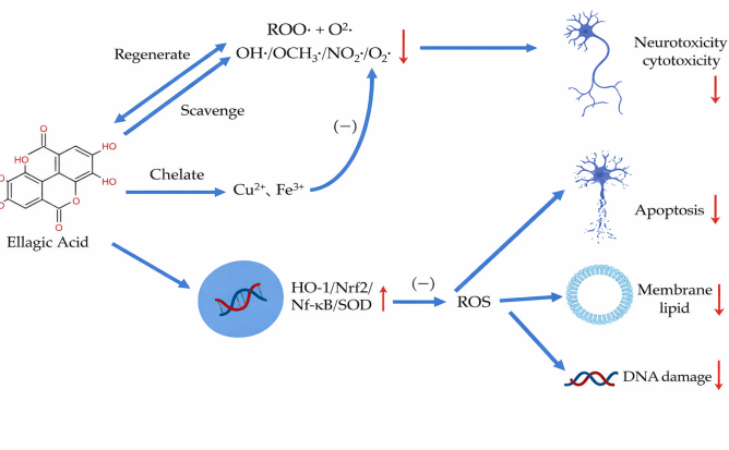
Fig. 3. Molecular mechanisms underlying the antioxidant effects of ellagic acid
The anti-inflammatory effects of EA involve suppression of pro-inflammatory cytokines (e.g., TNF-α, IL-6) and inhibition of key inflammatory mediators and pathways, such as COX-2, iNOS, and NF-κB, demonstrating benefits in conditions like asthma and cardiovascular diseases [3].
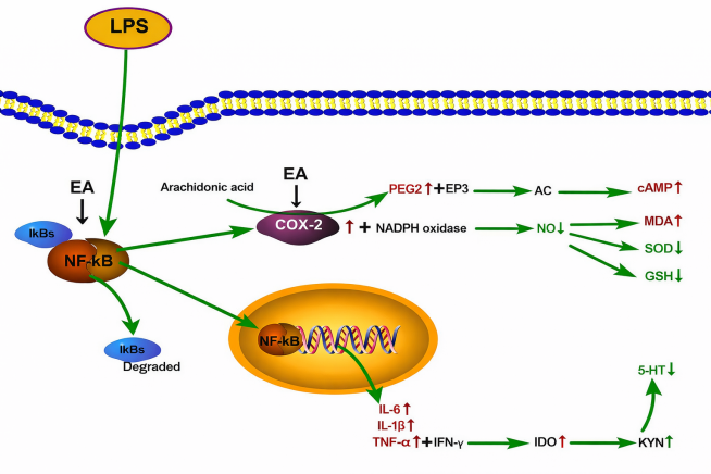
Fig. 4. Anti-inflammatory mechanisms of ellagic acid, highlighting NF-κB inhibition pathyway
EA also exhibits broad anti-infective and antihelminthic (anthelmintic) activities. In vitro studies have demonstrated its efficacy against helminth parasites, including nematodes such as Haemonchus contortus (via adult motility inhibition and egg hatch assays), Onchocerca ochengi, and drug-resistant strains of Caenorhabditis elegans. EA shows promising potential against anthelmintic-resistant nematodes, with low toxicity profiles. This activity is often linked to polyphenolic interference with parasite motility, viability, and reproductive processes, potentially through tannin-related mechanisms or direct disruption of parasite physiology. Additionally, EA contributes to broader antiparasitic effects in plant extracts against various helminths [4]. Other properties include neuroprotective effects in models of Alzheimer’s and Parkinson’s diseases through attenuation of oxidative stress and neurotoxic pathways; hepatoprotective and antidiabetic activities via improved glycaemic control, enhanced insulin secretion, and reduced advanced glycation end products; broad antimicrobial and anti-infective effects against bacterial, viral (e.g., HIV, influenza), and other parasitic (e.g., Plasmodium) pathogens; anti-obesity effects; and dermatological/cardiovascular benefits through photoprotective, anti-aging, anti-atherogenic, and cardioprotective mechanisms. These multifaceted actions are often interconnected through EA's core ability to mitigate oxidative stress and inflammation [5].
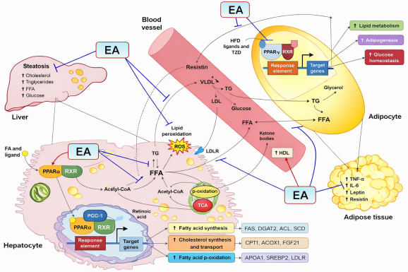
Fig. 5. Comprehensive schematic of ellagic acid's pharmacological mechanisms across multiple therapeutic areas.
1.3 Limitations of Conventional Ellagic Acid Delivery
Despite its broad pharmacological potential, the clinical translation of ellagic acid (EA) is markedly constrained by unfavourable physicochemical and pharmacokinetic properties that result in poor gastrointestinal absorption and low systemic bioavailability. Conventional oral administration of EA and its naturally occurring precursors, ellagitannins (ETs), therefore fails to reliably achieve therapeutically effective concentrations, limiting its utility as a chemo preventive or therapeutic agent. EA exhibits extremely low aqueous solubility and limited intestinal permeability, which severely restrict its gastrointestinal absorption. Reported solubility values of EA are as low as 9.7 μg/mL at physiological temperature, classifying it as a Biopharmaceutics Classification System (BCS) Class IV compound characterized by both low solubility and low permeability. Although EA solubility increases modestly under neutral or alkaline conditions, its phenolic structure renders it chemically unstable and susceptible to oxidative degradation at elevated pH. EA also demonstrates poor solubility under gastric conditions, further compromising oral absorption. Ellagitannins are relatively resistant to acidic hydrolysis and are predominantly degraded in the small intestine, resulting in delayed release of free EA. Following absorption, EA exhibits limited systemic availability due to inefficient intestinal uptake and extensive first-pass metabolism. Pharmacokinetic studies indicate rapid conversion of EA into methylated and glucuronide conjugates, leading to a short plasma half-life and low circulating concentrations. In humans, peak plasma concentrations of EA following consumption of EA-rich foods typically remain in the nanogram-per-millilitre range. Interindividual variability in EA metabolism represents an additional challenge to clinical translation. Unabsorbed EA and ETs are metabolized by colonic microbiota into urolithins, the production of which is highly dependent on individual gut microbial composition. This metabolism type-dependent variability contributes to inconsistent biological responses and unpredictable therapeutic outcomes. Furthermore, formulation-related challenges hinder clinical application, as EA is poorly soluble in most conventional solvents and exhibits limited stability in aqueous systems. Currently available commercial formulations, including tablets, capsules, and beverages, fail to adequately overcome these delivery limitations [6].
1.4 Need for Novel Delivery Systems for Ellagic Acid
The development of novel delivery systems for ellagic acid (EA) is primarily driven by its unfavorable physicochemical and pharmacokinetic characteristics, which significantly limit its therapeutic efficacy following conventional oral or topical administration.? Despite its well-documented pharmacological potential, EA exhibits extremely poor solubility, low permeability, rapid metabolism, and formulation instability, resulting in inadequate systemic exposure and inconsistent therapeutic outcomes. EA is classified as a Biopharmaceutics Classification System (BCS) Class IV compound due to its very low aqueous solubility and poor intestinal permeability. Its solubility has been reported to be as low as 9.7 μg/mL at physiological temperature, which severely restricts gastrointestinal absorption and leads to minimal plasma concentrations following oral administration. Clinical studies have demonstrated that even after consumption of EA-rich foods such as pomegranate juice, circulating EA levels remain extremely low, indicating that conventional dosage forms are insufficient to overcome absorption barriers. Furthermore, intestinal uptake of EA appears to be saturable, such that increasing the administered dose does not proportionally enhance bioavailability, thereby limiting its effectiveness as an in vivo chemo preventive or therapeutic agent. Besides decreased absorption, EA also has a low plasma half-life and a limited pharmacological action time, which is further worsened by a lack of chemical stability in solution, where hydrolytic and oxidative breakdowns occur during storage. Moreover, marked interindividual variability in gut microbiota–mediated metabolism of EA and ellagitannins leads to inconsistent formation of bioactive urolithins, resulting in variable therapeutic responses among individuals.
To fully exploit the broad therapeutic and prophylactic potential of EA—including its anticancer, antidiabetic, neuroprotective, and dermatological effects—advanced drug delivery strategies are essential. Novel micro- and nanotechnology-based systems have been proposed to enhance solubility and stability, improve intestinal and dermal permeability, reduce first-pass metabolism, and achieve sustained and targeted delivery. So, these approaches aim to maintain effective therapeutic concentrations of EA at target sites, thereby overcoming the limitations of conventional formulations and enabling its successful clinical translation [7].
The mechanism involves hydrogen bonding between the polar groups of the phytochemical and the phospholipid's choline head, while hydrophobic tails provide lipid compatibility. This confers dual solubility, better membrane permeation, and up to 20-fold bioavailability improvement. Unlike liposomes (where actives are encapsulated in aqueous cores), phytosomes feature chemical bonding for greater stability and efficacy.
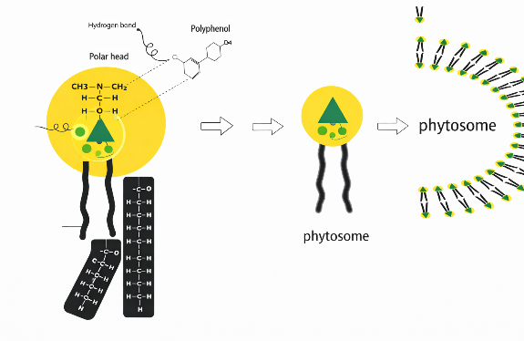
Fig. 6. Structural organization of a phytosome
2.1 Concept and Evolution of Phytosomes
Phytosomes (also known as herbosomes or phyto-phospholipid complexes) are an advanced vesicular drug delivery system designed to enhance the absorption of water-soluble phytochemicals, particularly polyphenolic compounds. The term "phytosome" derives from "phyto" meaning plant and "some" referring to cell-like structures.Phytosomes are formed through stoichiometric interactions between phospholipids, such as phosphatidylcholine, and polyphenolic phytochemicals in non-aqueous solvents, resulting in supramolecular complexes stabilized primarily by hydrogen bonding and hydrophobic interactions. These amphiphilic complexes readily integrate into cellular lipid bilayers, significantly improving gastrointestinal absorption compared to free phytochemicals.The concept originated in the 1980s from histochemical observations of natural affinities between plant polyphenols (e.g., anthocyanins) and membrane lipids, leading to the patenting of phytosome technology in 1989 by Indena S.p.A., an Italian firm. The primary goal is to overcome poor oral bioavailability of high-molecular-weight, poorly lipid-soluble flavonoids and tannins, which struggle to cross biological barriers.
Early developments used solvent precipitation methods, evolving to advanced techniques like supercritical fluid extraction and anti-solvent precipitation for compounds such as ellagic acid. Initial applications focused on nutraceuticals (e.g., liver support), while recent advancements incorporate targeted delivery for conditions like PCOS, skin disorders, and cancer. For instance, ellagic acid-loaded phytosomes enhance pharmacokinetics by improving water solubility, reducing metabolism, and amplifying antioxidant and anti-inflammatory effects.This technology bridges modern novel drug delivery systems with traditional herbal medicine, enabling controlled release, localized action, and reduced side effects [8].
2.2 Phospholipid–Phytoconstituent Interaction Mechanism
Phytosome creation relies on non-covalent interactions, primarily hydrogen bonds and van der Waals forces, between phospholipids and phytoconstituents, without altering the chemical structures of either component. Phospholipids like phosphatidylcholine (PC) and phosphatidylethanolamine (PE) feature a hydrophilic polar head and two hydrophobic fatty acid tails. Phenolic phytochemicals, such as ellagic acid (EA) with its hydroxyl and lactone groups, provide polar sites for hydrogen bond donation and acceptance.
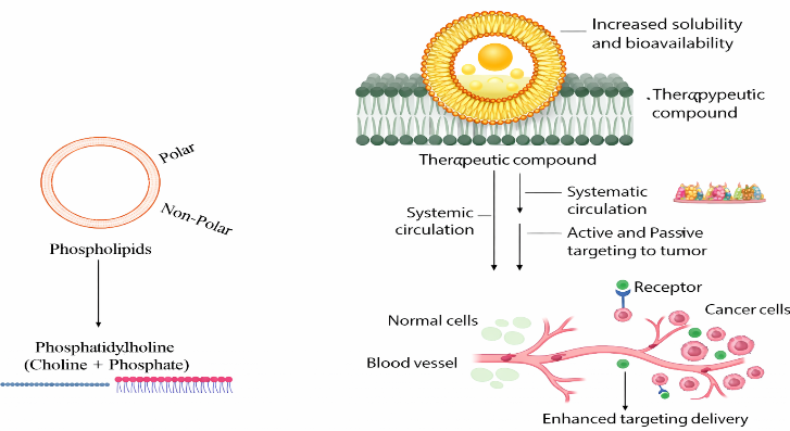
Fig. 7. Mechanism of phytosome-mediated enhancement of solubility, bioavailability, and targeted drug delivery
Complexation involves mixing the phytoconstituent and phospholipid in an aprotic solvent at a stoichiometric ratio of 1:1 to 1:4. Hydrogen bonds form between the phytoconstituent's hydroxyl groups and the phospholipid's polar head, while the lipid tails enclose the complex, yielding an amphiphilic, planar structure where the active is integrated into the membrane rather than encapsulated.FTIR spectroscopy reveals shifts in –OH and C=O stretching, confirming hydrogen bonds without covalent changes. NMR analysis shows phospholipid tails shielding the phytoconstituent's core, enhancing lipophilicity.For EA, its four hydroxyl groups and lactone rings interact with soya lecithin's phosphate moieties, forming stable precipitates via anti-solvent addition (e.g., n-hexane). This boosts solubility, protects against digestive degradation, and facilitates gut epithelial crossing into circulation.In water, phytosomes self-assemble into nanoscale vesicles (50–200 nm) with liposome-like properties but superior stability due to chemical bonding [9].
2.3 Comparison with Liposomes, Nanoemulsions, and Polymeric Nanoparticles
Phytosomes are especially well-suited for the delivery of phytoconstituents because they differ from other NDDS in terms of structure, preparation, and performance, but they share vesicular characteristics.
Table. 1. Comparative analysis of phytosomes and other nanocarrier-based delivery systems for phytoconstituent delivery
|
Delivery System |
Key Structural Features |
Preparation Method |
Advantages (Over Phytosomes) |
Limitations (Compared to Phytosomes) |
Relevant Applications in Phytoconstituent Delivery |
Ref |
|
Phytosomes |
Molecular complex via hydrogen bonds; phytoconstituent attached to phospholipid polar head; liposome-like vesicles (50–200 nm) |
Solvent evaporation or anti-solvent precipitation in aprotic medium with stoichiometric ratios |
N/A |
N/A |
Enhanced delivery of ellagic acid and resveratrol to skin/ovaries; superior bioavailability for polyphenols |
[10] |
|
Liposomes |
Concentric phospholipid bilayers with aqueous core; drug in core (hydrophilic) or bilayer (lipophilic); larger vesicles (100–1000 nm) |
Hydration of lipid films followed by sonication/extrusion |
Higher encapsulation for hydrophilic drugs; easier scale-up |
Prone to leakage and GI instability; no chemical bonding leads to burst release; lower permeability for polyphenols |
General herbal extracts; less effective for poorly soluble polyphenols like ellagic acid |
[10] |
|
Nanoemulsions |
Oil-in-water or water-in-oil droplets stabilized by surfactants (20–200 nm); non-vesicular |
High-energy emulsification (e.g., ultrasonication) or low-energy methods |
Better physical stability; faster production; suitable for oral/topical use |
Lacks vesicular structure/targeting; surfactant toxicity; poor release control for amphiphilic compounds |
Essential oils (e.g., eugenol); inferior for ellagic acid's polar-lipophilic balance |
[10] |
|
Polymeric Nanoparticles |
Solid polymeric matrix (e.g., PLGA, chitosan) entrapping drug (10–1000 nm); surface modifiable |
Emulsification-solvent evaporation or nanoprecipitation |
Sustained release (weeks); high loading; tunable targeting |
Potential toxicity; complex synthesis; lower biocompatibility; interference from degradation products |
Polysaccharide-based ellagic acid systems; phytosomes preferred for lipid mimicry without synthetic residues |
[10] |
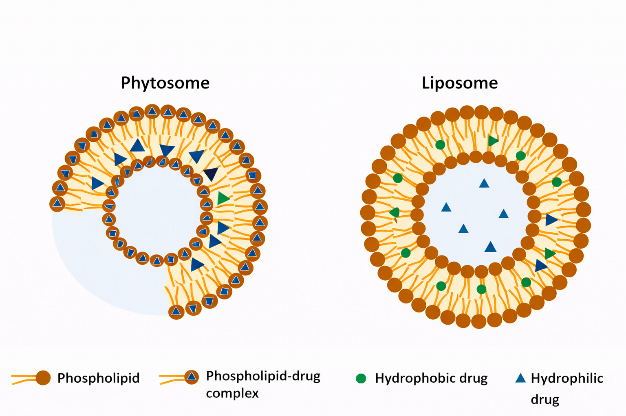
Fig. 8. Comparative schematic representation of phytosome and liposome
Due to stable hydrogen bonding, phytosomes offer superior stability and membrane fusion compared to liposomes, better physiological compatibility than polymeric nanoparticles, and enhanced cellular uptake over simpler nanoemulsions.
2.4 Advantages of Phytosomes (Enhanced Bioavailability, Membrane Permeability, Improved Stability)
For phytoconstituents such as ellagic acid (EA), which exhibit extremely low water solubility (<1 μg/mL), rapid metabolism, and poor oral bioavailability (typically 5–10%), phytosomes provide significant advantages over conventional herbal formulations.
Overall, these benefits position phytosomes as a powerful bridge between modern precision drug delivery and traditional herbal medicine, expanding therapeutic applications in dermatology, oncology, metabolic disorders, and beyond [11, 12].
Phytosomes represent an advanced vesicular drug delivery system designed to enhance the bioavailability of poorly soluble polyphenolic compounds like ellagic acid (EA). These complexes are formed through non-covalent interactions, primarily hydrogen bonding, between the polar head groups of phospholipids (such as phosphatidylcholine) and the hydroxyl groups of polyphenols. This amphiphilic nature allows phytosomes to traverse biological membranes more efficiently than free EA, which suffers from low aqueous solubility (<10 μg/mL), poor permeability, and rapid metabolism. The preparation methods leverage phospholipid complexation to create stable nano-sized carriers, often resulting in 5–10-fold improvements in solubility and 2–3-fold enhancements in oral bioavailability for similar polyphenols [13].
3.1 Solvent Evaporation Method
The solvent evaporation method, also known as rotary evaporation or thin-film formation, is one of the most conventional and scalable techniques for phytosome preparation. It involves dissolving EA (or EA-rich extracts) and phospholipids in a volatile organic solvent, followed by controlled evaporation to form a thin lipid film, which facilitates intimate molecular complexation.
Procedure:
Advantages and Outcomes:
This method yields high complexation efficiency (>90% in optimized cases) and stable complexes with particle sizes of 100–500 nm. Studies on analogous polyphenols (e.g., curcumin, silybin) show enhanced lipophilicity and antioxidant activity. For EA specifically, adaptations have produced complexes with improved dissolution rates. Variations include combining with hyaluronic acid for ocular delivery or pegylation for targeted therapy.
Limitations:
Potential residual solvent toxicity requires thorough removal; heat-sensitive compounds may degrade if temperatures exceed 60°C [14].
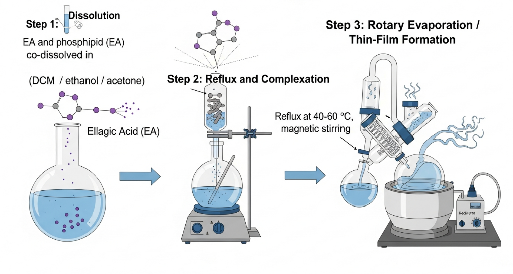
Fig. 9. Schematic representation of liposome preparation by rotatory evaporator i.e. Solvent Evaporation method
3.2 Anti-Solvent Precipitation Method
The anti-solvent precipitation method is favoured for producing smaller, more uniform nanoparticles with high entrapment efficiency, making it ideal for EA's poor solubility profile.
Procedure:
Advantages and Outcomes:
This technique often achieves particle sizes <100 nm, entrapment efficiencies >95%, and superior dissolution (up to 10-fold increase). Direct applications to EA include phospholipid complexes integrated into self-nanoemulsifying drug delivery systems (SNEDDS), demonstrating 2–3-fold higher ex vivo permeation and overcoming food effects on absorption. It is scalable and minimizes thermal degradation.
Limitations:
Requires careful anti-solvent selection to avoid incomplete precipitation; rapid addition can cause aggregation [15].

Fig. 10. Schematic representation of phytosome preparation by antisolvent precipitation
3.3 Thin-Film Hydration Method
This method, often overlapping with solvent evaporation, emphasizes hydration to form vesicular structures (phytosomal liposomes) and is commonly used for polyphenol extracts containing EA.
Procedure:
Advantages and Outcomes:
Produces stable vesicles with high entrapment (80–95%) and controlled release. Applied to EA-rich extracts (e.g., from Callistemon citrinus or pomegranate), it enhances stability and anti-obesity effects in vivo. Thin-layer sonication variants reduce sizes further for better penetration.
Limitations:
Multistep process; potential oxidation during hydration requires inert atmosphere [16].
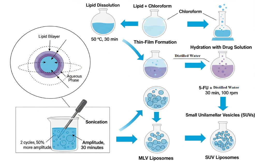
Fig. 11. Schematic representation of liposome preparation by thin-film hydration followed by sonication
3.4 Lyophilization (Freeze-Drying) Method
Lyophilization is typically an adjunct for drying and stabilizing phytosomes post-formation but can be a primary method when combined with co-solvency.
Procedure:
Advantages:
Enhances shelf-life (up to years) and prevents aggregation. Often paired with anti-solvent for EA complexes, yielding redispersible nanosuspensions.
This table [Table] compares the key preparation methods for ellagic acid (EA) phytosomes, summarizing process parameters, performance metrics, advantages, limitations, and applications [17].
Table. 2. Comparative Analysis of Phytosome Preparation Methods for Ellagic Acid
|
Method |
Key Process Parameters |
Entrapment Efficiency |
Particle Size |
Scalability |
Stability Enhancement |
Advantages |
Limitations |
Best Suited For |
|
Solvent Evaporation |
Drug-lipid ratio (1:1-1:4), solvent (aprotic like dichloromethane), temperature (40-60°C), reaction time (1-4h), evaporation pressure |
High (>90%) |
100-500 nm |
High |
Moderate |
Simple, high yield, adaptable for films |
Residual solvent risk, potential degradation |
General complexation, lab-to-industrial scale |
|
Anti-Solvent Precipitation |
Drug-lipid ratio (1:1-1:3), solvent (water-miscible like ethanol), anti-solvent (n-hexane/water), stirring speed (1000-2000 rpm), addition rate |
Very High (>95%) |
<100 nm |
High |
High |
Small particles, rapid process, superior dissolution |
Aggregation if not controlled, solvent selection critical |
High bioavailability needs, nano-formulations |
|
Thin-Film Hydration |
Like evaporation + hydration pH (6-7.4), volume, sonication/extrusion |
High (80-95%) |
100-200 nm |
Moderate |
High (vesicular) |
Forms stable liposomes, controlled release |
Multi-step, oxidation risk |
Vesicular delivery, targeted therapies |
|
Lyophilization (Adjunct) |
Cryoprotectants (5-10% w/v), freezing temp (-40--80°C), vacuum (0.1-0.5 mbar) |
Maintains prior |
Maintains prior |
High |
Very High |
Long-term storage, redispersible |
Requires pre-complex, time-consuming |
Storage-stable powders, combined with others |
3.5 Factors Affecting Phytosome Formation
Optimization is critical for maximizing complex yield, stability, and bioavailability. Key parameters include:
Statistical tools like factorial design or response surface methodology optimize these for >95% entrapment and <200 nm size.
3.6 Critical Process Parameters and Method Selection Rationale
Scalability and Reproducibility: Solvent evaporation and anti-solvent are GMP-compatible and scalable; thin-film hydration suits lab-scale vesicular forms.
Particle Size and Bioavailability: Anti-solvent yields smallest sizes (<100 nm), ideal for EA's intestinal absorption; solvent evaporation for controlled release.
Stability and Yield: Lyophilization ensures long-term stability; high yields (>90%) from all methods with optimization.
Rationale for EA Phytosomes: Anti-solvent preferred for rapid, high-bioavailability complexes (e.g., SNEDDS integration); solvent evaporation for simple, high-loading formulations. Emerging supercritical anti-solvent offers greener alternatives. Overall, these methods transform EA from a poorly bioavailable compound (absolute bioavailability <1%) into therapeutically viable forms, with pharmacokinetic studies showing prolonged plasma levels and enhanced tissue distribution.
4. FORMULATION STRATEGIES FOR ELLAGIC ACID PHYTOSOMES
4.1 Types of Phospholipids Used (Lecithin, Phosphatidylcholine)
Phytosomes are lipid-compatible molecular complexes designed to enhance the solubility and bioavailability of poorly water-soluble plant bioactives like ellagic acid (EA). Phospholipids such as lecithin and phosphatidylcholine (PC) serve as the structural backbone of these vesicular systems due to their amphiphilic nature, biocompatibility, and membrane-mimetic properties. Lecithin is a naturally derived mixture of phospholipids predominantly phosphatidylcholine, phosphatidylethanolamine, and phosphatidylinositol typically obtained from soy or egg sources. In EA phytosomes, soy lecithin forms hydrogen bonds with the hydroxyl groups of ellagic acid, enhancing complexation efficiency and promoting better dispersion in lipid environments. Studies have demonstrated that the hydrogen bonding between EA and lecithin improves both in vitro release and in vivo absorption by increasing membrane permeability and protecting the compound from enzymatic degradation. Phosphatidylcholine, the most abundant component of lecithin, is particularly preferred in phytosomal formulations due to its zwitterionic nature and strong ability to form stable bilayers that facilitate drug-lipid interactions. PC interacts with ellagic acid through electrostatic and hydrogen bonding forces, leading to the formation of a compact complex with improved physicochemical stability and lipid solubility. Phosphatidylcholine-based phytosomes have shown enhanced Cmax and AUC values for various polyphenols, indicating superior systemic bioavailability [18].
Table. 3. Comparison of commonly used phospholipids in ellagic acid phytosome formulations.
|
Parameter |
Lecithin |
Phosphatidylcholine |
|
Source |
Soy or egg |
Purified phospholipid (from lecithin) |
|
Composition |
Mixture of phospholipids |
Predominantly phosphatidylcholine |
|
Function |
Emulsifier, stabilizer |
Main structural lipid, enhances bilayer formation |
|
EA Binding |
Hydrogen bonding and hydrophobic interactions |
Strong polar and hydrophobic interactions |
|
Stability |
Moderate |
High (forms stable vesicles) |
4.2 Optimization Parameters (Drug–Lipid Ratio, Temperature, Hydration Time)
Optimizing formulation parameters is essential to achieve maximum complexation efficiency and desired physicochemical characteristics of EA phytosomes. The drug–lipid ratio, temperature, and hydration time significantly influence entrapment efficiency, particle size, and release profile.The drug–lipid ratio determines the degree of complex formation between EA and phospholipids. Studies have reported optimal ratios between 1:1 and 1:2 (EA: phospholipid, molar basis), where higher lipid content enhances complexation yield and dissolution but may increase particle size. It was found that an EA:lecithin ratio of 1:2 produced optimal solubility enhancement and sustained drug release up to 96% after 120 minutes. Beyond this ratio, excess lipid tends to form multilamellar aggregates, decreasing uniformity.Temperature plays a crucial role in facilitating solvent evaporation and molecular interaction between EA and the lipid phase. Moderate heating (40–60 °C) promotes phospholipid dispersion and complexation without causing degradation of ellagic acid, which is thermolabile. Maintaining the solvent removal step under controlled temperatures ensures consistent lipid matrix formation and avoids crystallization of free EA.Hydration time governs vesicle formation and stability. Longer hydration periods (≥30 min) allow for complete swelling of the lipid matrix, leading to well-defined, uniform vesicles with improved zeta potential and dispersion stability. Optimization tools such as response surface methodology (RSM) have been successfully used to study the combined effects of these parameters on encapsulation efficiency and release kinetics [19].
4.3 Challenges in Formulation and Scale-Up
Despite promising laboratory results, the scale-up and industrial production of ellagic acid phytosomes remain challenging. The process involves multiple critical steps solvent evaporation, hydration, homogenization, and drying each sensitive to minor deviations that can impact reproducibility.One of the major challenges is maintaining consistency in drug–lipid interactions on a large scale. Variations in phospholipid purity, solvent removal rates, and hydration uniformity can alter the final product’s particle size and entrapment efficiency (25). Additionally, ellagic acid’s poor solubility and multiple hydroxyl groups make it prone to oxidative degradation during processing and storage.Scale-up difficulties also arise due to differences in mixing efficiency and solvent recovery when transitioning from batch to continuous processing systems (27). Traditional methods such as rotary evaporation are not easily adaptable for high-throughput production, leading to inconsistent product quality. Emerging technologies like supercritical fluid extraction, microfluidization, and spray-drying have been investigated as alternatives to ensure better control and reproducibility.Moreover, regulatory challenges exist for phytosome-based formulations since they lie at the intersection of nutraceuticals and pharmaceuticals, often requiring additional bioequivalence and stability studies. Long-term stability testing, scalability assessments, and standardization of phospholipid sources remain key prerequisites for successful commercialization [20].
5. CHARACTERIZATION AND QUALITY EVALUATION
Characterization of ellagic acid (EA) phytosomes is essential to confirm successful complexation, ensure nanoscale properties, evaluate drug loading, assess structural changes, visualize morphology, understand release behavior, and verify stability. These techniques collectively demonstrate the advantages of phytosomal formulation over free EA, including improved solubility, altered crystallinity, and enhanced colloidal stability [21].
5.1 Physicochemical Characterization (Particle Size, PDI, Zeta Potential)
Dynamic light scattering (DLS) is the primary method for determining particle size, polydispersity index (PDI), and zeta potential in EA phytosomes.
These parameters confirm the formation of stable nanosystems and predict in vivo performance, with smaller sizes and lower PDI linked to higher oral absorption of EA [22].
5.2 Entrapment Efficiency Determination
Entrapment efficiency (EE) quantifies the amount of EA incorporated into the phytosome complex versus free drug. Common methods include ultracentrifugation, dialysis, or size-exclusion chromatography to separate unbound EA, followed by quantification via UV-Vis spectrophotometry (λ_max ~360 nm for EA) or HPLC.
EE is calculated as: EE (%) = (Total EA – Free EA) / Total EA × 100.
Reported values for EA phytosomes exceed 80–95%, significantly higher than conventional liposomes, due to strong hydrogen bonding and hydrophobic interactions. High EE minimizes drug loss and ensures therapeutic doses, contributing to dose reduction and reduced side effects [23].
5.3 Solid-State and Structural Characterization (FTIR, DSC, XRD)
These spectroscopic and thermal techniques provide evidence of molecular interactions and amorphous transformation in phytosomes.
These changes validate non-covalent complexation without chemical alteration of EA. The following table [Table] summarizes and visually depicts the advanced characterization methods for phytosomes or similar phospholipid complexes [24].
Table. 4. Comparative Analysis of Advanced Characterization Techniques for Ellagic Acid Phytosomes
|
Technique |
Purpose |
Methodology/Key Parameters |
Typical Findings for EA Phytosomes |
Advantages |
Limitations |
Ref |
|
Morphological Evaluation |
Visualize shape, size, surface, and structure |
TEM: Negative staining (phosphotungstic acid/uranyl acetate), high-resolution imaging; SEM: Gold/palladium sputtering, surface topology |
Spherical/vesicular particles, 50–200 nm, core-shell or multilamellar structure, smooth surface in lyophilized forms |
Direct visual confirmation, correlates with DLS data, reveals internal architecture |
Sample preparation artifacts (TEM drying), vacuum requirement (SEM), high cost |
[21] |
|
TEM vs. SEM Comparison |
Internal vs. external morphology |
TEM: Transmission through thin sample; SEM: Surface electron scattering |
TEM shows lamellarity and encapsulation; SEM highlights aggregation or roughness |
Complementary insights (internal + surface) |
TEM limited to ultra-thin samples; SEM lacks internal detail |
[22] |
|
In Vitro Drug Release Studies |
Assess release profile and mechanism |
Dialysis bag/Franz cell in SGF (pH 1.2) → SIF (pH 6.8), sink conditions, sampling at intervals, HPLC/UV quantification |
Sustained release (60–90% over 48–72 h) vs. <20% for free EA; follows Higuchi/Korsmeyer-Peppas kinetics |
Predicts in vivo behavior, evaluates sustained effect |
Does not fully mimic physiological conditions (enzymes, peristalsis) |
[25] |
|
Release Kinetics Models |
Determine release mechanism |
Zero-order, First-order, Higuchi, Korsmeyer-Peppas (n exponent) |
Diffusion-controlled (n ≈ 0.43–0.89), matrix erosion minimal |
Mechanistic understanding for optimization |
Multiple models may fit; requires validation |
[25] |
|
Stability Studies |
Evaluate physical/chemical stability over time |
ICH guidelines: Accelerated (40°C/75% RH, 6 months), Long-term (25°C/60% RH, 12 months); monitor size, PDI, zeta, EE, drug content |
Minimal changes (<10% variation) in lyophilized forms; superior to free EA suspensions |
Ensures shelf-life and quality for translation |
Time-consuming, requires controlled chambers |
[25] |
|
Short-term vs. Long-term |
Immediate vs. storage stability |
Short-term: Freeze-thaw, centrifugation; Long-term: ICH storage |
Lyophilized with cryoprotectants show best redispersibility and retention |
Comprehensive quality assurance |
Accelerated may not perfectly predict real-time |
[25] |
6. PHARMACOKINETICS AND THERAPEUTIC APPLICATIONS OF ELLAGIC ACID PHYTOSOMES
Ellagic acid is a polyphenolic compound that exists plentifully in natural sources such as pomegranate, berries, and nuts. In spite of its diverse pharmacological properties such as antioxidant, anticancer, anti-inflammatory, and cardioprotective activity, the use of ellagic acid could be hampered because it is poorly soluble in water, poorly permeable to the intestine, heavily undergoes the first-pass effect, and exhibits a short half-life. The use of a phytosome formulation could be a new strategy to reduce these pharmacokinetic limitations for the use of ellagic acid.
6.1 Improved Solubility, Permeability, and Oral Absorption
Phytosome technology combines ellagic acid with phospholipids, usually phosphatidylcholine. This process creates a lipid-friendly molecular complex instead of just a simple mix. This complex improves the lipophilicity of ellagic acid, helping it interact more effectively with biological membranes. As a result, the phytosome system increases its ability to disperse in water while also aiding transport across the intestinal lining. Better membrane affinity allows ellagic acid phytosomes to avoid problems with solubility and cuts down on degradation in the gastrointestinal tract. Research shows that phytosomal formulations greatly boost oral absorption efficiency compared to free ellagic acid, resulting in greater systemic exposure [26].
6.2 In Vivo Pharmacokinetic Enhancements
In vivo pharmacokinetic studies have shown that ellagic acid phytosomes lead to a significant increase in maximum plasma concentration (Cmax), area under the curve (AUC), and half-life compared to conventional formulations. These improvements result from better intestinal absorption, less efflux by P-glycoprotein transporters, and some protection from fast break down into less active metabolites. Additionally, phytosomes have longer circulation time and better tissue distribution, especially in organs like the liver, skin, and tumor tissues. This improved bioavailability keeps therapeutic plasma levels steady. It allows for lower doses to produce the desired pharmacological effect and may help reduce dose-related toxicity [27].
6.3 Preclinical and Clinical Evidence
Preclinical studies with animal models show strong evidence for the better effectiveness of ellagic acid phytosomes. These studies reveal significantly improved antioxidant, anti-inflammatory, and anticancer activities. In cancer models, phytosomal ellagic acid has shown better tumor growth inhibition, increased apoptosis, and lower oxidative stress compared to free ellagic acid. Anti-inflammatory studies indicate greater reduction of pro-inflammatory cytokines and oxidative mediators. Although there are still few clinical studies, early human research and nutraceutical applications point to improved bioavailability, better treatment outcomes, and good safety profiles. These findings underscore the potential of ellagic acid phytosomes to move from the lab to patient care [28].
6.4 Therapeutic Applications
The improved pharmacokinetic profile of ellagic acid phytosomes expands their use across different disease areas. In oncology, phytosomal ellagic acid shows strong anticancer effects by affecting cell cycle arrest, stopping new blood vessel formation, and triggering cell death. This makes it a promising addition to cancer prevention and treatment. Its powerful anti-inflammatory and antioxidant properties help manage chronic inflammatory conditions and diseases related to oxidative stress. The protective effects on the liver are especially notable, as phytosomes boost liver accumulation and shield liver cells from damage caused by toxins. In heart-related applications, ellagic acid phytosomes reduce lipid peroxidation, enhance blood vessel function, and slow down the progression of atherosclerosis. For skin health, they protect against UV damage, improve skin hydration, and decrease hyperpigmentation. Additionally, the anti-aging benefits of ellagic acid phytosomes relate to their ability to neutralize free radicals, support collagen strength, and slow down cell aging [28].
7. CURRENT CHALLENGES, FUTURE PERSPECTIVES, AND CLINICAL TRANSLATION
7.1 Scale-up and Industrial Feasibility
Scaling up phytosome production from laboratory-based solvent processes, such as rotary evaporation or anti-solvent precipitation, to a commercial capable process, must be able to produce uniform particle sizes (e.g. 200-300 nm) with good entrapment efficiency (>80%) and consistent release profiles while minimizing variances in zeta potential and polydispersity index (PDI). To produce phytosomes, the major challenges faced will be the optimisation of phospholipid ratios (e.g. 1:2 ellagic acid: soya lecithin) and solvent concentrations used in the manufacture process such that reproducibility is achieved. In preliminary formulations, due to phase separation and incomplete formation of complex structures, entrapment efficiencies ranged from 0 - 95%. Industrial feasibility of producing phytosomes was improved by the application of statistical method tools such as Box-Behnken Design and regression analysis to estimate desirability functions, resulting in a 92% or greater similarity across the checkpoints for the produced batches of phytosomes produced with a higher production volume capable of using phospholipids that were readily accessible; for example, using phosphatidylcholine. However, the high energy requirements and associated costs of Supercritical fluid extraction techniques (e.g. SAS, RESS) and resin recoveries will impose an economic hurdle for the implementation of these techniques in the large-scale production of ellagic acid phytosomes to be used for Dermatological or PCOS Efforts [29].
7.2 Regulatory Considerations for Phyto phospholipid Complexes
Regulatory barriers associated with Phyto phospholipid complexes are mainly due to the use of these systems as New Drug Delivery System (NDDS), requiring evidence of both safety and efficacy as well as enhanced bioavailability based on current regulatory frameworks like FDA's Generally Recognized as Safe (GRAS) for nutraceuticals and EMA guidelines for herbal medicinal products. Areas of concern include toxicological profiling (for example, conducting in vivo skin irritation tests in rabbits to demonstrate the absence of erythema with the use of resveratrol phytosomes) and stability data according to ICH guidelines, because the hydrogen bonding of phytosome molecules will likely cause pharmacokinetic variability. In addition, for ellagic acid phytosomes to gain regulatory approval, manufacturers must show data that demonstrates the decrease of first-pass metabolism in the liver and/or Mind-Area Concentration (AUC) but does not result in any genotoxicity and to provide evidence of the standardization of the raw material (such as purity of soya lecithin) to limit batch-to-batch variability. Intellectual property protection through patents, for example Indena's 1989 patent regarding a process to create phospholipid-phytoconstituent complexes, requires that the patent holder obtain bridging studies to support any clinical claims, especially concerning PCOS where nano formulations have raised concerns about reproductive toxicity [29].
7.3 Limitations of Current Studies
Research on ellagic acid phytosomes is largely confined to preclinical and in vitro models; findings are difficult to extrapolate to human PCOS or dermatological conditions because of the limited size of study populations; therefore, safety data regarding long-term use of these products is not yet available, and no RCTs exist. An additional limitation regarding bioavailability of ellagic acid is its poor solubility in water (<1µg/mL), and quick absorption by the gut microbiota into urolithins; therefore, it is estimated that only 5-10% would be absorbed systemically, although phytosomes did increase solubility of ellagic acid to 95.86% at 120 mins after ingestion. As previously stated, formulation issues (entrapment inefficiencies) and polydispersity (PDI>0.3) reduce the reproducibility of research results; furthermore, there has been no direct comparison of phytosomes with eugenol, a lipophilic active ingredient, as well as with those formulations that are commonly used to manage PCOS symptoms. In studies pertaining to PCOS, it is common practice to not consider clinical phenotype/disease stratification (i.e. Rotterdam Classification or NIH Criteria) as well potential comorbidities such as insulin intolerance, as well as assessments of side effects associated with standard pharmacological treatments (e.g. GI intolerance associated with metformin) have not been fully investigated in combination with phytosomes [30].
7.4 Future Directions (Targeted Delivery, Stimuli-Responsive Phytosomes, Hybrid Systems)
The development of targeted delivery systems using surface modified phytosomes (i.e., using folate for targeting to the ovary in women with PCOS) will support site specific uptake via receptor mediated endocytosis, while addressing the issue of hyper-androgenetic by way of 5-alpha reductase inhibition. Furthermore, the creation of stimuli responsive phytosomes that respond to different pH environments (e.g., low pH in the cancerous tumour microenvironment), or the production of matrix metalloproteinases (MMPs) during the inflammatory process associated with PCOS, will allow for controlled release of ellagic acid in conjunction with improved insulin sensitivity through the modulation of PI3K/Akt in those women experiencing PCOS, with a reduction of non-target effects. Hybrid systems developed with phytosomes combined with polymeric nanoparticles or liposome systems (i.e., the development of phytosome-liposome hybrid systems for co-loaded formulation of ellagic acid and apigenin) have potential for enhancing the synergistic bioavailability advantages of both classes of materials, with many studies already demonstrating a 2-5-fold increase in area under the curve when using nanotechnology in metabolic systems. Future research will also be focused on the development and/or utilization of ellagic acid prodrug formulations and co-crystals that are targeted to the human microbiome, along with the conduct of randomized controlled trials that are biomarker driven, in which will incorporate two endpoints, i.e., HOMA-IR and AMH, for the potential translation of research findings into clinical practice [30].
7.5 Commercialization and Nutraceutical Potential
The commercialization of ellagic acid phytosomes lends itself to nutraceuticals and cosmeceuticals. Marketing for phytosomes has been established with products such as Silymarin-phospholipid complexes (called Siliphos® for the protection of the liver) and Ginkgoselect Phytosome® for cognitive enhancement which showed 4-10 times the bioavailability of ellagic acid. In addition, eugenol-ellagic acid phytosomes could be used for the development of anti-aging creams with improved skin penetration (3-4 times through the stratum corneum), suggesting a significant potential for Over The Counter (OTC) formula development when conducting pharmacoeconomic analyses after accounting for the potential production costs. Furthermore, ellagic acid phytosomes could be developed as adjunctive nutraceuticals for the management of Polycystic Ovary Syndrome (PCOS) with apigenin or other novel compounds offering an added holistic support without chemical dependency. Currently the demand for herbal supplements is estimated to exceed $150 billion globally by 2025, however, these modifications will require claims validation through effective human pharmacokinetic studies from either the European Food Safety Authority (EFSA) or Food and Drug Administration (FDA). Patents that have been identified as having intellectual property enhancements include "Phospholipid complexes of olive fruit or olive leaf extracts" and provide many opportunities for research and development because they consider phytosomes as an extension/reinforcement for the use of Ayurvedic medicine towards sustainable products with lower side effects when combined with modern pharmaceuticals [30].
CONCLUSION
Among the various nanocarrier-based delivery systems evaluated for phytoconstituent delivery, phytosomes emerge as a superior and more rational approach, particularly for polyphenolic compounds such as ellagic acid and resveratrol. Unlike liposomes, nanoemulsions, and polymeric nanoparticles, phytosomes form a specific molecular complex through hydrogen bonding between the phytoconstituent and the phospholipid polar head, resulting in improved membrane permeability, stability, and bioavailability. While liposomes offer higher encapsulation of hydrophilic drugs and polymeric nanoparticles provide sustained release, both systems suffer from limitations such as leakage, burst release, potential toxicity, and complex manufacturing processes. Nanoemulsions, although physically stable and easy to produce, lack vesicular architecture and controlled targeting capability, making them less suitable for amphiphilic phytochemicals. Overall, phytosomes uniquely combine biocompatibility, structural simplicity, and biomimetic lipid compatibility, enabling efficient systemic and site-specific delivery without synthetic polymer residues. These advantages make phytosomes particularly well suited for chronic therapeutic applications, topical and oral formulations, and diseases where enhanced absorption of poorly soluble phytoconstituents is critical. Consequently, phytosome technology represents a preferred and clinically promising delivery platform for maximizing the therapeutic potential of plant-derived bioactive compounds.
REFERENCES


A. V. Vasanthi, B. Medha Gayatri, K. Kiranmai, G. Komal, V. Navya, S. Muni Sireesha, Dr. K. Vinutha, Phytosome - Enabled Delivery of Ellagic Acid: A Comprehensive Review of Formulation, Characterization, and Therapeutic Potential, Int. J. of Pharm. Sci., 2026, Vol 4, Issue 2, 2549-2574. https://doi.org/10.5281/zenodo.18668281
 10.5281/zenodo.18668281