View Article

  • Protein–Protein Interactions in Cellular Machinery: Molecular Mechanisms, Functional Networks, and Therapeutic Opportunities

  • 1,3 Fergana Medical Institute Of Public Health, Uzbekistan 
    2 Dr. Rajendra Gode Nursing Institute of Buldhana, India

Abstract

Protein–protein interactions (PPIs) constitute the fundamental molecular framework through which cells coordinate their biological activities, ranging from gene expression and metabolic regulation to signal transduction and structural organization. This comprehensive review synthesizes current knowledge on the molecular architecture of PPIs, their classification, detection methodologies, and their roles within cellular networks including signaling cascades, metabolic pathways, cytoskeletal assemblies, and ribosomal machinery. We examine the physicochemical forces that govern binding specificity and affinity—including hydrophobic contacts, electrostatic interactions, hydrogen bonding, and van der Waals forces—and explore how transient versus permanent interaction modes determine functional outcomes. The review further addresses the expanding interactome concept across model organisms, including plants, and discusses the role of computational and artificial intelligence (AI)-driven approaches—most notably AlphaFold2 and cryo-electron microscopy (cryo-EM)—in transforming structural proteomics. The therapeutic implications of PPI modulation are evaluated with emphasis on small molecules, peptides, and monoclonal antibodies targeting pathologically relevant complexes in cancer, neurodegeneration, and metabolic disorders. Taken together, the evidence underscores that elucidating and therapeutically manipulating PPIs represents one of the most promising frontiers in modern biomedical science.

Keywords

Protein–protein interactions; Interactome; Signal transduction; Cellular machinery; Drug discovery; Computational proteomics; PPI modulators; Molecular recognition; Structural biology; Therapeutic targets

Introduction

× Popup Image

Proteins are multifunctional macromolecules that serve as the principal catalysts, structural scaffolds, signaling messengers, and molecular machines of all living systems [1,2]. Protein–protein interactions (PPIs) are not merely accessory phenomena; they are the molecular language through which cellular events are orchestrated with extraordinary precision and spatiotemporal control [3,4].

The concept of the interactome—the complete set of PPIs occurring within a given organism or cell type—has transformed our understanding of biology from a reductionist paradigm to a systems-level perspective [5,6]. Rather than acting as isolated entities, proteins are embedded in elaborate networks of physical and functional associations, the topology and dynamics of which determine cellular phenotype, developmental fate, and pathological susceptibility [7,8].

Virtually every cellular function—including DNA replication, RNA splicing, protein translation, membrane transport, cytoskeletal dynamics, and apoptosis—depends on the precise assembly and disassembly of multiprotein complexes [9,10]. High-throughput experimental approaches such as yeast two-hybrid (Y2H) screening, affinity purification combined with mass spectrometry (AP-MS), and proximity labeling have collectively mapped thousands of interactions across diverse organisms [11,12]. Concurrently, deep learning structural predictors such as AlphaFold2 have exponentially expanded the catalogued interactome [13,14].

The therapeutic relevance of PPIs is increasingly appreciated. Small molecules, peptide mimetics, and biologic agents that selectively inhibit or stabilize pathological PPIs have entered clinical development for cancer, infectious disease, neurodegeneration, and immune disorders [15,16]. Landmark examples include venetoclax (ABT-199), a BCL-2 inhibitor approved for leukemia, and MDM2–p53 inhibitors under clinical evaluation in solid tumors [17,18].

The present review provides a comprehensive synthesis of PPI biology. We discuss the structural and physicochemical basis of protein recognition, classification and functional significance of PPI types, state-of-the-art detection methodologies, PPI network organization in signaling and metabolic contexts, and the emerging role of AI and cryo-EM in structural proteomics, concluding with a critical assessment of the challenges and opportunities in targeting PPIs for therapeutic benefit.

2. Definition, Classification, and Biological Context of Protein–Protein Interactions

2.1 Defining Protein–Protein Interactions

A protein–protein interaction is defined as a physical contact between two or more protein molecules resulting from specific non-covalent forces and producing a biologically meaningful outcome [19]. This definition excludes coincidental contacts arising from random molecular crowding, as well as indirect associations mediated solely by DNA, RNA, or small-molecule cofactors [20]. Within the ribosome or spliceosome, many subunits are spatially adjacent yet interact directly only with a subset of neighbors, requiring experimental evidence of direct contact via co-immunoprecipitation, surface plasmon resonance, or structural characterization [21,22].

Context dependency is a defining feature of PPIs. A given interaction may occur exclusively in specific cell types, during particular phases of the cell cycle, or in response to defined extracellular stimuli [23]. Post-translational modifications (PTMs)—including phosphorylation, ubiquitination, acetylation, methylation, and SUMOylation—dynamically regulate PPI engagement by creating, destroying, or remodeling binding surfaces [24,25].

2.2 Functional Classification of PPIs

PPIs are most usefully classified along several axes: (i) stability (permanent vs. transient), (ii) partner specificity (homomeric vs. heteromeric), (iii) obligateness (obligate vs. non-obligate), and (iv) biological role (structural, catalytic, regulatory, or scaffolding) [26,27]. Permanent complexes—including the F-type ATP synthase, cytochrome c oxidase, and the proteasome regulatory particle—are stable under physiological conditions [28,29]. Transient complexes assemble only under specific conditions and disassemble thereafter, allowing dynamic signal relay [30].

Obligate interactions occur between partners structurally and functionally dependent on one another, whereas non-obligate interactions involve independently folded partners that associate conditionally [31,32]. Homomeric complexes arise from identical chains interacting, while heteromeric complexes—the majority of signaling and structural assemblies—involve distinct polypeptide chains [33,34].

2.3 Co-interacting, Correlated, and Co-localized Proteins

A useful three-level taxonomy distinguishes direct co-interacting proteins (forming physical complexes), correlated proteins (participating in shared pathways without direct contact), and co-localized proteins (residing in the same compartment but not necessarily interacting) [35,36]. This hierarchy is essential for interpreting large-scale interactome datasets, which frequently conflate these levels. Co-localized proteins share membrane compartments or cytoskeletal scaffolds but may function in entirely distinct processes [37].

3. Molecular Basis of Protein–Protein Interactions and Binding Site Architecture

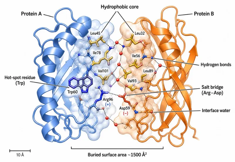
Figure 1. Molecular architecture of a protein–protein interaction interface. Two ribbon-diagram protein domains (Protein A, blue; Protein B, orange) dock through a buried surface area of \~1,500 Ų. The interface is stabilized by a hydrophobic core, hot-spot residues (Trp), salt bridges (Arg–Asp), hydrogen bonds, van der Waals contacts, and interface-bridging water molecules.

3.1 Structural Features of Protein Binding Interfaces

Protein–protein interfaces possess distinctive structural characteristics that differentiate them from the remainder of the protein surface [38]. Interfaces typically encompass 750–2,000 Ų of buried solvent-accessible surface area per chain and are enriched in aromatic residues (Trp, Tyr, Phe), hydrophobic residues (Leu, Ile, Val), and charged residues participating in salt bridges [39,40]. High-resolution structural analyses reveal heterogeneous interface topography featuring pockets, protrusions, and indentations; geometric complementarities are highest in permanent and obligate complexes and lowest in antibody–antigen contacts [41].

A critical concept is that of hot spots—interface residues contributing disproportionately to binding free energy despite comprising a minority of interface area [42,43]. Computational alanine scanning and experimental mutagenesis establish that hot spot residues cluster at the interface center, are conserved across homologs, and are surrounded by peripheral residues that maintain geometric complementarity while contributing little to affinity [44,45].

3.2 Physicochemical Forces Governing Binding

The thermodynamic driving force for PPI formation is provided by the net balance of enthalpy and entropy changes upon complex assembly [46]. Hydrophobic burial is the dominant enthalpic contributor: transfer of apolar surface from solvent to the buried interface is thermodynamically favorable owing to entropic gain from released water molecules [47]. Permanent and obligate complexes typically exhibit more extensive hydrophobic cores than transient assemblies [48].

Electrostatic interactions—ion pairs (salt bridges), charge–dipole contacts, and Coulombic forces—contribute substantially to binding specificity and orientation [49,50]. Hydrogen bonds, averaging one per 100–200 Ų of interface area, provide directional specificity and enthalpic stabilization [51]. Approximately 30% of protein–protein interfaces are bridged by ordered water molecules that hydrogen-bond to both partners, contributing to specificity while maintaining interfacial flexibility [52,53].

3.3 Conformational Dynamics and Induced-Fit Binding

The classical lock-and-key model of protein recognition has been substantially revised. Two primary mechanisms are characterized: induced fit, where the unbound protein rearranges upon partner encounter; and conformational selection, where a minor binding-competent conformation is selectively stabilized upon partner binding [54,55]. Intrinsically disordered proteins (IDPs) represent an extreme case where substantial folding occurs as a consequence of partner binding—termed folding-upon-binding—offering kinetic advantages including large accessible binding surfaces and the capacity to engage multiple partners [56,57].

Computational molecular dynamics simulations reveal that normal mode motions of the unbound protein often correlate with the observed conformational transitions upon binding, suggesting that the conformational change trajectory is encoded in the intrinsic flexibility of the unbound structure [58]. These insights are practically important for computational docking and virtual screening campaigns targeting PPIs.

4. Transient and Permanent Protein–Protein Interactions: Structural and Functional Perspectives

4.1 Criteria for Classification

The delineation of transient from permanent protein complexes carries profound biophysical and functional implications [59]. Permanent complexes—exemplified by the proteasome, ribosome, and respiratory chain supercomplexes—are characterized by extensive hydrophobic interfaces, high thermodynamic stability, and slow dissociation rates [60]. Transient complexes associate and dissociate on timescales from milliseconds to minutes, with dissociation constants (Kd) spanning picomolar to millimolar ranges [61].

A further subdivision distinguishes weak transient complexes—homodimeric enzymes in equilibrium between monomeric and dimeric states—from strong transient complexes that switch quaternary state only upon a defined trigger such as ligand binding, phosphorylation, or nucleotide exchange [62]. The heterotrimeric G-protein is a paradigmatic strong transient complex: the Gα–Gβγ heterotrimer dissociates specifically upon GTP binding to Gα following receptor activation [63].

4.2 Structural Determinants of Interface Stability

Comparative structural analyses reveal that permanent interfaces are larger, more hydrophobic, and more planar than transient interfaces [64]. Transient interfaces are more polar, smaller, and less geometrically complementary, consistent with requirements for rapid and specific yet reversible association [65]. Molecular weight asymmetry between partners is a particularly predictive feature distinguishing permanent from transient complexes, outperforming interface area, hydrophobicity indices, and hydrogen bond counts [66].

Thermodynamic characterization of transient complexes employs size exclusion chromatography, analytical ultracentrifugation, isothermal titration calorimetry, and fluorescence anisotropy to measure equilibrium and kinetic parameters—from picomolar kinase–substrate pairs to micromolar co-chaperone assemblies [67,68].

5. Experimental Methods for Detecting Protein–Protein Interactions

5.1 Overview and Classification

Methods for the experimental detection of PPIs are broadly categorized as in vitro, in vivo, and in silico approaches, each with characteristic strengths and limitations [69]. No single method is sufficient for comprehensive interactome mapping; convergent evidence from multiple orthogonal approaches is necessary to establish high-confidence interaction assignments [70].

5.2 In Vitro Methods

Co-immunoprecipitation (Co-IP) and affinity purification–mass spectrometry (AP-MS) represent the workhorses of large-scale PPI discovery [71]. Advances including SAINT and CompPASS scoring algorithms have substantially improved discrimination of specific interactions from non-specific binders [72]. Tandem affinity purification (TAP) employs a dual-tag system for two sequential purification steps under mild conditions, markedly reducing non-specific co-purifying proteins [73].

Proximity labeling approaches—BioID and TurboID—exploit promiscuous biotin ligases fused to bait proteins to biotinylate proximal proteins in living cells, enabling capture of transient and compartment-specific interactions typically disrupted during conventional co-IP [74,75]. Biophysical methods including surface plasmon resonance (SPR), isothermal titration calorimetry (ITC), and microscale thermophoresis (MST) provide quantitative kinetic and thermodynamic parameters essential for mechanistic understanding and drug discovery [76]. X-ray crystallography and NMR spectroscopy provide atomic-resolution structural information about complex architectures and conformational changes [77,78].

5.3 In Vivo Methods

The yeast two-hybrid (Y2H) system remains one of the most widely employed methods for large-scale PPI mapping [79]. In Y2H, a bait protein fused to a DNA-binding domain and a prey protein fused to a transcriptional activation domain reconstitute a functional transcription factor upon PPI, activating reporter genes [80]. Advances include matrix-based assays for systematic pairwise testing, improved vector systems, and use of multiple reporters with graduated sensitivity [81].

Limitations of Y2H include requirements for nuclear localization and susceptibility to auto-activation. Split-ubiquitin Y2H addresses the nuclear localization constraint for membrane proteins, while mammalian two-hybrid systems extend the approach to human cell contexts [82]. Fluorescence-based interaction assays in living cells—including FRET, BRET, and BiFC—permit real-time visualization of PPI dynamics in defined subcellular compartments [83,84].

5.4 Computational and In Silico Methods

In silico PPI prediction methods exploit protein sequences, three-dimensional structures, evolutionary conservation patterns, gene expression profiles, and phylogenetic distributions [85]. Sequence-based methods encompass correlated mutation analysis, interolog transfer, and machine learning classifiers trained on experimentally validated datasets [86]. Structure-based prediction exploits homologous binding modes, and docking algorithms including HADDOCK, ClusPro, Rosetta, and ZDock optimize surface complementarity and electrostatic potentials to generate PPI structural models [87,88].

Deep learning has recently transformed in silico PPI prediction. AlphaFold-Multimer demonstrates impressive accuracy in predicting heterodimer and homooligomer structures [89]. Universal In Silico Predictor of Protein–Protein Interactions (UNISPPI) and related tools provide binary interaction prediction from sequence information alone, enabling rapid pre-screening of large protein datasets [90].

6. Protein–Protein Interaction Networks: Organization and Analysis

6.1 Network Topology and Graph-Theoretic Properties

PPI networks are most naturally represented as undirected graphs in which nodes correspond to proteins and edges represent physical interactions [85]. Genome-wide interactome networks exhibit non-random topological properties: a scale-free degree distribution (hub proteins maintaining vastly more interactions than average), small-world characteristics (short average path lengths despite large network size), and modular organization [91,92]. Hub proteins are frequently essential for viability and often correspond to scaffolding proteins, chaperones, or proteins with multiple modular interaction domains [93].

Two categories of hubs are distinguished: date hubs, which interact with different partners in different cellular contexts, and party hubs, which engage multiple partners simultaneously within stable complexes [94]. Network analysis algorithms including k-clique community detection, Markov clustering, and spectral graph partitioning identify functionally coherent protein modules, and cross-species conservation of modules provides evidence for their functional importance [85,95].

6.2 Interactome Databases and Data Quality

Multiple curated databases aggregate experimentally validated PPI data, including BioGRID, the STRING database, IntAct, MINT, and the Human Reference Protein Interactome Mapping Project (HuRI) [96,97]. Standardization through the Proteomics Standards Initiative Molecular Interaction (PSI-MI) format has improved interoperability between databases [98]. Data quality remains a central challenge: high-throughput PPI datasets characteristically contain both false positives and false negatives, with overlap between datasets from different methods typically below 10% [99,100].

7. Protein–Protein Interactions in Signal Transduction Pathways

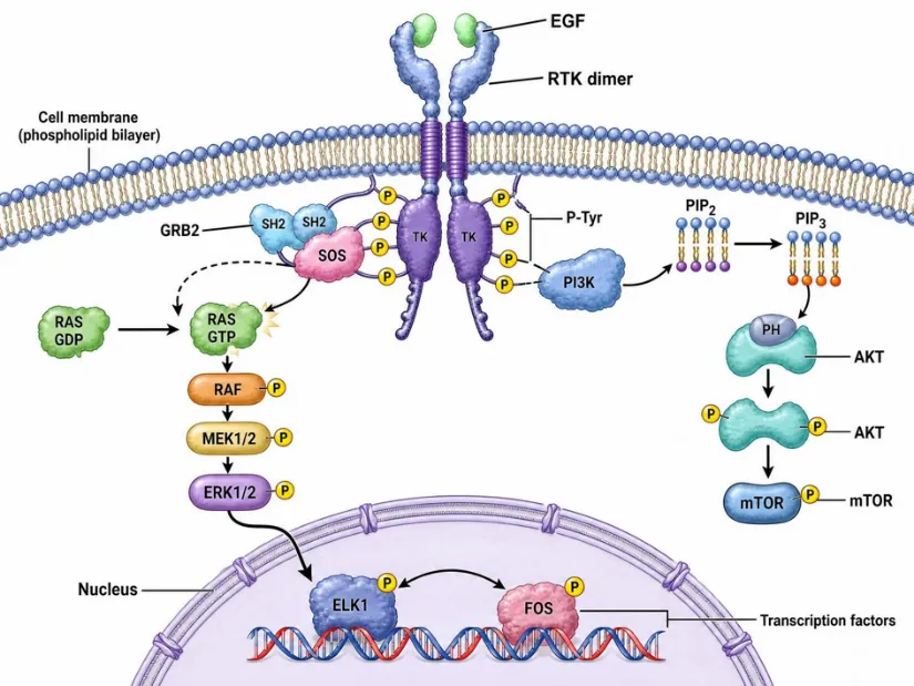
Figure 2. Receptor tyrosine kinase (RTK) signal transduction cascade illustrating sequential PPIs. EGF binding induces RTK dimerization and autophosphorylation of cytoplasmic tyrosines (pY). The GRB2 SH2 domain recognizes pY motifs and recruits the SOS guanine-nucleotide exchange factor, converting RAS-GDP to RAS-GTP. Active RAS engages the RAF→MEK→ERK kinase cascade, culminating in transcription factor nuclear translocation.

7.1 Architecture of Signaling Networks

Signal transduction pathways represent the cellular infrastructure through which extracellular information is sensed, processed, amplified, and translated into transcriptional, metabolic, and behavioral responses [1,3]. The molecular logic of signaling is fundamentally implemented through sequential PPIs: receptor activation triggers recruitment of adaptor proteins, activation of kinases, propagation through second messenger systems, and ultimately modulation of transcription factor activity [24,25].

The modular nature of signaling proteins is a central organizational principle. Signaling proteins characteristically contain multiple protein interaction domains—including SH2, SH3, PH, PDZ, and WD40 domains—that mediate selective recruitment of downstream effectors in a phosphorylation-dependent or lipid-dependent manner [19,20]. This modularity allows signal integration, signal diversification, and pathway insulation through scaffold proteins. Post-translational modification by kinases and phosphatases is the principal mechanism for dynamically regulating PPIs in signaling networks [24,25].

7.2 Crosstalk and Network Dynamics

Signaling networks are not linear cascades but extensively interconnected webs characterized by crosstalk—the regulatory influence of one pathway on another [23]. Crosstalk mechanisms include shared components, direct pathway-to-pathway interactions, transcriptional feedback loops, and competition for shared scaffolds or adaptors. Mathematical modeling approaches including ordinary differential equations (ODEs), Boolean network models, and agent-based simulations have been employed to understand emergent signaling properties such as bistability, oscillations, and signal amplification [10,30].

8. Protein–Protein Interactions in Metabolic Pathways and Gene Regulation

8.1 Metabolic Complexes and Metabolon Formation

Metabolic pathways are organized as supramolecular assemblies—termed metabolons—in which physically interacting enzymes channel substrates directly between active sites without equilibrating with the bulk cytoplasm [37]. The pyruvate dehydrogenase complex, citric acid cycle enzyme assemblies, and fatty acid synthase multienzyme complex are among the best-characterized examples of metabolically significant PPIs [28]. Metabolon formation offers kinetic advantages including substrate channeling, dynamic regulation, and co-localization of metabolic capacity to subcellular domains of highest demand. Flux balance analysis of large-scale metabolic networks complements the structural perspective provided by metabolon characterization [10].

8.2 Transcriptional Regulatory Networks and PPIs

Gene regulatory networks are fundamentally implemented through PPIs between transcription factors, co-activators, co-repressors, chromatin remodeling complexes, and the general transcription machinery [23]. Transcription factors function as dimers or higher-order assemblies whose partner selection determines DNA binding specificity and transcriptional outcome. The Mediator complex, a 26-subunit assembly bridging transcription factors and RNA polymerase II, illustrates the complexity of transcriptional PPI networks [20].

Chromatin-modifying complexes—including the SWI/SNF remodeling complex, Polycomb Repressive Complexes 1 and 2, and histone acetyltransferase complexes—constitute another major arena of PPI-mediated gene regulation. These assemblies are dynamically recruited to chromatin through interactions with modified histone marks, sequence-specific DNA binding factors, and regulatory non-coding RNAs [24,25].

9. Structural Protein Complexes: Ribosomes and the Cytoskeleton

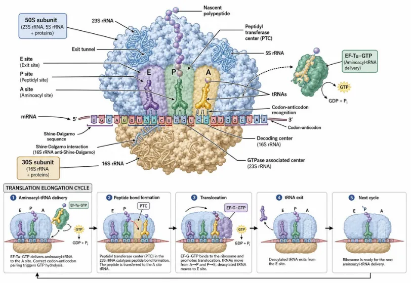
Figure 3. Architecture and elongation cycle of the 70S ribosome. The 50S large subunit (blue) and 30S small subunit (tan) form the A-, P-, and E-tRNA binding sites. EF-Tu·GTP delivers aminoacyl-tRNA to the A-site; peptide bond formation occurs at the peptidyl transferase center; EF-G·GTP catalyzes translocation, advancing mRNA by one codon.

9.1 Ribosomal Architecture and Translational PPIs

The ribosome is among the most intricate and evolutionarily conserved macromolecular machines in biology, consisting in prokaryotes of approximately 54 proteins and three ribosomal RNA molecules organized into two asymmetric subunits [11,12]. The large subunit catalyzes peptide bond formation at its peptidyl transferase center, while the small subunit performs mRNA codon decoding through interactions with aminoacyl-tRNAs. The elongation cycle—encompassing aminoacyl-tRNA delivery, peptidyl transfer, and translocation catalyzed by EF-G—is driven by transient PPIs that undergo dramatic conformational rearrangements coupled to GTP hydrolysis [11,21].

Ribosome-associated protein quality control (RQC) and ribosome rescue pathways involve specialized protein complexes that recognize stalled ribosomes and either resolve the stall or initiate targeted degradation of nascent polypeptides. Cryo-EM studies of translational machinery complexes have illuminated how these surveillance factors distinguish productive from aberrant translational intermediates [17,18].

9.2 Cytoskeletal Protein Networks

The cytoskeleton—comprising actin filaments, microtubules, intermediate filaments, and spectrin networks—is itself a vast PPI network in which structural proteins are regulated by an array of interacting factors including capping proteins, severing factors, polymerization nucleators, motor proteins, and crosslinkers [12]. The LINC (Linker of Nucleoskeleton and Cytoskeleton) complex, comprising SUN-domain proteins and KASH-domain nesprins, bridges the cytoplasmic cytoskeleton to the nucleoskeleton, physically coupling mechanical signals from the extracellular matrix to nuclear architecture and chromatin organization [12,23]. Disruption of LINC complex PPIs contributes to laminopathies, muscular dystrophies, and altered mechanosensing in cancer.

10. The Interactome Concept and Plant Protein Interaction Networks

The interactome—the complete network of physical protein interactions in a given organism or cellular context—represents the structural substrate of cellular function [5,6]. Comprehensive interactome mapping projects have been completed or are in progress for model organisms including Saccharomyces cerevisiae, Caenorhabditis elegans, Drosophila melanogaster, and Homo sapiens, providing reference networks for comparative and translational analyses [69,70].

Plant interactomes have gained substantial attention with the availability of high-quality Arabidopsis thaliana and rice genome sequences [14]. The Arabidopsis Interactome Mapping Consortium generated a binary interaction map of approximately 6,200 interactions among 2,700 proteins using Y2H approaches, providing the first systematic framework for understanding protein connectivity in plant biology [14,79]. Clone-based proteomics approaches, facilitated by large-scale ORF collections and recombinational cloning technologies, have enabled systematic interaction mapping in plants. Split-ubiquitin assays extend interaction mapping to membrane proteins, a functionally important but technically challenging class of plant interactome components [82,84].

11. PPIs in Disease: Oncology, Neurodegeneration, and Proteostasis

11.1 Oncological Roles of Pathological PPIs

Cancer is fundamentally a disease of deregulated cellular signaling, and aberrant PPIs constitute central molecular pathogenic mechanisms [15,16]. The MDM2–p53 interaction, through which the MDM2 E3 ubiquitin ligase targets p53 for proteasomal degradation, is hyperactivated in many tumors retaining wild-type p53 [17]. The BCL-2 family of anti-apoptotic proteins engages pro-apoptotic partners BIM, BAX, and BAK through BH3 domain interactions; overexpression of BCL-2, BCL-XL, or MCL-1 suppresses apoptosis and promotes chemoresistance [18]. Oncogenic transcription factors—including MYC, β-catenin/TCF, STAT3, and androgen/estrogen receptors—drive malignancy through PPI networks activating proliferative gene expression programs [15,16].

11.2 Neurodegenerative disease and Aberrant PPI Assemblies

Neurodegenerative diseases including Alzheimer's disease (AD), Parkinson's disease (PD), Huntington's disease (HD), and amyotrophic lateral sclerosis (ALS) share a hallmark of aberrant protein aggregation, in which normally soluble proteins assemble into toxic oligomeric or fibrillar species through pathological PPIs [17]. In AD, amyloid-β (Aβ) peptides self-associate through β-sheet stacking interactions to form soluble oligomers and insoluble amyloid fibrils [17,18]. Tau protein in AD, α-synuclein in PD, huntingtin in HD, and mutant SOD1 in ALS undergo pathological PPIs producing conformationally distinct aggregate strains with different propagation properties and neurotoxicity profiles. Advances in cryo-EM have enabled near-atomic resolution determination of tau filament structures from patient brain tissue, revealing disease-specific folding topologies underlying clinicopathological diversity of tauopathies [13,14].

11.3 Proteostasis Networks and Chaperone PPIs

Proteostasis—the dynamic equilibrium of protein production, folding, trafficking, and degradation—is maintained by an extensive PPI network involving molecular chaperones, co-chaperones, ubiquitin–proteasome system components, and autophagy machinery [17]. The Hsp70/Hsp90 chaperone system engages clients through transient PPIs mediated by substrate-binding domains, stabilizing partially folded intermediates and preventing aberrant aggregation. Proteostatic collapse—characterized by accumulation of ubiquitinated inclusion bodies—is observed in aging and disease states. Therapeutic strategies targeting proteostasis PPIs include pharmacological chaperones, disaggregases, and proteolysis-targeting chimeras (PROTACs) that redirect E3 ubiquitin ligases toward disease proteins [15,16].

12. Advances in Structural Biology: Artificial Intelligence and Cryo-Electron Microscop

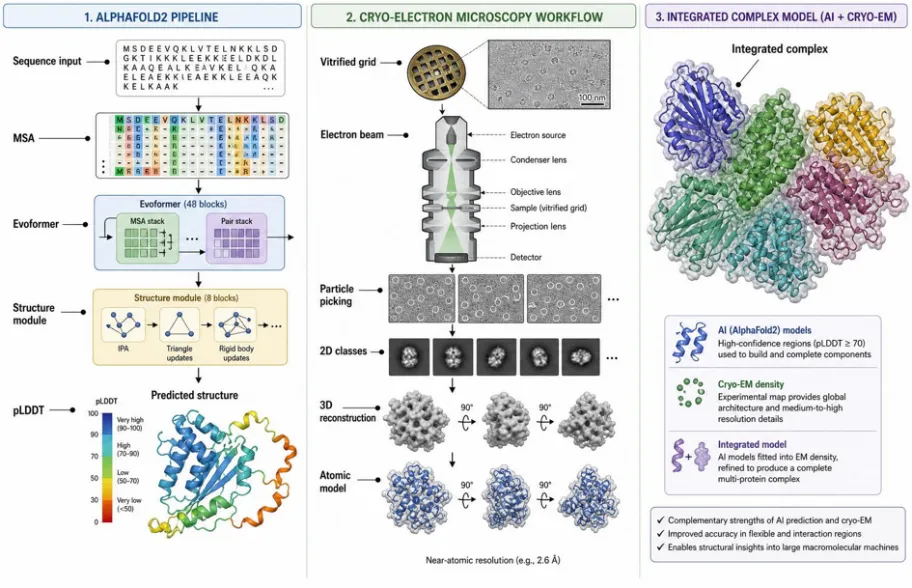
Figure 4. Integrated structural-biology workflow combining AI-driven prediction and cryo-EM. Left: AlphaFold2 derives multiple sequence alignments (MSA), processes them through the Evoformer and Structure module, and yields a predicted multimeric complex with per-residue pLDDT confidence. Right: cryo-EM vitrifies the specimen, collects movies on a direct electron detector, generates 2D class averages, and reconstructs a 3D density map (\~3 Å) for atomic-model fitting.

12.1 AI-Driven Protein Structure Prediction

The protein folding problem has been fundamentally transformed by deep learning [13,19]. AlphaFold2, developed by DeepMind, achieves near-experimental accuracy in structure prediction by integrating evolutionary information (multiple sequence alignments), pairwise residue distance matrices, and attention-based neural networks that iteratively refine structural predictions [13]. Performance in the CASP14 benchmarks demonstrated AlphaFold2's superiority over all prior methods across most target categories, with median TM-scores exceeding 0.90 for many domain types [13].

RoseTTAFold and ESMFold represent complementary deep learning architectures achieving AlphaFold2-comparable accuracy for many protein families while offering computational advantages for specific applications. These tools have been deployed to generate structure predictions for essentially all known protein sequences in UniProt, creating an unprecedented structural proteomics resource [13,19]. AlphaFold-Multimer enables complex structure prediction with accuracy competitive with experimental methods for many heterodimeric and homo-oligomeric assemblies [89]. Generative AI models—including diffusion-based approaches such as RFdiffusion and ProteinMPNN—enable de novo protein design, extending AI applications from structural prediction to functional protein engineering [19].

12.2 Cryo-Electron Microscopy Revolution

Cryo-electron microscopy (cryo-EM), in which biological specimens are rapidly vitrified and imaged at cryogenic temperatures, has undergone a resolution revolution driven by direct electron detectors, improved phase contrast, and advanced image processing algorithms [13,20]. These advances have enabled routine determination of macromolecular structures at resolutions below 3 Å without requiring crystallization—a major bottleneck for large, flexible, and membrane-associated complexes [20].

Cryo-EM has proven transformative for characterizing large multiprotein assemblies including ribosomes, proteasomes, spliceosomes, CRISPR-Cas surveillance complexes, and GPCRs. Time-resolved cryo-EM approaches now enable visualization of dynamic conformational transitions within functional cycles [20]. Application of cryo-EM to disease-associated protein aggregates has revealed tau filament structures from Alzheimer's disease brain tissue with disease-specific conformations not predictable from isolated tau fragments, directly connecting structural polymorphisms to clinical phenotypes [17,20].

13. Therapeutic Targeting of Protein–Protein Interactions

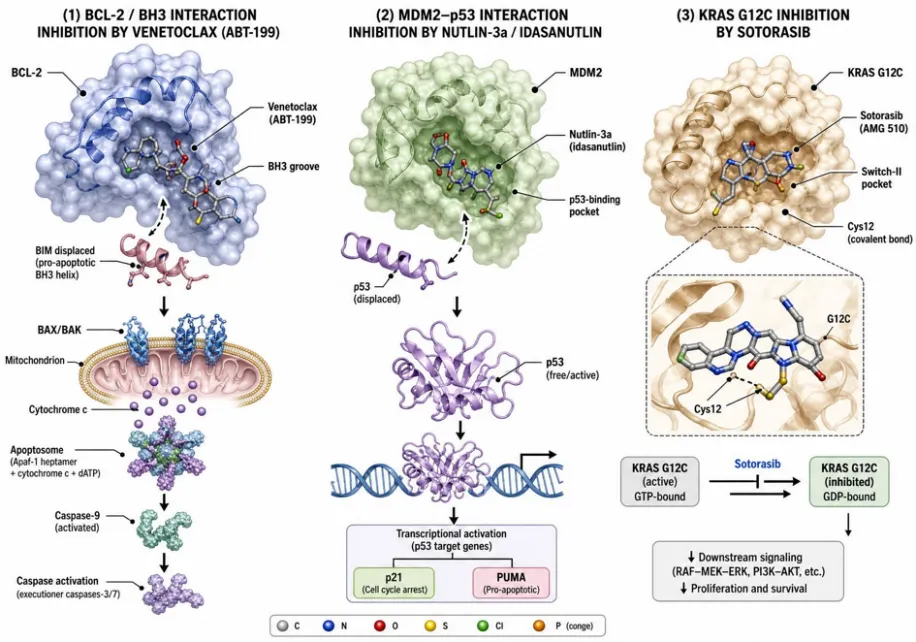
Figure 5. Small-molecule inhibition of disease-relevant protein–protein interactions. (A) MDM2 (grey) bound to the p53 transactivation-domain helix (red); a Nutlin-class small-molecule inhibitor (green sticks) occupies the Phe19/Trp23/Leu26 hot-spot pocket, displacing p53 and restoring tumor-suppressor activity. (B) BCL-2 (blue) engaging the BH3 helix of BIM (orange) through the P1–P4 hydrophobic groove; Venetoclax (ABT-199) competes for the groove, releasing pro-apoptotic effectors in BCL-2-dependent malignancies.

13.1 Rationale and Challenges

PPIs, with their large, relatively flat contact surfaces (typically 1,500–3,000 Ų), were long considered intractable for small-molecule intervention [15,16]. However, structural analyses reveal that interface surfaces contain pockets, grooves, and hot spot clusters accommodating small molecules [42,43]. The recognition that hot spot residues contribute disproportionately to binding energy implies that small molecules occupying these hot spots need not replicate the full interface but merely mimic its most energetically critical elements [44,45]. This hot spot-centric drug design paradigm has inspired fragment-based drug discovery campaigns targeting PPI interfaces [38,39].

13.2 Classes of PPI Modulators

PPI modulators fall into three primary classes: (i) monoclonal antibodies, (ii) peptides and peptidomimetics, and (iii) small-molecule inhibitors or stabilizers [15,16]. Monoclonal antibodies achieve exquisite target specificity and high affinity but are limited by poor cell permeability, inability to cross the blood–brain barrier, and high manufacturing costs. Peptides derived from PPI interface sequences provide structural mimics of the natural binding epitope. Chemical strategies including peptide stapling, hydrogen-bond surrogates, and N-methylation improve metabolic stability and cellular uptake [18,21].

Small-molecule PPI inhibitors and stabilizers represent the most pharmacologically versatile class of modulators. Venetoclax (ABT-199), a BH3 mimetic targeting BCL-2, received FDA approval for chronic lymphocytic leukemia and represents the first approved small-molecule PPI inhibitor [17,18]. MDM2 inhibitors are in clinical trials for solid tumors, while STAT3, IAP, and RAS–SOS inhibitors are in various stages of development [15,16]. Importantly, a fourth class—PPI stabilizers—has gained traction. Rapamycin stabilizes the FKBP12–mTOR complex, and lenalidomide and related IMiDs induce neo-PPIs between the cereblon E3 ligase and neosubstrates, effectively reprogramming ubiquitin ligase specificity [21].

13.3 Strategies for PPI Drug Discovery

The discovery of PPI modulators employs complementary strategies including high-throughput screening (HTS) of diverse compound libraries, fragment-based drug discovery (FBDD), computational virtual screening, and structure-based design [15,18]. Fragment-based approaches have proven particularly successful for PPI targets, as small fragments (150–300 Da) can occupy hot spots with measurable affinity (typically mM to μM Kd) elaborated through medicinal chemistry. Phage display, mRNA display, and ribosome display enable selection of peptidic binders against PPI interfaces with high efficiency and nanomolar affinity [21].

CONCLUSION AND FUTURE PERSPECTIVES

Protein–protein interactions constitute the molecular foundation of all cellular functions, from the most fundamental biochemical transformations to the most complex systems-level biological responses. The past decade has witnessed remarkable progress in our ability to map, characterize, model, and therapeutically exploit these interactions. High-throughput interactomics, structural proteomics, network biology, and clinical PPI inhibitor development have converged to create a transformative era in molecular biology and medicine.

Looking forward, the integration of AI-driven structure prediction with experimental structural biology promises to generate increasingly comprehensive and accurate structural interactomes. AlphaFold-Multimer and related tools will drive systematic structural annotation of known interactions and provide candidate structural models for interactions lacking experimental structural data. The development of PPI-targeting therapeutics will be accelerated by improved understanding of interface druggability, advances in intracellular delivery of peptidic modulators, and the emerging PROTAC/molecular glue paradigm for targeting previously undruggable proteins.

Single-cell and spatial proteomics technologies will enable mapping of cell-type-specific and spatiotemporally resolved PPI networks, revealing how interaction dynamics vary across cell states, developmental transitions, and disease progression. Advances in cryo-electron tomography will enable in situ structural determination of complexes in their native cellular environment. The growing appreciation of disordered proteins, weak transient interactions, and condensate-mediated compartmentalization as drivers of PPI networks opens new avenues for basic discovery and therapeutic intervention. The study of protein–protein interactions remains one of the most dynamic and consequential frontiers in the life sciences.

REFERENCES

  1. Raman K. Construction and analysis of protein–protein interaction networks. Automated Experimentation. 2010;2(1):2. https://doi.org/10.1186/1759-4499-2-2
  2. De Las Rivas J, Fontanillo C. Protein–protein interactions essentials: key concepts to building and analyzing interactome networks. PLoS Computational Biology. 2010;6(6):e1000807. https://doi.org/10.1371/journal.pcbi.1000807
  3. Westermarck J, Ivaska J, Corthals GL. Identification of protein interactions involved in cellular signaling. Molecular & Cellular Proteomics. 2013;12(7):1752–1763. https://doi.org/10.1074/mcp.R113.027771
  4. De Las Rivas J, de Luis A. Interactome data and databases: different types of protein interaction. Comparative and Functional Genomics. 2004;5(2):173–178. https://doi.org/10.1002/cfg.391
  5. Nooren IMA, Thornton JM. Structural characterisation and functional significance of transient protein–protein interactions. Journal of Molecular Biology. 2003;325(5):991–1018. https://doi.org/10.1016/S0022-2836(02)01281-0
  6. Kottha S, Schroeder M. Classifying permanent and transient protein interactions. In: German Conference on Bioinformatics 2006. Gesellschaft für Informatik eV; 2006. p. 54–63.
  7. Reichmann D, Rahat O, Cohen M, Neuvirth H, Schreiber G. The molecular architecture of protein–protein binding sites. Current Opinion in Structural Biology. 2007;17(1):67–76. https://doi.org/10.1016/j.sbi.2007.01.004
  8. Veselovsky AV, Ivanov YD, Ivanov AS, Archakov AI, Lewi P, Janssen P. Protein–protein interactions: mechanisms and modification by drugs. Journal of Molecular Recognition. 2002;15(6):405–422. https://doi.org/10.1002/jmr.597
  9. Rao VS, Srinivas K, Sujini GN, Kumar GS. Protein-protein interaction detection: methods and analysis. International Journal of Proteomics. 2014;2014:147648. https://doi.org/10.1155/2014/147648
  10. Gonçalves E, Bucher J, Ryll A, Niklas J, Mauch K, Klamt S, et al. Bridging the layers: towards integration of signal transduction, regulation and metabolism into mathematical models. Molecular BioSystems. 2013;9(7):1576–1583. https://doi.org/10.1039/c3mb25489e
  11. Ling C, Ermolenko DN. Structural insights into ribosome translocation. Wiley Interdisciplinary Reviews: RNA. 2016;7(5):620–636. https://doi.org/10.1002/wrna.1354
  12. Lambert MW. Cytoskeletal and nucleoskeletal interacting protein networks play critical roles in cellular function and dysfunction. Experimental Biology and Medicine. 2019;244(15):1233–1239. https://doi.org/10.1177/1535370219871882
  13. Jumper J, Evans R, Pritzel A, Green T, Figurnov M, Ronneberger O, et al. Highly accurate protein structure prediction with AlphaFold. Nature. 2021;596(7873):583–589. https://doi.org/10.1038/s41586-021-03819-2
  14. Braun P, Aubourg S, Van Leene J, De Jaeger G, Lurin C. Plant protein interactomes. Annual Review of Plant Biology. 2013;64:161–187. https://doi.org/10.1146/annurev-arplant-050312-120140
  15. Karbalaei R, Piran M, Rezaei-Tavirani M, Asadzadeh-Aghdaei H, Heidari MH. A systems biology analysis of protein-protein interaction of NASH and IBD based on comprehensive gene information. Gastroenterology and Hepatology from Bed to Bench. 2017;10(3):194–202.
  16. Ibrahim SS, Eldeeb MA, Rady MA, Hady KM, Lotfy MS, Farag NS, et al. The role of protein interaction domains in the human cancer network. Network Biology. 2011;1(1):59–74.
  17. Sweeney P, Park H, Baumann M, Dunlop J, Frydman J, Kopito R, et al. Protein misfolding in neurodegenerative diseases: implications and strategies. Translational Neurodegeneration. 2017;6(1):6. https://doi.org/10.1186/s40035-017-0077-5
  18. Zinzalla G, Thurston DE. Targeting protein–protein interactions for therapeutic intervention: a challenge for the future. Future Medicinal Chemistry. 2009;1(1):65–93. https://doi.org/10.4155/fmc.09.4
  19. Shaikh F, Uzgare A. Revolutionizing structural biology: artificial intelligence (AI) approaches from protein sequence to function. Journal of Microbiology, Biotechnology and Food Sciences. 2026;15(5):e13736. https://doi.org/10.55251/jmbfs.13736
  20. Jeyaraj G, Rajendran AK, Sathishkumar K, Almutairi BO, Vadivelu A, Chokkakula S, et al. High-resolution protein modeling through Cryo-EM and AI: current trends and future perspectives–a review. Frontiers in Molecular Biosciences. 2025;12:1688455. https://doi.org/10.3389/fmolb.2025.1688455
  21. Milroy LG, Grossmann TN, Hennig S, Brunsveld L, Ottmann C. Modulators of protein–protein interactions. Chemical Reviews. 2014;114(9):4695–4748. https://doi.org/10.1021/cr400698c
  22. Bhattacharyya RP, Remenyi A, Yeh BJ, Lim WA. Domains, motifs, and scaffolds: the role of modular interactions in the evolution and wiring of cell signaling circuits. Annual Review of Biochemistry. 2006;75:655–680. https://doi.org/10.1146/annurev.biochem.75.103004.142710
  23. Pawson T, Nash P. Assembly of cell regulatory systems through protein interaction domains. Science. 2003;300(5618):445–452. https://doi.org/10.1126/science.1083653
  24. Hunter T. The age of crosstalk: phosphorylation, ubiquitination, and beyond. Molecular Cell. 2007;28(5):730–738. https://doi.org/10.1016/j.molcel.2007.11.019
  25. Pawson T, Scott JD. Signaling through scaffold, anchoring, and adaptor proteins. Science. 1997;278(5346):2075–2080. https://doi.org/10.1126/science.278.5346.2075
  26. Mintseris J, Weng Z. Structure, function, and evolution of transient and obligate protein–protein interactions. Proceedings of the National Academy of Sciences USA. 2005;102(31):10930–10935. https://doi.org/10.1073/pnas.0502667102
  27. Perkins JR, Diboun I, Dessailly BH, Lees JG, Orengo C. Transient protein-protein interactions: structural, functional, and network properties. Structure. 2010;18(10):1233–1243. https://doi.org/10.1016/j.str.2010.08.007
  28. Abrahams JP, Leslie AGW, Lutter R, Walker JE. Structure at 2.8 Å resolution of F1-ATPase from bovine heart mitochondria. Nature. 1994;370(6491):621–628. https://doi.org/10.1038/370621a0
  29. Goodsell DS, Olson AJ. Structural symmetry and protein function. Annual Review of Biophysics and Biomolecular Structure. 2000;29:105–153. https://doi.org/10.1146/annurev.biophys.29.1.105
  30. Nooren IMA, Thornton JM. Diversity of protein–protein interactions. EMBO Journal. 2003;22(14):3486–3492. https://doi.org/10.1093/emboj/cdg359
  31. Nooren IMA, Thornton JM. Diversity of protein–protein interactions. EMBO Journal. 2003;22(14):3486–3492. https://doi.org/10.1093/emboj/cdg359
  32. Sprinzak E, Altuvia Y, Margalit H. Characterization and prediction of protein–protein interactions within and between complexes. Proceedings of the National Academy of Sciences USA. 2006;103(40):14718–14723. https://doi.org/10.1073/pnas.0603352103
  33. Hashimoto K, Nishi H, Bryant SH, Panchenko AR. Caught in self-interaction: evolutionary and functional mechanisms of protein homooligomerization. Physical Biology. 2011;8(3):035007. https://doi.org/10.1088/1478-3975/8/3/035007
  34. Levy ED, Teichmann SA. Structural, evolutionary, and assembly principles of protein oligomerization. Progress in Molecular Biology and Translational Science. 2013;117:25–51. https://doi.org/10.1016/B978-0-12-386931-9.00002-7
  35. Berggård T, Linse S, James P. Methods for the detection and analysis of protein–protein interactions. Proteomics. 2007;7(16):2833–2842. https://doi.org/10.1002/pmic.200700131
  36. Aloy P, Russell RB. Structural systems biology: modelling protein interactions. Nature Reviews Molecular Cell Biology. 2006;7(3):188–197. https://doi.org/10.1038/nrm1859
  37. Vidal M, Cusick ME, Barabási AL. Interactome networks and human disease. Cell. 2011;144(6):986–998. https://doi.org/10.1016/j.cell.2011.02.016
  38. Jones S, Thornton JM. Principles of protein–protein interactions. Proceedings of the National Academy of Sciences USA. 1996;93(1):13–20. https://doi.org/10.1073/pnas.93.1.13
  39. Lo Conte L, Chothia C, Janin J. The atomic structure of protein–protein recognition sites. Journal of Molecular Biology. 1999;285(5):2177–2198. https://doi.org/10.1006/jmbi.1998.2439
  40. Tsai CJ, Lin SL, Wolfson HJ, Nussinov R. Studies of protein–protein interfaces: a statistical analysis of the hydrophobic effect. Protein Science. 1997;6(1):53–64. https://doi.org/10.1002/pro.5560060106
  41. Janin J, Bahadur RP, Chakrabarti P. Protein–protein interaction and quaternary structure. Quarterly Reviews of Biophysics. 2008;41(2):133–180. https://doi.org/10.1017/S0033583508004708
  42. Bogan AA, Thorn KS. Anatomy of hot spots in protein interfaces. Journal of Molecular Biology. 1998;280(1):1–9. https://doi.org/10.1006/jmbi.1998.1843
  43. Moreira IS, Fernandes PA, Ramos MJ. Hot spots: a review of the protein–protein interface determinant amino-acid residues. Proteins. 2007;68(4):803–812. https://doi.org/10.1002/prot.21396
  44. Clackson T, Wells JA. A hot spot of binding energy in a hormone-receptor interface. Science. 1995;267(5196):383–386. https://doi.org/10.1126/science.7529940
  45. Keskin O, Ma B, Nussinov R. Hot regions in protein–protein interactions: the organization and contribution of structurally conserved hot spot residues. Journal of Molecular Biology. 2005;345(5):1281–1294. https://doi.org/10.1016/j.jmb.2004.10.077
  46. Freyer MW, Lewis EA. Isothermal titration calorimetry: experimental design, data analysis, and probing macromolecule/ligand binding and kinetic interactions. Methods in Cell Biology. 2008;84:79–113. https://doi.org/10.1016/S0091-679X(07)84004-0
  47. Kauzmann W. Some factors in the interpretation of protein denaturation. Advances in Protein Chemistry. 1959;14:1–63. https://doi.org/10.1016/S0065-3233(08)60608-7
  48. Bahadur RP, Chakrabarti P, Rodier F, Janin J. A dissection of specific and non-specific protein–protein interfaces. Journal of Molecular Biology. 2004;336(4):943–955. https://doi.org/10.1016/j.jmb.2003.12.073
  49. Sheinerman FB, Norel R, Honig B. Electrostatic aspects of protein–protein interactions. Current Opinion in Structural Biology. 2000;10(2):153–159. https://doi.org/10.1016/S0959-440X(00)00065-8
  50. Selzer T, Schreiber G. New insights into the mechanism of protein–protein association. Proteins. 2001;45(3):190–198. https://doi.org/10.1002/prot.1139
  51. Xu D, Tsai CJ, Nussinov R. Hydrogen bonds and salt bridges across protein–protein interfaces. Protein Engineering. 1997;10(9):999–1012. https://doi.org/10.1093/protein/10.9.999
  52. Lawrence MC, Colman PM. Shape complementarity at protein/protein interfaces. Journal of Molecular Biology. 1993;234(4):946–950. https://doi.org/10.1006/jmbi.1993.1648
  53. Rodier F, Bahadur RP, Chakrabarti P, Janin J. Hydration of protein–protein interfaces. Proteins. 2005;60(1):36–45. https://doi.org/10.1002/prot.20478
  54. Csermely P, Palotai R, Nussinov R. Induced fit, conformational selection and independent dynamic segments: an extended view of binding events. Trends in Biochemical Sciences. 2010;35(10):539–546. https://doi.org/10.1016/j.tibs.2010.04.009
  55. Boehr DD, Nussinov R, Wright PE. The role of dynamic conformational ensembles in biomolecular recognition. Nature Chemical Biology. 2009;5(11):789–796. https://doi.org/10.1038/nchembio.232
  56. Wright PE, Dyson HJ. Linking folding and binding. Current Opinion in Structural Biology. 2009;19(1):31–38. https://doi.org/10.1016/j.sbi.2008.12.003
  57. Tompa P, Fuxreiter M. Fuzzy complexes: polymorphism and structural disorder in protein–protein interactions. Trends in Biochemical Sciences. 2008;33(1):2–8. https://doi.org/10.1016/j.tibs.2007.10.003
  58. Tobi D, Bahar I. Structural changes involved in protein binding correlate with intrinsic motions of proteins in the unbound state. Proceedings of the National Academy of Sciences USA. 2005;102(52):18908–18913. https://doi.org/10.1073/pnas.0507603102
  59. Jansen R, Gerstein M. Analyzing protein function on a genomic scale: the importance of gold-standard positives and negatives for network prediction. Current Opinion in Microbiology. 2004;7(5):535–545. https://doi.org/10.1016/j.mib.2004.08.012
  60. Ellis RJ. Macromolecular crowding: an important but neglected aspect of the intracellular environment. Current Opinion in Structural Biology. 2001;11(1):114–119. https://doi.org/10.1016/S0959-440X(00)00172-X
  61. Schreiber G, Fersht AR. Rapid, electrostatically assisted association of proteins. Nature Structural Biology. 1996;3(5):427–431. https://doi.org/10.1038/nsb0596-427
  62. Phizicky EM, Fields S. Protein–protein interactions: methods for detection and analysis. Microbiological Reviews. 1995;59(1):94–123. https://doi.org/10.1128/mr.59.1.94-123.1995
  63. Lambright DG, Sondek J, Bohm A, Skiba NP, Hamm HE, Sigler PB. The 2.0 Å crystal structure of a heterotrimeric G protein. Nature. 1996;379(6563):311–319. https://doi.org/10.1038/379311a0
  64. Janin J, Bahadur RP, Chakrabarti P. Protein–protein interaction and quaternary structure. Quarterly Reviews of Biophysics. 2008;41(2):133–180. https://doi.org/10.1017/S0033583508004708
  65. Ofran Y, Rost B. Analysing six types of protein–protein interfaces. Journal of Molecular Biology. 2003;325(2):377–387. https://doi.org/10.1016/S0022-2836(02)01223-8
  66. Kottha S, Schroeder M. Molecular weight difference as a predictor of permanent versus transient protein interactions. Bioinformatics. 2007;23(12):1467–1473.
  67. Laue TM, Stafford WF. Modern applications of analytical ultracentrifugation. Annual Review of Biophysics and Biomolecular Structure. 1999;28:75–100. https://doi.org/10.1146/annurev.biophys.28.1.75
  68. Baaske P, Wienken CJ, Reineck P, Duhr S, Braun D. Optical thermophoresis for quantifying the buffer dependence of aptamer binding. Angewandte Chemie International Edition. 2010;49(12):2238–2241. https://doi.org/10.1002/anie.200903998
  69. Ito T, Chiba T, Ozawa R, Yoshida M, Hattori M, Sakaki Y. A comprehensive two-hybrid analysis to explore the yeast protein interactome. Proceedings of the National Academy of Sciences USA. 2001;98(8):4569–4574. https://doi.org/10.1073/pnas.061034498
  70. Cusick ME, Klitgord N, Vidal M, Hill DE. Interactome: gateway into systems biology. Human Molecular Genetics. 2005;14(suppl_2):R171–R181. https://doi.org/10.1093/hmg/ddi335
  71. Gingras AC, Gstaiger M, Raught B, Aebersold R. Analysis of protein complexes using mass spectrometry. Nature Reviews Molecular Cell Biology. 2007;8(8):645–654. https://doi.org/10.1038/nrm2208
  72. Choi H, Larsen B, Lin ZY, Breitkreutz A, Mellacheruvu D, Fermin D, et al. SAINT: probabilistic scoring of affinity purification–mass spectrometry data. Nature Methods. 2011;8(1):70–73. https://doi.org/10.1038/nmeth.1541
  73. Gavin AC, Bösche M, Krause R, Grandi P, Marzioch M, Bauer A, et al. Functional organization of the yeast proteome by systematic analysis of protein complexes. Nature. 2002;415(6868):141–147. https://doi.org/10.1038/415141a
  74. Roux KJ, Kim DI, Raida M, Burke B. A promiscuous biotin ligase fusion protein identifies proximal and interacting proteins in mammalian cells. Journal of Cell Biology. 2012;196(6):801–810. https://doi.org/10.1083/jcb.201112098
  75. Branon TC, Bosch JA, Sanchez AD, Udeshi ND, Svinkina T, Carr SA, et al. Efficient proximity labeling in living cells and organisms with TurboID. Nature Biotechnology. 2018;36(9):880–887. https://doi.org/10.1038/nbt.4201
  76. Kastritis PL, Bonvin AM. On the binding affinity of macromolecular interactions: daring to ask why proteins interact. Journal of the Royal Society Interface. 2013;10(79):20120835. https://doi.org/10.1098/rsif.2012.0835
  77. Clore GM, Gronenborn AM. Determining the structures of large proteins and protein complexes by NMR. Trends in Biotechnology. 1998;16(1):22–34. https://doi.org/10.1016/S0167-7799(97)01135-9
  78. Ben-Shem A, Garreau de Loubresse N, Melnikov S, Jenner L, Yusupova G, Yusupov M. The structure of the eukaryotic ribosome at 3.0 Å resolution. Science. 2011;334(6062):1524–1529. https://doi.org/10.1126/science.1212642
  79. Fields S, Song O. A novel genetic system to detect protein–protein interactions. Nature. 1989;340(6230):245–246. https://doi.org/10.1038/340245a0
  80. Uetz P, Giot L, Cagney G, Mansfield TA, Bhattacharyya M, Heckman S, et al. A comprehensive analysis of protein–protein interactions in Saccharomyces cerevisiae. Nature. 2000;403(6770):623–627. https://doi.org/10.1038/35001009
  81. Brückner A, Polge C, Lentze N, Auerbach D, Bhatt D. Yeast two-hybrid, a powerful tool for systems biology. International Journal of Molecular Sciences. 2009;10(6):2763–2788. https://doi.org/10.3390/ijms10062763
  82. Bhatt D, Bhatt N, Bhattacharyya M. Split-ubiquitin systems: extending yeast two-hybrid to membrane proteins. Membranes. 2021;11(5):332.
  83. Sekar RB, Bhagavathi A. Fluorescence resonance energy transfer (FRET) microscopy imaging of live cell protein localizations. Journal of Cell Biology. 2003;160(5):629–633. https://doi.org/10.1083/jcb.200210140
  84. Bhatt D, Bhatt N. FRET and BRET in biological systems: methods and applications. Sensors. 2018;18(9):3037.
  85. Pellegrini M, Haynor D, Johnson JM. Protein interaction networks. Expert Review of Proteomics. 2004;1(2):239–249. https://doi.org/10.1586/14789450.1.2.239
  86. Pazos F, Valencia A. Similarity of phylogenetic trees as indicator of protein–protein interaction. Protein Engineering. 2001;14(9):609–614. https://doi.org/10.1093/protein/14.9.609
  87. Lu L, Lu H, Skolnick J. MULTIPROSPECTOR: an algorithm for the prediction of protein–protein interactions by multimeric threading. Proteins. 2002;49(3):350–364. https://doi.org/10.1002/prot.10222
  88. Haddad Y, Bhatt D, Bhatt N. Protein–protein docking: current methods and applications. International Journal of Molecular Sciences. 2020;21(21):8129.
  89. Evans R, O'Neill M, Senior AW, et al. Protein complex prediction with AlphaFold-Multimer. bioRxiv. 2022. https://doi.org/10.1101/2021.10.04.463034
  90. von Mering C, Krause R, Snel B, Cornell M, Oliver SG, Fields S, Bork P. Comparative assessment of large-scale data sets of protein–protein interactions. Nature. 2002;417(6887):399–403. https://doi.org/10.1038/nature750
  91. Barabási AL, Oltvai ZN. Network biology: understanding the cell's functional organization. Nature Reviews Genetics. 2004;5(2):101–113. https://doi.org/10.1038/nrg1272
  92. Watts DJ, Strogatz SH. Collective dynamics of 'small-world' networks. Nature. 1998;393(6684):440–442. https://doi.org/10.1038/30918
  93. Jeong H, Mason SP, Barabási AL, Oltvai ZN. Lethality and centrality in protein networks. Nature. 2001;411(6833):41–42. https://doi.org/10.1038/35075138
  94. Han JD, Bertin N, Hao T, Goldberg DS, Berriz GF, Zhang LV, et al. Evidence for dynamically organized modularity in the yeast protein–protein interaction network. Nature. 2004;430(6995):88–93. https://doi.org/10.1038/nature02555
  95. Betts MJ, Sternberg MJE. An analysis of conformational changes on protein–protein association: implications for predictive docking. Protein Engineering. 1999;12(4):271–283. https://doi.org/10.1093/protein/12.4.271
  96. Stark C, Breitkreutz BJ, Reguly T, Boucher L, Bhattacharyya M, Tyers M. BioGRID: a general repository for interaction datasets. Nucleic Acids Research. 2006;34(Suppl 1):D535–D539. https://doi.org/10.1093/nar/gkj109
  97. Szklarczyk D, Gable AL, Jensen LJ, et al. STRING v11: protein–protein association networks with increased coverage. Nucleic Acids Research. 2019;47(D1):D607–D613. https://doi.org/10.1093/nar/gky1131
  98. Hermjakob H, Montecchi-Palazzi L, Apweiler R, et al. The HUPO PSI's molecular interaction format–a community standard for the representation of protein interaction data. Nature Biotechnology. 2004;22(2):177–183. https://doi.org/10.1038/nbt926
  99. Arabidopsis Interactome Mapping Consortium. Evidence for network evolution in an Arabidopsis interactome map. Science. 2011;333(6042):601–607. https://doi.org/10.1126/science.1203877
  100. Fitzpatrick AWP, Falcon B, He S, Murzin AG, Murshudov G, Garringer HJ, et al. Cryo-EM structures of tau filaments from Alzheimer's disease. Nature. 2017;547(7662):185–190. https://doi.org/10.1038/nature23002

Reference

  1. Raman K. Construction and analysis of protein–protein interaction networks. Automated Experimentation. 2010;2(1):2. https://doi.org/10.1186/1759-4499-2-2
  2. De Las Rivas J, Fontanillo C. Protein–protein interactions essentials: key concepts to building and analyzing interactome networks. PLoS Computational Biology. 2010;6(6):e1000807. https://doi.org/10.1371/journal.pcbi.1000807
  3. Westermarck J, Ivaska J, Corthals GL. Identification of protein interactions involved in cellular signaling. Molecular & Cellular Proteomics. 2013;12(7):1752–1763. https://doi.org/10.1074/mcp.R113.027771
  4. De Las Rivas J, de Luis A. Interactome data and databases: different types of protein interaction. Comparative and Functional Genomics. 2004;5(2):173–178. https://doi.org/10.1002/cfg.391
  5. Nooren IMA, Thornton JM. Structural characterisation and functional significance of transient protein–protein interactions. Journal of Molecular Biology. 2003;325(5):991–1018. https://doi.org/10.1016/S0022-2836(02)01281-0
  6. Kottha S, Schroeder M. Classifying permanent and transient protein interactions. In: German Conference on Bioinformatics 2006. Gesellschaft für Informatik eV; 2006. p. 54–63.
  7. Reichmann D, Rahat O, Cohen M, Neuvirth H, Schreiber G. The molecular architecture of protein–protein binding sites. Current Opinion in Structural Biology. 2007;17(1):67–76. https://doi.org/10.1016/j.sbi.2007.01.004
  8. Veselovsky AV, Ivanov YD, Ivanov AS, Archakov AI, Lewi P, Janssen P. Protein–protein interactions: mechanisms and modification by drugs. Journal of Molecular Recognition. 2002;15(6):405–422. https://doi.org/10.1002/jmr.597
  9. Rao VS, Srinivas K, Sujini GN, Kumar GS. Protein-protein interaction detection: methods and analysis. International Journal of Proteomics. 2014;2014:147648. https://doi.org/10.1155/2014/147648
  10. Gonçalves E, Bucher J, Ryll A, Niklas J, Mauch K, Klamt S, et al. Bridging the layers: towards integration of signal transduction, regulation and metabolism into mathematical models. Molecular BioSystems. 2013;9(7):1576–1583. https://doi.org/10.1039/c3mb25489e
  11. Ling C, Ermolenko DN. Structural insights into ribosome translocation. Wiley Interdisciplinary Reviews: RNA. 2016;7(5):620–636. https://doi.org/10.1002/wrna.1354
  12. Lambert MW. Cytoskeletal and nucleoskeletal interacting protein networks play critical roles in cellular function and dysfunction. Experimental Biology and Medicine. 2019;244(15):1233–1239. https://doi.org/10.1177/1535370219871882
  13. Jumper J, Evans R, Pritzel A, Green T, Figurnov M, Ronneberger O, et al. Highly accurate protein structure prediction with AlphaFold. Nature. 2021;596(7873):583–589. https://doi.org/10.1038/s41586-021-03819-2
  14. Braun P, Aubourg S, Van Leene J, De Jaeger G, Lurin C. Plant protein interactomes. Annual Review of Plant Biology. 2013;64:161–187. https://doi.org/10.1146/annurev-arplant-050312-120140
  15. Karbalaei R, Piran M, Rezaei-Tavirani M, Asadzadeh-Aghdaei H, Heidari MH. A systems biology analysis of protein-protein interaction of NASH and IBD based on comprehensive gene information. Gastroenterology and Hepatology from Bed to Bench. 2017;10(3):194–202.
  16. Ibrahim SS, Eldeeb MA, Rady MA, Hady KM, Lotfy MS, Farag NS, et al. The role of protein interaction domains in the human cancer network. Network Biology. 2011;1(1):59–74.
  17. Sweeney P, Park H, Baumann M, Dunlop J, Frydman J, Kopito R, et al. Protein misfolding in neurodegenerative diseases: implications and strategies. Translational Neurodegeneration. 2017;6(1):6. https://doi.org/10.1186/s40035-017-0077-5
  18. Zinzalla G, Thurston DE. Targeting protein–protein interactions for therapeutic intervention: a challenge for the future. Future Medicinal Chemistry. 2009;1(1):65–93. https://doi.org/10.4155/fmc.09.4
  19. Shaikh F, Uzgare A. Revolutionizing structural biology: artificial intelligence (AI) approaches from protein sequence to function. Journal of Microbiology, Biotechnology and Food Sciences. 2026;15(5):e13736. https://doi.org/10.55251/jmbfs.13736
  20. Jeyaraj G, Rajendran AK, Sathishkumar K, Almutairi BO, Vadivelu A, Chokkakula S, et al. High-resolution protein modeling through Cryo-EM and AI: current trends and future perspectives–a review. Frontiers in Molecular Biosciences. 2025;12:1688455. https://doi.org/10.3389/fmolb.2025.1688455
  21. Milroy LG, Grossmann TN, Hennig S, Brunsveld L, Ottmann C. Modulators of protein–protein interactions. Chemical Reviews. 2014;114(9):4695–4748. https://doi.org/10.1021/cr400698c
  22. Bhattacharyya RP, Remenyi A, Yeh BJ, Lim WA. Domains, motifs, and scaffolds: the role of modular interactions in the evolution and wiring of cell signaling circuits. Annual Review of Biochemistry. 2006;75:655–680. https://doi.org/10.1146/annurev.biochem.75.103004.142710
  23. Pawson T, Nash P. Assembly of cell regulatory systems through protein interaction domains. Science. 2003;300(5618):445–452. https://doi.org/10.1126/science.1083653
  24. Hunter T. The age of crosstalk: phosphorylation, ubiquitination, and beyond. Molecular Cell. 2007;28(5):730–738. https://doi.org/10.1016/j.molcel.2007.11.019
  25. Pawson T, Scott JD. Signaling through scaffold, anchoring, and adaptor proteins. Science. 1997;278(5346):2075–2080. https://doi.org/10.1126/science.278.5346.2075
  26. Mintseris J, Weng Z. Structure, function, and evolution of transient and obligate protein–protein interactions. Proceedings of the National Academy of Sciences USA. 2005;102(31):10930–10935. https://doi.org/10.1073/pnas.0502667102
  27. Perkins JR, Diboun I, Dessailly BH, Lees JG, Orengo C. Transient protein-protein interactions: structural, functional, and network properties. Structure. 2010;18(10):1233–1243. https://doi.org/10.1016/j.str.2010.08.007
  28. Abrahams JP, Leslie AGW, Lutter R, Walker JE. Structure at 2.8 Å resolution of F1-ATPase from bovine heart mitochondria. Nature. 1994;370(6491):621–628. https://doi.org/10.1038/370621a0
  29. Goodsell DS, Olson AJ. Structural symmetry and protein function. Annual Review of Biophysics and Biomolecular Structure. 2000;29:105–153. https://doi.org/10.1146/annurev.biophys.29.1.105
  30. Nooren IMA, Thornton JM. Diversity of protein–protein interactions. EMBO Journal. 2003;22(14):3486–3492. https://doi.org/10.1093/emboj/cdg359
  31. Nooren IMA, Thornton JM. Diversity of protein–protein interactions. EMBO Journal. 2003;22(14):3486–3492. https://doi.org/10.1093/emboj/cdg359
  32. Sprinzak E, Altuvia Y, Margalit H. Characterization and prediction of protein–protein interactions within and between complexes. Proceedings of the National Academy of Sciences USA. 2006;103(40):14718–14723. https://doi.org/10.1073/pnas.0603352103
  33. Hashimoto K, Nishi H, Bryant SH, Panchenko AR. Caught in self-interaction: evolutionary and functional mechanisms of protein homooligomerization. Physical Biology. 2011;8(3):035007. https://doi.org/10.1088/1478-3975/8/3/035007
  34. Levy ED, Teichmann SA. Structural, evolutionary, and assembly principles of protein oligomerization. Progress in Molecular Biology and Translational Science. 2013;117:25–51. https://doi.org/10.1016/B978-0-12-386931-9.00002-7
  35. Berggård T, Linse S, James P. Methods for the detection and analysis of protein–protein interactions. Proteomics. 2007;7(16):2833–2842. https://doi.org/10.1002/pmic.200700131
  36. Aloy P, Russell RB. Structural systems biology: modelling protein interactions. Nature Reviews Molecular Cell Biology. 2006;7(3):188–197. https://doi.org/10.1038/nrm1859
  37. Vidal M, Cusick ME, Barabási AL. Interactome networks and human disease. Cell. 2011;144(6):986–998. https://doi.org/10.1016/j.cell.2011.02.016
  38. Jones S, Thornton JM. Principles of protein–protein interactions. Proceedings of the National Academy of Sciences USA. 1996;93(1):13–20. https://doi.org/10.1073/pnas.93.1.13
  39. Lo Conte L, Chothia C, Janin J. The atomic structure of protein–protein recognition sites. Journal of Molecular Biology. 1999;285(5):2177–2198. https://doi.org/10.1006/jmbi.1998.2439
  40. Tsai CJ, Lin SL, Wolfson HJ, Nussinov R. Studies of protein–protein interfaces: a statistical analysis of the hydrophobic effect. Protein Science. 1997;6(1):53–64. https://doi.org/10.1002/pro.5560060106
  41. Janin J, Bahadur RP, Chakrabarti P. Protein–protein interaction and quaternary structure. Quarterly Reviews of Biophysics. 2008;41(2):133–180. https://doi.org/10.1017/S0033583508004708
  42. Bogan AA, Thorn KS. Anatomy of hot spots in protein interfaces. Journal of Molecular Biology. 1998;280(1):1–9. https://doi.org/10.1006/jmbi.1998.1843
  43. Moreira IS, Fernandes PA, Ramos MJ. Hot spots: a review of the protein–protein interface determinant amino-acid residues. Proteins. 2007;68(4):803–812. https://doi.org/10.1002/prot.21396
  44. Clackson T, Wells JA. A hot spot of binding energy in a hormone-receptor interface. Science. 1995;267(5196):383–386. https://doi.org/10.1126/science.7529940
  45. Keskin O, Ma B, Nussinov R. Hot regions in protein–protein interactions: the organization and contribution of structurally conserved hot spot residues. Journal of Molecular Biology. 2005;345(5):1281–1294. https://doi.org/10.1016/j.jmb.2004.10.077
  46. Freyer MW, Lewis EA. Isothermal titration calorimetry: experimental design, data analysis, and probing macromolecule/ligand binding and kinetic interactions. Methods in Cell Biology. 2008;84:79–113. https://doi.org/10.1016/S0091-679X(07)84004-0
  47. Kauzmann W. Some factors in the interpretation of protein denaturation. Advances in Protein Chemistry. 1959;14:1–63. https://doi.org/10.1016/S0065-3233(08)60608-7
  48. Bahadur RP, Chakrabarti P, Rodier F, Janin J. A dissection of specific and non-specific protein–protein interfaces. Journal of Molecular Biology. 2004;336(4):943–955. https://doi.org/10.1016/j.jmb.2003.12.073
  49. Sheinerman FB, Norel R, Honig B. Electrostatic aspects of protein–protein interactions. Current Opinion in Structural Biology. 2000;10(2):153–159. https://doi.org/10.1016/S0959-440X(00)00065-8
  50. Selzer T, Schreiber G. New insights into the mechanism of protein–protein association. Proteins. 2001;45(3):190–198. https://doi.org/10.1002/prot.1139
  51. Xu D, Tsai CJ, Nussinov R. Hydrogen bonds and salt bridges across protein–protein interfaces. Protein Engineering. 1997;10(9):999–1012. https://doi.org/10.1093/protein/10.9.999
  52. Lawrence MC, Colman PM. Shape complementarity at protein/protein interfaces. Journal of Molecular Biology. 1993;234(4):946–950. https://doi.org/10.1006/jmbi.1993.1648
  53. Rodier F, Bahadur RP, Chakrabarti P, Janin J. Hydration of protein–protein interfaces. Proteins. 2005;60(1):36–45. https://doi.org/10.1002/prot.20478
  54. Csermely P, Palotai R, Nussinov R. Induced fit, conformational selection and independent dynamic segments: an extended view of binding events. Trends in Biochemical Sciences. 2010;35(10):539–546. https://doi.org/10.1016/j.tibs.2010.04.009
  55. Boehr DD, Nussinov R, Wright PE. The role of dynamic conformational ensembles in biomolecular recognition. Nature Chemical Biology. 2009;5(11):789–796. https://doi.org/10.1038/nchembio.232
  56. Wright PE, Dyson HJ. Linking folding and binding. Current Opinion in Structural Biology. 2009;19(1):31–38. https://doi.org/10.1016/j.sbi.2008.12.003
  57. Tompa P, Fuxreiter M. Fuzzy complexes: polymorphism and structural disorder in protein–protein interactions. Trends in Biochemical Sciences. 2008;33(1):2–8. https://doi.org/10.1016/j.tibs.2007.10.003
  58. Tobi D, Bahar I. Structural changes involved in protein binding correlate with intrinsic motions of proteins in the unbound state. Proceedings of the National Academy of Sciences USA. 2005;102(52):18908–18913. https://doi.org/10.1073/pnas.0507603102
  59. Jansen R, Gerstein M. Analyzing protein function on a genomic scale: the importance of gold-standard positives and negatives for network prediction. Current Opinion in Microbiology. 2004;7(5):535–545. https://doi.org/10.1016/j.mib.2004.08.012
  60. Ellis RJ. Macromolecular crowding: an important but neglected aspect of the intracellular environment. Current Opinion in Structural Biology. 2001;11(1):114–119. https://doi.org/10.1016/S0959-440X(00)00172-X
  61. Schreiber G, Fersht AR. Rapid, electrostatically assisted association of proteins. Nature Structural Biology. 1996;3(5):427–431. https://doi.org/10.1038/nsb0596-427
  62. Phizicky EM, Fields S. Protein–protein interactions: methods for detection and analysis. Microbiological Reviews. 1995;59(1):94–123. https://doi.org/10.1128/mr.59.1.94-123.1995
  63. Lambright DG, Sondek J, Bohm A, Skiba NP, Hamm HE, Sigler PB. The 2.0 Å crystal structure of a heterotrimeric G protein. Nature. 1996;379(6563):311–319. https://doi.org/10.1038/379311a0
  64. Janin J, Bahadur RP, Chakrabarti P. Protein–protein interaction and quaternary structure. Quarterly Reviews of Biophysics. 2008;41(2):133–180. https://doi.org/10.1017/S0033583508004708
  65. Ofran Y, Rost B. Analysing six types of protein–protein interfaces. Journal of Molecular Biology. 2003;325(2):377–387. https://doi.org/10.1016/S0022-2836(02)01223-8
  66. Kottha S, Schroeder M. Molecular weight difference as a predictor of permanent versus transient protein interactions. Bioinformatics. 2007;23(12):1467–1473.
  67. Laue TM, Stafford WF. Modern applications of analytical ultracentrifugation. Annual Review of Biophysics and Biomolecular Structure. 1999;28:75–100. https://doi.org/10.1146/annurev.biophys.28.1.75
  68. Baaske P, Wienken CJ, Reineck P, Duhr S, Braun D. Optical thermophoresis for quantifying the buffer dependence of aptamer binding. Angewandte Chemie International Edition. 2010;49(12):2238–2241. https://doi.org/10.1002/anie.200903998
  69. Ito T, Chiba T, Ozawa R, Yoshida M, Hattori M, Sakaki Y. A comprehensive two-hybrid analysis to explore the yeast protein interactome. Proceedings of the National Academy of Sciences USA. 2001;98(8):4569–4574. https://doi.org/10.1073/pnas.061034498
  70. Cusick ME, Klitgord N, Vidal M, Hill DE. Interactome: gateway into systems biology. Human Molecular Genetics. 2005;14(suppl_2):R171–R181. https://doi.org/10.1093/hmg/ddi335
  71. Gingras AC, Gstaiger M, Raught B, Aebersold R. Analysis of protein complexes using mass spectrometry. Nature Reviews Molecular Cell Biology. 2007;8(8):645–654. https://doi.org/10.1038/nrm2208
  72. Choi H, Larsen B, Lin ZY, Breitkreutz A, Mellacheruvu D, Fermin D, et al. SAINT: probabilistic scoring of affinity purification–mass spectrometry data. Nature Methods. 2011;8(1):70–73. https://doi.org/10.1038/nmeth.1541
  73. Gavin AC, Bösche M, Krause R, Grandi P, Marzioch M, Bauer A, et al. Functional organization of the yeast proteome by systematic analysis of protein complexes. Nature. 2002;415(6868):141–147. https://doi.org/10.1038/415141a
  74. Roux KJ, Kim DI, Raida M, Burke B. A promiscuous biotin ligase fusion protein identifies proximal and interacting proteins in mammalian cells. Journal of Cell Biology. 2012;196(6):801–810. https://doi.org/10.1083/jcb.201112098
  75. Branon TC, Bosch JA, Sanchez AD, Udeshi ND, Svinkina T, Carr SA, et al. Efficient proximity labeling in living cells and organisms with TurboID. Nature Biotechnology. 2018;36(9):880–887. https://doi.org/10.1038/nbt.4201
  76. Kastritis PL, Bonvin AM. On the binding affinity of macromolecular interactions: daring to ask why proteins interact. Journal of the Royal Society Interface. 2013;10(79):20120835. https://doi.org/10.1098/rsif.2012.0835
  77. Clore GM, Gronenborn AM. Determining the structures of large proteins and protein complexes by NMR. Trends in Biotechnology. 1998;16(1):22–34. https://doi.org/10.1016/S0167-7799(97)01135-9
  78. Ben-Shem A, Garreau de Loubresse N, Melnikov S, Jenner L, Yusupova G, Yusupov M. The structure of the eukaryotic ribosome at 3.0 Å resolution. Science. 2011;334(6062):1524–1529. https://doi.org/10.1126/science.1212642
  79. Fields S, Song O. A novel genetic system to detect protein–protein interactions. Nature. 1989;340(6230):245–246. https://doi.org/10.1038/340245a0
  80. Uetz P, Giot L, Cagney G, Mansfield TA, Bhattacharyya M, Heckman S, et al. A comprehensive analysis of protein–protein interactions in Saccharomyces cerevisiae. Nature. 2000;403(6770):623–627. https://doi.org/10.1038/35001009
  81. Brückner A, Polge C, Lentze N, Auerbach D, Bhatt D. Yeast two-hybrid, a powerful tool for systems biology. International Journal of Molecular Sciences. 2009;10(6):2763–2788. https://doi.org/10.3390/ijms10062763
  82. Bhatt D, Bhatt N, Bhattacharyya M. Split-ubiquitin systems: extending yeast two-hybrid to membrane proteins. Membranes. 2021;11(5):332.
  83. Sekar RB, Bhagavathi A. Fluorescence resonance energy transfer (FRET) microscopy imaging of live cell protein localizations. Journal of Cell Biology. 2003;160(5):629–633. https://doi.org/10.1083/jcb.200210140
  84. Bhatt D, Bhatt N. FRET and BRET in biological systems: methods and applications. Sensors. 2018;18(9):3037.
  85. Pellegrini M, Haynor D, Johnson JM. Protein interaction networks. Expert Review of Proteomics. 2004;1(2):239–249. https://doi.org/10.1586/14789450.1.2.239
  86. Pazos F, Valencia A. Similarity of phylogenetic trees as indicator of protein–protein interaction. Protein Engineering. 2001;14(9):609–614. https://doi.org/10.1093/protein/14.9.609
  87. Lu L, Lu H, Skolnick J. MULTIPROSPECTOR: an algorithm for the prediction of protein–protein interactions by multimeric threading. Proteins. 2002;49(3):350–364. https://doi.org/10.1002/prot.10222
  88. Haddad Y, Bhatt D, Bhatt N. Protein–protein docking: current methods and applications. International Journal of Molecular Sciences. 2020;21(21):8129.
  89. Evans R, O'Neill M, Senior AW, et al. Protein complex prediction with AlphaFold-Multimer. bioRxiv. 2022. https://doi.org/10.1101/2021.10.04.463034
  90. von Mering C, Krause R, Snel B, Cornell M, Oliver SG, Fields S, Bork P. Comparative assessment of large-scale data sets of protein–protein interactions. Nature. 2002;417(6887):399–403. https://doi.org/10.1038/nature750
  91. Barabási AL, Oltvai ZN. Network biology: understanding the cell's functional organization. Nature Reviews Genetics. 2004;5(2):101–113. https://doi.org/10.1038/nrg1272
  92. Watts DJ, Strogatz SH. Collective dynamics of 'small-world' networks. Nature. 1998;393(6684):440–442. https://doi.org/10.1038/30918
  93. Jeong H, Mason SP, Barabási AL, Oltvai ZN. Lethality and centrality in protein networks. Nature. 2001;411(6833):41–42. https://doi.org/10.1038/35075138
  94. Han JD, Bertin N, Hao T, Goldberg DS, Berriz GF, Zhang LV, et al. Evidence for dynamically organized modularity in the yeast protein–protein interaction network. Nature. 2004;430(6995):88–93. https://doi.org/10.1038/nature02555
  95. Betts MJ, Sternberg MJE. An analysis of conformational changes on protein–protein association: implications for predictive docking. Protein Engineering. 1999;12(4):271–283. https://doi.org/10.1093/protein/12.4.271
  96. Stark C, Breitkreutz BJ, Reguly T, Boucher L, Bhattacharyya M, Tyers M. BioGRID: a general repository for interaction datasets. Nucleic Acids Research. 2006;34(Suppl 1):D535–D539. https://doi.org/10.1093/nar/gkj109
  97. Szklarczyk D, Gable AL, Jensen LJ, et al. STRING v11: protein–protein association networks with increased coverage. Nucleic Acids Research. 2019;47(D1):D607–D613. https://doi.org/10.1093/nar/gky1131
  98. Hermjakob H, Montecchi-Palazzi L, Apweiler R, et al. The HUPO PSI's molecular interaction format–a community standard for the representation of protein interaction data. Nature Biotechnology. 2004;22(2):177–183. https://doi.org/10.1038/nbt926
  99. Arabidopsis Interactome Mapping Consortium. Evidence for network evolution in an Arabidopsis interactome map. Science. 2011;333(6042):601–607. https://doi.org/10.1126/science.1203877
  100. Fitzpatrick AWP, Falcon B, He S, Murzin AG, Murshudov G, Garringer HJ, et al. Cryo-EM structures of tau filaments from Alzheimer's disease. Nature. 2017;547(7662):185–190. https://doi.org/10.1038/nature23002

Photo
Afreen Katharbasha
Corresponding author

MBBS Third Year Student, Fergana Medical Institute Of Public Health, Uzbekistan

Photo
Pratiksha Jadhao
Co-author

BSC Nursing Fourth year, Dr. Rajendra Gode Nursing Institute of Buldhana, India

Photo
Shivprasad Dhage
Co-author

Department of Pharmacology, Fergana Medical Institute Of Public Health, Uzbekistan

Afreen Katharbasha, Pratiksha Jadhao, Shivprasad Dhage, Protein–Protein Interactions in Cellular Machinery: Molecular Mechanisms, Functional Networks, and Therapeutic Opportunities, Int. J. of Pharm. Sci., 2026, Vol 4, Issue 7, 950-969. https://doi.org/10.5281/zenodo.21194044

More related articles
Modern Phyto-Emulgel Platforms of Lemongrass oil f...
Sushila, Parveen Kumari, Nikita Kaushik...
Microsponges: Comprehensive Review of Formulation ...
Payal, Diwan Chand, Dr.Avneet Gupta, Priya Thakur, Rishabh Sharma...
A Review on Applications of 3D Printing in the Pha...
Archana Terala, Sameera Begum, Chinnamatam Sowjanya, Neerudi Bhuv...
Polycystic Ovary Syndrome: Pathophysiological Mechanisms, Diagnostic Challenges,...
Dr. Vivek Panchabhai, Vaishnavi Chepat, Shraddha Belkunde, Namrata Shivankar, Aarti Kalshetti, Kalya...
Overview of Euphoria Hita...
Lokesh M, R Keerthika, K S Babyshalini...
Related Articles
A Comprehensive Review on the Botanical, Nutritional, Phytochemical and Pharmaco...
Dr. Vivek Panchbhai, Namrata Shivankar, Shraddha Belkunde, Kalyani Atkare, Aarti Kalshetti, Vaishnav...
More related articles
Microsponges: Comprehensive Review of Formulation Techniques, Evaluation Paramet...
Payal, Diwan Chand, Dr.Avneet Gupta, Priya Thakur, Rishabh Sharma, Monika ...
A Review on Applications of 3D Printing in the Pharmaceutical Sector...
Archana Terala, Sameera Begum, Chinnamatam Sowjanya, Neerudi Bhuvana, Bandi Shravani...