
We use cookies to ensure our website works properly and to personalise your experience. Cookies policy
Department of Pharmacy, University College of Technology, Osmania University, Hyderabad, Telangana 500007, India.
Targeted drug delivery, was first developed by Paul Ehrlich in 1909, with the development of vesicular carriers such as niosomes. Niosomes are microscopic, and bilayered vesicles which are composed of non-ionic surfactants and cholesterol, and stabilized with additives like dicetyl phosphate. Their amphiphilic nature allows them to encapsulate both hydrophilic and hydrophobic molecules, Compared to liposomes, niosomes are more chemically stable, has lower production costs, easy to store, and reduces toxicity, which makes them as an alternatives for pharmaceutical and cosmetic applications. Niosomes has a wide range of applications through multiple routes of administration like oral, parenteral, ocular, topical, and transdermal which provides them a controlled, sustained, and site-specific drug release. Niosomal gels, elastic vesicles, and proniosomes have challenges like leakage and instability of the dosage form, which enhances the dermal penetration, drug retention, and therapeutic efficacy which requires a long-term therapy, it improves the bioavailability and reduces systemic toxicity. Their nanoscale size and biocompatibility enables the potential use in central nervous system by targeting the drug transport across the blood–brain barrier. Recent research highlights the potential use of niosomes as they are capable of integrating the targeting ligands, imaging agents, or responsive polymers to achieve combined diagnostic and therapeutic functions. As these challenges includes large-scale production, regulatory approval, and long-term safety. Overall, niosomes have a great advancement in novel drug delivery, along with combining stability, cost-effectiveness, and adaptability. They enhance solubility, permeability, and therapeutic index while minimizing side effects which makes them one of the Nano carrier systems for future applications in pharmaceuticals and also in personalized medicine.
Paul Ehrlich in 1909 initiated and started the development of targeted drug delivery. He envisaged a mechanism for the delivery of drugs that would directly target diseased cell. Drug targeting can be characterized as the ability to direct a therapeutic agent directly to the intended site of action with little or no non-target tissue involvement. In noisome, amphiphilic vesicles are from non-ionic surfactant like Span-60 i.e. normally stabilized by adding cholesterol and a small amount of anionic surfactant or dicetyl phosphate. The first non-ionic surfactant vesicles were prepared and marketed by L’Oréal’s company which is used cosmetic applications1. Niosomes such as targeting, ophthalmic, topical, parental, etc. can be used for various types of drug delivery.
Niosomes are microscopic lamellar structures and smaller in size with a size range between 10 or 20nm to1000 nm. Their small size can easily pass through any transdermal routes of administration. The niosome is made up of surfactants that are non-immunogenic, biodegradable and biocompatible and they are better than liposomes and their higher surfactant chemical stability than phospholipids which are easily hydrolysed by the ester bond and it is cost-effective. 2Niosome contains different charge on their surface due to the different charge like (+) and (-), it shows flocculation or aggregation, which reduces ionic surfactant that is added for maintaining same charge in formulation. Span-60 is non-ionic surfactant which is used in the formulation of niosome. 3
Niosome are does not required any special condition for preparation and storage conditions like liposome. For formulation of niosome in its non-ionic surfactant it shows more stability than phospholipid and is used in preparation of liposome. In liposomes, phospholipid contain ester bond and due to this bond, phospholipid undergoes hydrolysis. Niosome shows the controlled release of drug in blood circulation at pre-determine time and at pre-determine rate. Some niosomes are water soluble carrier particle which are coated with surfactant or in dry form of noisome which are also called as pro niosome. The pro-niosome reduces the many problems of such as its physical stability. 4Pro-niosome are also another drug delivery formulation, which shows good transdermal penetration property because it contains surfactant, which acts as a penetration enhancer and it also non-toxic, biodegradable and they can be entrapped both hydrophilic and lipophilic drug in it.
The niosomal formulation decreases the systemic non-selective toxicity of anti-cancer drug. Cholesterol is the important structure of niosome which gives rigidity to vesicle but when cholesterol is added in more quantity in vesicle t it not only affects the fluidity but also penetration and permeability of drug.5 Niosomal formulation is administered by several route such as transdermal, parenteral, oral, ocular and subcutaneous route. In targeted drug delivery several carriers are used such as immunoglobulin, plasma protein, microsphere, synthetic polymers, and sometimes erythrocytes.6
Targeted drug delivery is a concept which aims to concentrate a drug in the tissues by lowering the relative concentration. Which results, in the localisation of the drug at the desired location. As a result, the medication has no effect on the underlying tissues. Synthetic polymers, liposomes, microspheres, erythrocytes, and niosomes have been targeted using various carriers. 7Niosomes are vesicular Nano carriers which have amphiphilic molecules in a lamellar (bilayer) structure surrounded by an aqueous compartment which Contains both hydrophobic (tails) and hydrophilic (heads) that are self-assembling, which gets aggregated into a variety of shapes like micelles or into a planar lamellar bilayer. the bilayer structure is oriented away from the aqueous solvent, while the hydrophilic heads remain in contact with it. 8The composition of the vesicles, size, tapped volume, surface charge, and concentration can be modified. Various forces act within the molecules, including repulsive forces arising from electrostatic interactions between charged groups of surfactant molecules. 9
In the past years, research has been focused on the elaboration of various drug delivery systems, which aims to overcome the limitations of conventional dosage forms and to ensure an improved bioavailability, reduced side effects, controlled drug release, and targeted delivery. The elaboration of different types of vesicular carriers such as niosomes, transferosomes, ethosomes, pharmacosomes, etc., which retain the characteristic lamellar structure, but differ in the type of structural components. Over the years, niosomes have been investigated as a promising drug delivery platform for various routes of administration oral, parenteral, dermal/ transdermal, ocular, and pulmonary. 10
Niosomes are novel drug delivery system, which entrapped the hydrophilic drug into core cavity and hydrophobic drug into the non-polar region present within the bilayer, hence both these hydrophilic and hydrophobic drugs can be incorporated into niosome. 11The main reason of developing a niosomal system is chemical stability, biodegradability, biocompatibility, low production cost, easy storage, easy handling, and low toxicity. Niosomes can be administrated through various routes such as oral route, parenteral route, topical route, ocular route etc.
For many decades, medication of an acute disorder or a chronic illness has been developed by delivering the drugs to the patients through various pharmaceutical dosage forms like tablets, capsules, pills, creams, ointments, liquids, aerosols, parenteral and suppositories as carriers. 12Niosomes are one of the best carriers. There are various techniques to obtain a controlled release system, of niosomes. 13The multilamellar or unilamellar structure of niosomes are formed by mixing non-ionic surfactant, cholesterol, and diethyl ether along with subsequent hydration in aqueous media.
Niosomes are nanometric size or multilamellar or tiny lamellar vesicle which is formed by the hydration of synthetic surfactant (non-ionic) with or without the addition of sterols (cholesterol) or other lipids. They are more stable than liposome due to their structural components14. They are less toxic and more stable than liposome because as it contains non-ionic surfactant. The particle size of noisome ranges in between 20 nm -100 nm. They are biodegradable, non-immunogenic and biocompatible surfactants with 10nm -100nm size range.
The vesicles is formed in amphiphilic which is a non-ionic surfactant that is normally stabilized by adding cholesterol and a little quantity of anionic surfactant such as dicetyl phosphate, it helps in vesicle stabilization. Niosomes have hydrophilic and hydrophobic moieties, which allows them to accept medicinal molecules with a variety of solubilities15. Niosomes are used for a variety of drug delivery methods, which includes targeted, ocular, topical, parenteral. Niosomes have drug carrier, which minimizes the medication side effects and improve therapeutic efficacy .16
Niosomes are a new and novel drug delivery system (NDDS) which delivers the medication at a determined rate to treat an illness which improves absorption and distributes the active substance to the target location. Niosomes can readily pass through all routes of action via the skin17. The niosomes undergoes less metabolism and removal by the reticular endothelial system due to its Nano size. Niosomes are non-ionic in nature, have low toxicity, and improves drug therapeutic efficiency by targeting certain cells.
Table. 1 Advantages and Disadvantages of Niosomes
|
Advantages of Niosomes |
Disadvantages of Niosomes |
|
|
18CLASSIFICATION/ TYPES OF NIOSOMES

Figure.1 Classification of Niosomes
It consists of several bilayers surrounding the aqueous lipid compartment separately. The size of these vesicles is 0.5-10 μm diameter. These vesicles are the most widely used niosomes. And are highly suited as a drug carrier for lipophilic compounds. They are easy to prepare and they are more stable for long period of time. These vesicles are more suitable for lipid drug compound. This types of niosomes are mainly prepared by thin film hydration method or hand shaking method. (Size=>0.05 μm)
Niosomes of this type have a high aqueous or lipid compartment ratio, so that the larger volume of bio-active materials can be entrapped with very economical use of membrane lipids. These can be prepared by using reverse phase evaporation or the detergent solubilisation process. (Size=>0.10 μm).
The small uni-lamellar vesicles are prepared from multilamellar vesicles by sonication method, French press extrusion electrostatic stabilization is the inclusion of diacetyl phosphate in 5,6 - carboxyfluorescein loaded Span 60 based niosomes. (Size=0.025-0.05 μm)
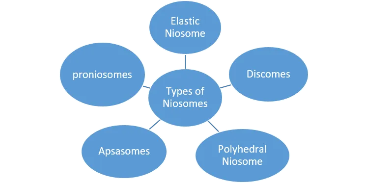
Figure. 2 Types of Niosomes
19PROPERTIES OF NIOSOMES
20,21Comparison between Niosomes and Liposomes
Table.2 Comparision between Niosomes and Liposomes
|
Niosomes |
Liposomes |
|
It uses Non-ionic surfactant-based vesicles |
It uses Phospholipid-based vesicles |
|
It is formed from non-ionic surfactants and cholesterol |
It is Composed of natural or synthetic phospholipids and cholesterol |
|
They are more chemically more stable |
They are less stable |
|
Relatively inexpensive (surfactants are cheaper) |
Expensive (high-purity phospholipids are costly) |
|
Biocompatible and less biodegradable compared to liposomes |
Highly biodegradable and biocompatible |
|
Easier to prepare, requires simpler conditions |
Requires more sophisticated techniques for preparation |
|
Can encapsulate both hydrophilic and lipophilic drugs |
Can encapsulate both hydrophilic and lipophilic drugs |
|
Low toxic due to non-ionic surfactants |
They have very low toxicity as the natural phospholipids resemble cell membranes |
|
Longer shelf life due to better stability |
Shorter shelf life due to phospholipid degradation |
|
Widely used in drug delivery, cosmetics, and vaccines |
Used in drug delivery, gene therapy, and as carriers in cancer treatment |
Structure of Niosomes
22The niosome are circular bilayer structure of non-ionic surfactant like span-60 surfactant which has the ability to form micelle. When surfactant concentration goes above the critical micelle concentration (CMC) then it forms micelles in formation, but non-ionic surfactant has ability to form circular bilayer structure instead of micelles. The cholesterol is also added in formulation to give rigidity to vesicle and ionic surfactant reduces aggregation. The structure of niosome can be uni-lamellar or multi-lamellar depending on which method used for preparation of niosome.
23There are all types of drugs can be incorporate in structure of niosome such as hydrophilic, lipophilic and amphiphilic drugs. Because of their peculiar structure as vesicular systems, niosomes can encapsulate both hydrophilic and lipophilic substances. Lipophilic substances are entrapped by partitioning into the lipophilic domain of the bilayers, whereas hydrophilic substances are adsorbed on the bilayer surfaces.

Figure.3 Structure of Niosome
CHARACTERIZATION OF NIOSOMES
1. Bilayer Rigidity and Homogeneity: Niosomes are determined by the bilayer's rigidity. In homogeneity, dispersion can occur within niosome and can be defined by: PNMR, Differential Calorimetry Scanning (DSC) and Fourier Red Spectroscopy Transform-Infra (FT-IR) techniques. 24
2. Size and Shape: Different methods are used to calculate the mean diameter, such as the process of laser light scattering, as well as electron microscopy, molecular sieve chromatography, photon correlation microscopy, optical microscopy.
3. Stability Study: Niosomal formulations are subject to stability studies by processing for a duration of three months at 4 ° C, 25 ° C and 37 ° C in a thermostatic oven. After a month, the drug quality of all the formulations is tested by entrapping efficiency the parameter of output.
4. Scanning Electron Microscopy: 25In a scanning electron microscope (SEM) (JSM 6100 JEOL, Tokyo, Japan) the niosomes were detected. The niosomes were mounted directly onto the SEM sample stub using double-sided sticking tape and coated under a reduced pressure of 0.001 mmHg with 200 nm thick gold film. At sufficient magnification, photographs were taken.
5. Vesicle Charge: The surface charge of the vesicle plays a significant role in the actions of in vivo and in vitro niosomes. Charged niosomes are more stable than uncharged vesicles against aggregation and fusion. To obtain an approx. surface potential, micro electrophoresis can be used to calculate the zeta potential of individual niosomes. The use of pH-sensitive fluorophores is an alternative approach. More recently, for calculating the zeta potential of niosomes, dynamic light scattering was used.
6. Niosomal Drug Loading and Encapsulation Efficiency: 26The niosomal aqueous suspension was ultracentric, supernatant was removed and sediment is washed twice with distilled water to remove the adsorbent material to assess drug loading and encapsulation capacity.
The entrapment efficiency (EE) was then calculated using formula:
7. In-vitro Release: 27In-vitro release rate study carried out by the use of
1. Dialysis Tubing: A dialysis bag is washed with distilled water. The prepared vesicle suspension is piped into a bag consisting of the dialysis of the tubing and sealed after, then the bag containing the vesicles is put in a 250 ml beaker with a steady shaking at 25 ° C in 200 ml of buffer solution. The buffer is an analysis of the drug content of a suitable system of research at different time intervals.28
2. Reverse dialysis: 29A number of small dialysis is put in proniosomes as containing 1ml of the dissolution medium. The pro-niosomes are then transferred to the process of dissolution. Direct pro-niosome dilution is possible.
Limitation: It is not possible to quantify the rapid release using this method.
3. Franz diffusion cell: 30Using Franz diffusion cell, the study of in vitro diffusion can be performed. Pro-niosomes are placed in a Franz diffusion cell's donor chamber filled with cellophane membrane. The pro-niosomes are then dialyzed at room temperature against an acceptable dissolution medium; and then the samples are removed from the medium at suitable time intervals and tested for drug content using specific methods such as U.V spectroscopy, HPLC, etc.
31COMPOSITION OF NIOSOMES
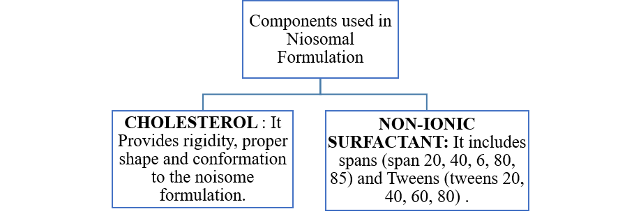
Figure. 4 Composition of Niosome
32Formulation/ Approaches to Niosomes
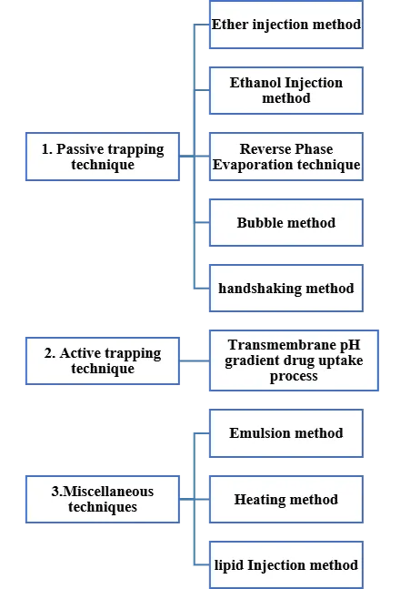
Figure.5 Formulation approaches to Niosomes
Evaluation of Niosomes
EE (%)=Entrapped drug/ Total/drug added×100
39,40 Factors affecting Niosomal Formulation
1. Nature of encapsulated drug
2. Nature and type of surfactant
3. Cholesterol content and charge
4. Resistance to osmotic stress
5. Temperature of hydration
6. Bilayer formation
7. Number of lamellae
8. membrane composition and Rigidity
Recent Advances in Transdermal Niosomal Drug Delivery
Research on niosomes for transdermal drug delivery has expanded in recent years, with emphasis on optimizing preparation methods, exploring novel excipients, and improving final formulations. One important development is the introduction of elastic vesicles, in which edge activators (e.g., ethanol) impart flexibility to the bilayer. 41This increased deformability allows the vesicles to pass more efficiently into deeper skin layers, enhancing drug penetration.
42A drawback of conventional niosomes is their liquid state, which may lead to leakage after topical application. This issue can be resolved by formulating niosomal gels, where gelling agents are added to the dispersion. Such gels provide better retention on the skin and maintain sustained drug release over time. The evolution of niosomes has also led to the creation of proniosomes, which are dry, free-flowing systems that must be hydrated before use to generate niosomes. 43When applied under occlusion, hydration from the skin converts proniosomes into vesicles in situ. This approach minimizes issues such as fusion, leakage, and instability, making proniosomes a more stable alternative for dermal delivery.
44Studies over the past decade have reported encouraging outcomes with NSAIDs delivered via niosomal systems. Oral administration of these drugs often results in gastrointestinal irritation and extensive first-pass metabolism, reducing systemic bioavailability to nearly half. Transdermal formulations bypass these drawbacks and are especially useful for long-term therapy in rheumatic conditions, provided the drug can sufficiently penetrate the skin.45 For example, it evaluates the role of penetration enhancers in proniosomal formulations of nisoldipine, a calcium channel blocker with poor oral bioavailability due to hepatic metabolism. By incorporating lecithin, oleic acid, and propylene glycol, they achieved significant enhancement in transdermal drug permeation. The combination of oleic acid with propylene glycol produced the greatest improvement compared to other enhancers tested. 46In conclusion, elastic vesicles, niosomal gels, and proniosomes represent the latest advances in transdermal niosomal technology. These systems provide better drug stability, enhanced penetration, and improved patient compliance compared to conventional niosomes.
Clinical Trails investigated on Niosomal formulations47
Table.3 Niosomal formulations in Clinical Trials
|
Drug / Active |
Condition / Disease |
Dosage Form |
Dose (if reported) |
Route of Administration |
|
Dexmedetomidine (DEX-NANO) |
Post-operative pain in pediatric cancer patients (bone marrow biopsy/aspiration) |
Niosomal rectal formulation |
Not specified (nano-rectal preparation) |
Rectal |
|
Papilocare (HA + ß-glucan in niosomes) |
HPV-related cervical lesions, cervico-vaginal mucosa repair |
Vaginal gel |
Applied once daily (per trial protocol) |
Vaginal |
|
Zinc sulfate (niosomal 2%) + cryotherapy |
Common warts (verruca vulgaris) |
Topical / intralesional preparation |
2% zinc sulfate niosomal solution |
Topical / Intralesional |
|
Curcumin (CUR-NIO, 0.1%) |
Psoriasis (localized skin lesions) |
Niosomal gel |
0.1% curcumin applied topically |
Topical |
|
Benzoyl peroxide + Clindamycin (niosomal) |
Acne vulgaris |
Niosomal lotion |
- |
Topical (facial) |
|
Kopexil 1% vs Minoxidil 2% (niosomal) |
Androgenic alopecia (hair loss) |
Niosomal lotion |
Kopexil 1% lotion / Minoxidil 2% lotion |
Topical (scalp) |
Challenges and Future Perspectives in Niosomal Drug Delivery
48One of the major challenges in niosomal drug delivery is ensuring biocompatibility while minimizing potential toxicity. Although niosomes are generally regarded as safe, issues such as cytotoxicity, immune responses, and the long-term effects of repeated administration need to be carefully evaluated. Future research must therefore emphasize the optimization of lipid composition, surface modification techniques, and formulation parameters to improve safety and enhance tolerability for clinical applications. Alongside these concerns, regulatory hurdles also present a significant barrier to the successful translation of niosomal formulations from laboratory research to patient care. Approval requires comprehensive preclinical and clinical data to establish safety, efficacy, and quality. 49To overcome these obstacles, efforts should be directed toward standardizing evaluation criteria, streamlining approval processes, and strengthening collaborations between academic researchers, industry stakeholders, and regulatory authorities, thereby accelerating the pathway toward commercialization.
50Looking ahead, several promising directions are shaping the future of niosomal drug delivery. The emergence of multifunctional niosomes marks a significant advancement, as these carriers are designed to simultaneously perform multiple tasks such as targeted drug delivery, imaging, diagnostics, and therapeutic monitoring. Their versatility lies in the incorporation of functional elements like targeting ligands, imaging agents, and stimuli-responsive polymers, which enable site-specific action and controlled drug release. These systems have shown great potential in cancer therapy, infectious diseases, central nervous system disorders, and cardiovascular conditions, where improved delivery efficiency which can lead to superior treatment outcomes. 51However, the complexity of design, potential toxicity of added functional components, and regulatory concerns for combination products remain challenges that need to be addressed.
Another important direction is the integration of personalized medicine approaches, where treatments are tailored to the unique genetic, physiological, and environmental characteristics of individual patients. Niosomes, owing to their flexibility and tunable properties, are well-suited to support precision medicine by enabling customized drug delivery regimens that optimize efficacy while minimizing side effects. Similarly, the integration of nanotechnology into niosomal systems is opening new opportunities to enhance their performance. 52The incorporation of nanomaterials, hybrid Nano systems, and advanced nanoscale fabrication techniques allows precise control over vesicle size, morphology, and release properties, while surface engineering strategies such as PEGylation or ligand attachment can improve targeting and circulation time. Furthermore, advanced analytical tools such as atomic force microscopy, dynamic light scattering, and electron microscopy provide critical insights into niosome structure and behaviour, thereby supporting the design and quality control of more efficient systems53.
Finally, the advancement of niosomal drug delivery technologies depends heavily on collaboration and financial support.54 Strong partnerships between academia, industry, and government agencies are essential to bridge the gap between laboratory research and clinical application. Interdisciplinary projects, technology transfer initiatives, and joint funding programs will play a pivotal role in sustaining innovation and ensuring that promising niosomal formulations can successfully progress to commercial and therapeutic use.55
Drugs in Niosomal Delivery Systems: Route of administration, Indications, Dosage Forms, and Dose.
Table.4 Different Niosomal Drug Delivery Systems
|
Route of Administration |
Drugs (in Niosomes) |
Disease/Condition |
Dosage Form |
Dose (In Studies/Trials) |
|
Intravenous |
Doxorubicin |
Breast cancer, lymphomas |
Niosomal injection |
50–75 mg/m² every 3 weeks |
|
|
Camptothecin |
Colon & ovarian cancer |
Niosomal IV injection |
10–15 mg/m²/day (clinical trial basis) |
|
|
Insulin |
Diabetes mellitus |
Niosomal IV injection |
0.1–0.2 units/kg |
|
|
Zidovudine |
HIV/AIDS |
Niosomal injection |
1–2 mg/kg every 4 hours |
|
|
Cisplatin |
Solid tumours (lung, ovarian, testicular) |
Niosomal injection |
50–100 mg/m² every 3–4 weeks |
|
|
Rifampicin |
Tuberculosis |
Niosomal IV formulation |
10 mg/kg daily |
|
Inhalation |
All-trans retinoic acid |
Lung cancer, acute promyelocytic leukaemia (APL) |
Niosomal aerosol/inhalation suspension |
45 mg/m²/day |
|
Transdermal |
Piroxicam |
Rheumatoid arthritis, osteoarthritis |
Niosomal gel/patch |
20 mg/day (topical equivalent) |
|
|
Estradiol |
Hormone replacement therapy |
Niosomal patch/gel |
0.05–0.1 mg/day |
|
|
Nimesulide |
Inflammation, pain |
Niosomal gel |
100 mg/day equivalent |
|
Ocular |
Timolol maleate |
Glaucoma, ocular hypertension |
Niosomal eye drops |
0.25–0.5% solution, 1 drop twice daily |
|
|
Cyclopentolate |
Mydriasis (eye examination) |
Niosomal eye drops |
1% solution, 1–2 drops |
|
Nasal |
Sumatriptan |
Migraine |
Niosomal nasal spray |
20 mg single dose (repeat after 2 hrs if needed) |
|
|
Influenza vaccines |
Influenza prevention |
Niosomal nasal vaccine |
15 µg hemagglutinin per strain |
CONCLUSION
Niosomes, formed from non-ionic surfactants, are emerging as one of the most effective vesicular drug delivery systems due to their safety, stability, low cost, and ability to encapsulate both hydrophilic and hydrophobic drugs. They enhance solubility, permeability, and bioavailability while reducing toxicity and side effects. Compared to liposomes, niosomes are more chemically stable, economical, and easier to manufacture, making them a promising alternative. They can be prepared using methods such as hand shaking, sonication, and ether injection, and have applications in targeted, ocular, topical, parenteral, and transdermal drug delivery. Niosomal gels, in particular, show improved skin penetration, retention, and therapeutic efficacy, making them suitable for treating fungal infections, eye diseases, and acne. Their “reservoir effect” allows prolonged drug release at the site of action.
Recent Clinical trials on niosomal formulation serves as a next generation drug delivery system with potential significance in localized therapy, oncology and dermatology as they have ability to combine safety, efficacy and patient acceptability which serves as the promising platform for future pharmaceutical innovation. However larger, well‑controlled clinical studies are also an essential components to establish long‑term safety, comparative effectiveness, and regulatory acceptance.
The niosomal drug delivery system have a diverse route of administration like intravenous anticancer agents like doxorubicin, cisplatin, as they have ability to encapsulate both lipophilic drugs and hydrophilic molecules. They can also be given through non-invasive routes like transdermal patches, gels, ocular drops, inhalation aerosols, and nasal sprays which offers targeted drug delivery, improves drug pharmacokinetic parameters including bioavailability and reduces toxicity and overall enhances patient compliance.
Niosomes are not only limited to parenteral delivery, but they represent a platform technology which has the capability of addressing in cancer therapy, infectious diseases, hormonal replacement, pain management and vaccination. They can be the next generation drug delivery system as they have ability to cross the biological barriers especially in the brain and provides a controlled drug release system.
Niosomes are consistently compared with liposomes as they have similar vesicular drug delivery system, they have similar structural and functional characteristics and pharmaceutical applications. These are not compared with aquasomes or electrosomes as they are significantly different in structure, composition and function. As aquasomes have a three layered solid nanoparticle structure and electrosomes have lipid vesicles and are electrically charged as they have ion channels or enzymes they cannot be compared with other nano particulate carriers.
In summary, niosomes represent a versatile, stable, and cost-effective drug delivery system with significant advantages over conventional dosage forms. Their ability to improve targeting and therapeutic outcomes makes them one of the most promising nanocarriers in modern pharmaceutical and healthcare applications.
REFERENCES

Mohd Abdul Muneim Farzaan, Sushmitha Bujji, Recent Progress in Niosomes for Enhanced Bioavailability and Site-Specific Drug Delivery, Int. J. of Pharm. Sci., 2026, Vol 4, Issue 7, 1001-1015. https://doi.org/10.5281/zenodo.21194465
 10.5281/zenodo.21194465