View Article

  • Study of the Combined Effect of Azadirachta indica, Ocimum sanctum, and Curcuma longa Extracts on Common Drug Resistant Microbial Species in India: Evaluation of Antibacterial and Antifungal Activities

  • Department of Pharmaceutics, Maharashtra Institute of Pharmacy (B. Pharm), Betala, Bramhapuri, Maharashtra, India - 441206.

Abstract

The need for alternative sources of antimicrobial agents, such as medicinal plants and polyherbal combinations, has grown due to antimicrobial resistance. The current study assessed the antibacterial efficacy of ethanolic extracts of Curcuma longa (turmeric), Ocimum sanctum (tulsi), and Azadirachta indica (neem) against specific bacterial and fungal infections, both separately and in combination.Soxhlet extraction was used to authenticate and extract plant materials using 95% ethanol. Neem, Tulsi, and Turmeric extract yield percentages were 18.5%, 15.8%, and 21.4%, respectively. Alkaloids, flavonoids, tannins, phenolic chemicals, and glycosides were found in all three extracts, according to preliminary phytochemical screening. Antifungal activity was measured using the agar well diffusion method against Candida albicans and Aspergillus niger, whereas antibacterial activity was examined against Staphylococcus aureus, Escherichia coli, and Pseudomonas aeruginosa. The combined extract generated 19.5 and 18 mm inhibition zones against Candida albicans and Candida niger, respectively. For Neem and Tulsi, the reported MIC values were 1250 µg/mL; for Turmeric, they were 625 µg/mL; and for the combined extract, they were ?156.25 µg/mL. Neem, Tulsi, and Turmeric have FIC values of 0.125, 0.125, and 0.25, respectively, when the reported FIC values were recalculated, yielding a FICI of 0.50.According to the used FICI criterion, the results show increased antibacterial activity of the combined extract and offer preliminary evidence consistent with a synergistic interaction. To validate the relationship, more organism-specific MIC and checkerboard research is needed

Keywords

Study of the Combined Effect of Azadirachta indica, Ocimum sanctum, and Curcuma longa Extracts on Common Drug-Resistant Microbial Species in India: Evaluation of Antibacterial and Antifungal Activities

Introduction

× Popup Image

Due to antimicrobial resistance (AMR), which decreases the efficacy of traditional antimicrobial agents and necessitates the use of other sources of antimicrobial compounds, AMR has emerged as a significant global health concern. Flavonoids, tannins, phenolics, and alkaloids are examples of physiologically active secondary metabolites found in medicinal plants that may have antibacterial properties. [1,2] Neem, or Azadirachta indica, has been extensively studied for its therapeutic qualities and includes a variety of bioactive components that have been shown to have antibacterial activity. [3,8] Another traditional medicinal plant with a number of pharmacologically active components and documented antibacterial qualities is Ocimum sanctum, or Tulsi. [4,9] Similar biological activity, including antibacterial properties, have been shown by Curcuma longa (turmeric) and its primary bioactive component, curcumin. [5, 10, 15]

The use of combinations of natural products may provide enhanced antimicrobial effects through complementary or synergistic interactions among their phytoconstituents. Previous studies have highlighted the potential of combining plant-derived products to improve antimicrobial efficacy. [6,7] Several medicinal plants and spices have also demonstrated activity against pathogenic and multidrug-resistant microorganisms, supporting their investigation as potential sources of antimicrobial agents. [11–14,16,17]

2. MATERIALS AND METHODS

2.1 Collection and Authentication of Plant Materials

Fresh healthy leaves of Azadirachta indica (Neem), fresh leaves of Ocimum sanctum (Tulsi), and mature rhizomes of Curcuma longa (Turmeric) were collected during May–September from authenticated herbal gardens and local medicinal-plant suppliers in Maharashtra, India. Diseased, damaged, and insect-infested materials were excluded. The plant materials were washed with tap water followed by distilled water and air-dried before processing.

The collected plant materials were authenticated by Dr. Arun K. Zingare, Ph.D. Supervisor (Botany), M. B. Patel College of Arts, Commerce and Science, Deori. Voucher/herbarium specimens were preserved for future reference. The assigned herbarium numbers were 01 for Neem, 02 for Tulsi, and 03 for Turmeric.


Table 1. Authentication details of plant materials

Plant

Botanical name

Family

Part used

Herbarium No.

Neem

Azadirachta indica

Meliaceae

Leaves

01

Tulsi

Ocimum sanctum

Lamiaceae

Leaves

02

Turmeric

Curcuma longa

Zingiberaceae

Rhizomes

03


2.2 Preparation of Ethanolic Extracts

The dried plant materials were powdered and extracted separately by continuous Soxhlet extraction using 95% ethanol. For each extraction, 100 g of powdered plant material was extracted with 500 mL ethanol using a Soxhlet apparatus. The solvent was subsequently recovered and the extracts were concentrated and dried. The percentage yield was calculated as:

Percentage yield=Weight of dried extractWeight of plant powder×100

 

The extraction procedure and quantities were based on the experimental procedure documented in the thesis.

2.3 Preliminary Phytochemical Screening

Approximately 1 g of each dried ethanolic extract was dissolved in 10 mL ethanol and filtered. The filtrates were subjected to standard qualitative phytochemical tests for alkaloids, flavonoids, tannins, phenolic compounds, and glycosides.

The reported screening showed positive reactions for alkaloids, flavonoids, tannins, phenolic compounds, and glycosides in all three extracts.

2.4 Antibacterial Activity

Antibacterial activity was evaluated by the agar well diffusion method against Staphylococcus aureus, Escherichia coli, and Pseudomonas aeruginosa. Nutrient agar containing peptone (5 g/L), beef extract (3 g/L), sodium chloride (5 g/L), and agar (15 g/L) was prepared and adjusted to pH 7.2. The medium was sterilized at 121°C and 15 lb pressure for 15 min.

Fresh bacterial cultures were grown in nutrient broth at 37°C for 24 h and adjusted to 0.5 McFarland turbidity, corresponding to approximately 1.5 × 10^8 CFU/mL.

Wells of approximately 6 mm diameter were prepared in inoculated nutrient agar plates. Approximately 100 µL of each individual extract and the combined extract was introduced into the respective wells. After diffusion, plates were incubated at 37°C for 24 h. Zones of inhibition were measured in millimetres. Experiments were performed in triplicate and results were expressed as mean values with standard deviation.

2.5 Antifungal Activity

Antifungal activity was evaluated against Candida albicans and Aspergillus niger using Sabouraud Dextrose Agar (SDA). The medium consisted of peptone (10 g/L), dextrose (40 g/L), agar (15 g/L), and distilled water, with the pH adjusted to approximately 5.6. The medium was sterilized at 121°C and 15 lb pressure for 15 min.

Fungal cultures were subcultured on SDA slants and suspended in sterile saline. The suspensions were uniformly inoculated onto SDA plates. Wells approximately 6 mm in diameter were prepared, and 100 µL of each extract or combined extract was added. Plates were allowed to stand for approximately 30 min for diffusion and were then incubated at 28°C for 48 h. Zones of inhibition were measured in millimetres.

2.6 Preparation of Extract Stock Solutions

Stock solutions were prepared by dissolving 100 mg of dried extract in 10 mL solvent to obtain a concentration of 10 mg/mL, equivalent to 10,000 µg/mL. The thesis records sterile distilled water as the solvent for the stock solution.

The prepared stocks were used for antimicrobial and MIC studies.

2.7 Determination of Minimum Inhibitory Concentration

MIC was determined by the tube dilution method using serial two-fold dilutions of the extract. The tested concentrations were 5000, 2500, 1250, 625, 312.5, and 156.25 µg/mL. The MIC was defined as the lowest concentration showing no visible turbidity.

The MIC experiment in the thesis was conducted using a mixed bacterial inoculum containing S. aureus, E. coli, and P. aeruginosa.

The individual MIC values reported were:

  • Neem: 1250 µg/mL
  • Tulsi: 1250 µg/mL
  • Turmeric: 625 µg/mL
  • Combined extract: ≤156.25 µg/mL

For the combined extract, all tested concentrations down to 156.25 µg/mL were reported as clear. Because 156.25 µg/mL was the lowest concentration tested, the scientifically conservative expression is ≤156.25 µg/mL, rather than an exact MIC of 156.25 µg/mL.

2.8 FIC Index Calculation

The fractional inhibitory concentration was calculated using:

FICA=MICA in combinationMICA alone
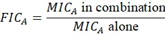
 

and:

FICI=FICNeem+FICTulsi+FICTurmeric

 

Thus:

FICI=0.125+0.125+0.25
FICI=0.50



The thesis used ≤0.5 as the criterion for synergistic interaction.

Important methodological qualification: because the thesis reports a single combined MIC against a mixed bacterial inoculum and does not provide organism-specific combination concentrations, the FICI should be considered preliminary evidence consistent with synergism, rather than definitive confirmation.

2.9 Statistical Analysis

Antimicrobial experiments were performed in triplicate. Results were expressed as mean ± SD. One-way analysis of variance (ANOVA) was used for comparison, with p < 0.05 considered statistically significant according to the thesis methodology.

Because replicate-level raw measurements were not available in the manuscript source, the reported SD values are retained as thesis-reported values and should ideally be verified against the original laboratory observations before journal submission.

3. RESULTS

3.1 Extraction Yield

The extraction yields differed among the three medicinal plants. Turmeric showed the highest yield (21.4%), followed by Neem (18.5%) and Tulsi (15.8%).


Table 2. Percentage yield of ethanolic extracts

Plant

Powder taken (g)

Extract obtained (g)

Yield (%)

A. indica

100

18.5

18.5

O. sanctum

100

15.8

15.8

C. longa

100

21.4

21.4


The relatively higher extractive yield of Turmeric indicates greater recovery of ethanol-soluble constituents under the selected extraction conditions.

3.2 Preliminary Phytochemical Screening

All three ethanolic extracts showed positive reactions for the major phytochemical groups evaluated.


Table 3. Preliminary phytochemical screening

Phytoconstituent

Neem

Tulsi

Turmeric

Alkaloids

+

+

+

Flavonoids

+

+

+

Tannins

+

+

+

Phenolic compounds

+

+

+

Glycosides

+

+

+


The presence of these classes of secondary metabolites provides a possible chemical basis for the observed antimicrobial activity, although qualitative screening does not establish the identity, concentration, or contribution of individual compounds.

3.3 Antibacterial Activity

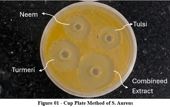
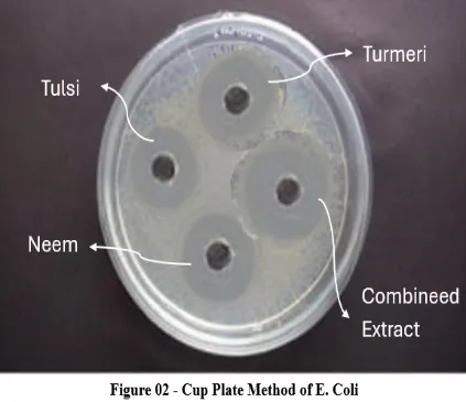
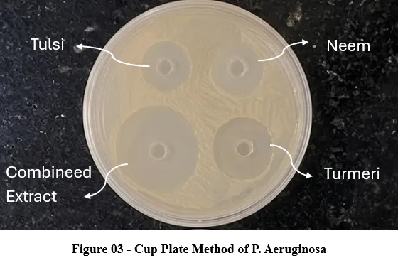
The individual extracts demonstrated antibacterial activity against all three tested organisms. Turmeric showed the greatest individual antibacterial activity, whereas the combined extract produced the largest zones of inhibition.


Table 4. Antibacterial activity of individual and combined extracts

Test organism

Neem (mm)

Tulsi (mm)

Turmeric (mm)

Combined (mm)

S. aureus

10.0

9.5

14.0

21.0

E. coli

9.5

8.0

13.0

20.0

P. aeruginosa

8.0

7.5

13.0

20.5


The combined extract produced the highest inhibition against all three bacterial organisms. The reported overall mean inhibition zones were 9.16 ± 0.5 mm for Neem, 8.3 ± 0.4 mm for Tulsi, 13.33 ± 0.3 mm for Turmeric, and 20.5 ± 0.6 mm for the combined extract, with reported statistical significance at p < 0.05.
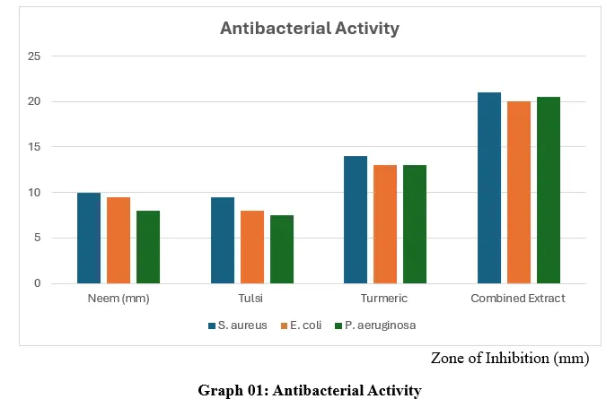


These findings demonstrate enhanced antibacterial activity of the combined preparation compared with the individual extracts. However, inhibition-zone enhancement alone should not be interpreted as definitive proof of synergism.


3.4 Antifungal Activity

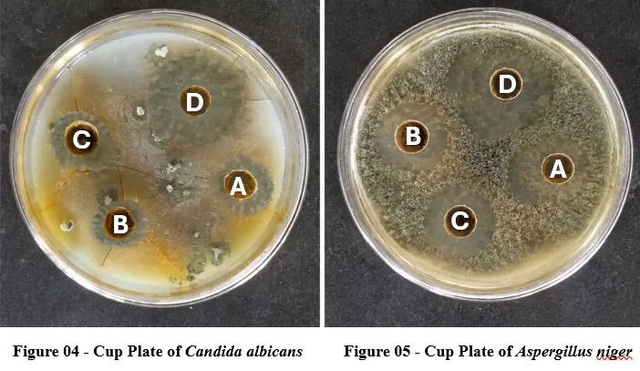
All tested extracts showed antifungal activity against C. albicans and A. niger. Among the individual extracts, Neem produced the greatest inhibition zones.


Table 5. Antifungal activity of individual and combined extracts

Test organism

Neem (mm)

Tulsi (mm)

Turmeric (mm)

Combined (mm)

C. albicans

12.5

10.0

10.5

19.5

A. niger

11.0

10.0

9.0

18.0


The combined extract produced 19.5 mm inhibition against C. albicans and 18 mm against A. niger, compared with 12.5 and 11 mm, respectively, for Neem, the most active individual extract.

Relative to Neem, the combined extract increased the inhibition zone by approximately 56.0% against C. albicans and 63.6% against A. niger.

The thesis reported overall mean zones of 11.75 ± 0.4 mm for Neem, 10.0 ± 0.3 mm for Tulsi, 9.75 ± 0.2 mm for Turmeric, and 18.75 ± 0.5 mm for the combined extract, with p < 0.05.

3.5 Minimum Inhibitory Concentration

The MIC results showed greater inhibitory potency of the combined preparation than the individual extracts.


Table 6. Comparative MIC values

Extract

MIC reported (µg/mL)

Neem

1250

Tulsi

1250

Turmeric

625

Combined extract

≤156.25


Neem and Tulsi showed growth inhibition up to 1250 µg/mL, while turbidity appeared at 625 µg/mL and lower concentrations.

Turmeric showed a lower MIC of 625 µg/mL. The combined extract remained clear at all tested concentrations, including 156.25 µg/mL.

The combined preparation therefore required substantially less extract to inhibit the tested mixed bacterial inoculum than the individual extracts.

3.6 Corrected FIC Index


Table 7. Corrected FIC calculation

Extract

MIC alone (µg/mL)

MIC in combination used for calculation (µg/mL)

Corrected FIC

Neem

1250

156.25

0.125

Tulsi

1250

156.25

0.125

Turmeric

625

156.25

0.250

FICI

—

—

0.500


The original thesis reported Neem FIC as 0.25, but 156.25/1250 equals 0.125. Correcting this arithmetic produces a total FICI of 0.50.

According to the criterion used in the thesis, a FICI ≤0.5 was interpreted as synergistic.

Therefore, the corrected FICI of 0.50 is consistent with synergistic interaction under the predefined criterion.

Nevertheless, because the MIC experiment was performed against a mixed bacterial inoculum, this result should be regarded as preliminary evidence rather than definitive confirmation of organism-specific synergism.

DISCUSSION

The present study investigated the antimicrobial potential of ethanolic extracts of Neem, Tulsi, and Turmeric, both individually and in combination. The study was motivated by the increasing need for alternative antimicrobial sources in the context of growing antimicrobial resistance. Plant-derived products contain diverse secondary metabolites that can exhibit antimicrobial effects through multiple mechanisms.

Soxhlet extraction with 95% ethanol produced yields of 18.5%, 15.8%, and 21.4% for Neem, Tulsi, and Turmeric, respectively. Turmeric gave the highest yield under the selected extraction conditions. Differences in extractive yield may reflect variation in plant part, chemical composition, polarity of constituents, moisture content, and extraction efficiency. The present yield results are therefore useful for comparing recovery under the specific experimental conditions but should not be interpreted as direct measures of antimicrobial potency.

Preliminary phytochemical screening demonstrated alkaloids, flavonoids, tannins, phenolic compounds, and glycosides in all three extracts. These findings provide a broad phytochemical basis for the observed antimicrobial effects. However, qualitative phytochemical screening cannot establish which individual compounds are responsible for activity. Quantitative chromatographic or spectroscopic characterization would therefore be required to correlate specific constituents with antimicrobial effects.

The antibacterial results demonstrated clear differences among the extracts. Turmeric was the most active individual extract, producing inhibition zones of 14 mm against S. aureus and 13 mm against both E. coli and P. aeruginosa. Neem and Tulsi showed lower inhibition zones. The stronger activity of Turmeric may reflect its characteristic bioactive constituents, although the present study did not chemically quantify these compounds.

The combined extract produced considerably larger inhibition zones than the individual preparations. Zones of 21, 20, and 20.5 mm were observed against S. aureus, E. coli, and P. aeruginosa, respectively. This consistent increase across both Gram-positive and Gram-negative bacteria suggests that combining the extracts may improve overall antimicrobial activity. Plant combinations can theoretically provide complementary mechanisms of action because their constituents may interact with multiple microbial targets. Such multi-component interactions have been discussed in the phytopharmaceutical literature.

The antifungal results followed a similar pattern. Neem demonstrated the strongest individual antifungal activity, whereas the combined preparation produced the greatest inhibition against both C. albicans and A. niger. The increase from 12.5 to 19.5 mm against C. albicans and from 11 to 18 mm against A. niger indicates substantially enhanced activity of the combined extract.

The MIC findings further support the enhanced antimicrobial activity of the combination. Neem and Tulsi showed MIC values of 1250 µg/mL, while Turmeric showed 625 µg/mL. The combined extract remained inhibitory at the lowest tested concentration of 156.25 µg/mL. Because this was the lowest concentration tested, its MIC should conservatively be reported as ≤156.25 µg/mL rather than exactly 156.25 µg/mL.

The FIC calculation required correction. The thesis stated:

FICNeem=156.251250=0.25
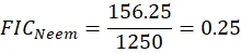
 

but the correct arithmetic is:

156.251250=0.125
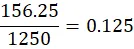
 

Thus, the corrected FIC values are 0.125 for Neem, 0.125 for Tulsi, and 0.25 for Turmeric, giving:

FICI=0.125+0.125+0.25=0.50

 

The corrected value is therefore consistent with the thesis's predefined criterion for synergistic interaction.

However, an important methodological limitation must be acknowledged. The MIC assay was conducted using a mixed bacterial inoculum containing S. aureus, E. coli, and P. aeruginosa. Consequently, the reported MIC represents inhibition of the mixed inoculum rather than a species-specific MIC. Similarly, the FICI calculation is based on the reported overall combination value rather than separate organism-specific checkerboard matrices. FICI methodology is useful for investigating antimicrobial interactions, but interpretation depends strongly on experimental design and reproducibility. (PubMed)

Therefore, the present findings should be described as enhanced antimicrobial activity with preliminary evidence consistent with synergistic interaction, rather than definitive confirmation of synergism.

A further limitation is that the study used preliminary phytochemical screening rather than quantitative chemical profiling. Future work should identify and quantify the principal active constituents using chromatographic techniques such as HPLC, LC-MS, or GC-MS where appropriate. Species-specific MIC testing and checkerboard assays should also be conducted separately against each microorganism. Time-kill experiments could provide additional evidence regarding the dynamics of the interaction.

CONCLUSION

The present study demonstrated that ethanolic extracts of Azadirachta indica, Ocimum sanctum, and Curcuma longa possess antimicrobial activity against selected bacterial and fungal pathogens. Turmeric exhibited the highest antibacterial activity among the individual extracts, whereas Neem showed the greatest individual antifungal activity.

The combined extract demonstrated markedly enhanced antimicrobial activity, producing larger inhibition zones against all tested microorganisms than the individual extracts. The combined preparation also showed a substantially lower reported MIC (≤156.25 µg/mL) than Neem, Tulsi, or Turmeric individually.

Correction of the FIC arithmetic yielded FIC values of 0.125, 0.125, and 0.25 for Neem, Tulsi, and Turmeric, respectively, resulting in a FICI of 0.50. According to the criterion used in the study, this value is consistent with a synergistic interaction.

However, because the MIC/FIC experiment was performed using a mixed bacterial inoculum and did not include organism-specific checkerboard testing, the result should be regarded as preliminary evidence of synergism rather than definitive confirmation. Further species-specific MIC, checkerboard FICI, time-kill, quantitative phytochemical, toxicity, and in-vivo studies are recommended to establish the reproducibility, mechanism, safety, and therapeutic relevance of the polyherbal combination.

REFERENCES
  1. Ventola CL. The antibiotic resistance crisis: causes and threats. Pharmacy and Therapeutics. 2015;40(4):277-283.
  2. Cowan MM. Plant products as antimicrobial agents. Clinical Microbiology Reviews. 1999;12(4):564-582.
  3. Biswas K, Chattopadhyay I, Banerjee RK, Bandyopadhyay U. Biological activities and medicinal properties of Neem (Azadirachta indica). Current Science. 2002;82(11):1336-1345.
  4. Prakash P, Gupta N. Therapeutic uses of Ocimum sanctum Linn. (Tulsi) with a note on eugenol and its pharmacological actions: a short review. Indian Journal of Physiology and Pharmacology. 2005;49(2):125-131.
  5. Gupta SC, Patchva S, Aggarwal BB. Therapeutic roles of curcumin: lessons learned from clinical trials. In: Aggarwal BB, Surh YJ, Shishodia S, editors. The molecular targets and therapeutic uses of curcumin in health and disease. New York: Springer; 2013. p. 1-75.
  6. Hemaiswarya S, Kruthiventi AK, Doble M. Synergism between natural products and antibiotics against infectious diseases. Phytomedicine. 2008;15(8):639-652.
  7. Wagner H, Ulrich-Merzenich G. Synergy research: approaching a new generation of phytopharmaceuticals. Phytomedicine. 2009;16(2-3):97-110.
  8. Subapriya R, Nagini S. Medicinal properties of Neem leaves: a review. Current Medicinal Chemistry – Anti-Cancer Agents. 2005;5(2):149-156.
  9. Singh P, Mishra G, Srivastava S, Jha KK, Khosa RL. Traditional uses, phytochemistry and pharmacological properties of Ocimum sanctum: a review. International Journal of Pharmaceutical Sciences and Research. 2010;1(1):44-48.
  10. Aggarwal BB, Sundaram C, Malani N, Ichikawa H. Curcumin: the Indian solid gold. In: Aggarwal BB, Surh YJ, Shishodia S, editors. Advances in Experimental Medicine and Biology. New York: Springer; 2007. Vol. 595, p. 1-75.
  11. Alzoreky NS, Nakahara K. Antibacterial activity of extracts from some edible plants commonly consumed in Asia. International Journal of Food Microbiology. 2003;80(3):223-230.
  12. Arora DS, Kaur J. Antimicrobial activity of spices. International Journal of Antimicrobial Agents. 1999;12(3):257-262.
  13. Ahmad I, Beg AZ. Antimicrobial and phytochemical studies on 45 Indian medicinal plants against multi-drug resistant human pathogens. Journal of Ethnopharmacology. 2001;74(2):113-123.
  14. Rai MK, Pandit R, Gaikwad S, Kövics G. Antimicrobial peptides as natural bio-preservatives against food spoilage microorganisms. Journal of Food Science and Technology. 2016;56(3):120-129.
  15. Harish R, Shanmugam S. Antibacterial activity of Curcuma longa against human pathogens. Journal of Medicinal Plants Research. 2012;6(8):1481-1483.
  16. Raut JS, Karuppayil SM. A status review on the medicinal properties of essential oils. Industrial Crops and Products. 2014;62:250-264.
  17. Chaudhary H, Kaur G. Antibacterial activity of Neem (Azadirachta indica) leaf extracts against pathogenic bacteria. International Journal of Pharmaceutical Sciences Review and Research. 2016;36(2):221-225.
  18. Mondal S, Varma S, Bamola VD, Naik SN, Mirdha BR, Padhi MM, et al. Double-blinded randomized controlled trial for immunomodulatory effects of Tulsi (Ocimum sanctum) among healthy volunteers. Journal of Ethnopharmacology. 2011;136(3):452-456.
  19. Tyagi P, Singh M, Kumari H, Kumari A, Mukhopadhyay K. Bactericidal activity of curcumin against clinical isolates of Staphylococcus aureus. World Journal of Microbiology and Biotechnology. 2015;31(1):1-10.
  20. Bansal S, Choudhary S, Sharma M, Kumar S, Lohan S, Bhardwaj V, et al. Synergistic antibacterial activity of natural plant extracts. Journal of Applied Pharmaceutical Science. 2014;4(8):92-96.
  21. Pandey G, Madhuri S. Pharmacological activities of Ocimum sanctum (Tulsi): a review. International Journal of Pharmaceutical Sciences Review and Research. 2010;5(1):61-66.
  22. Pelczar MJ, Chan ECS, Krieg NR. Microbiology: concepts and applications. New York: McGraw-Hill Education; 1993. p. 45-678.
  23. Hugo WB, Russell AD. Pharmaceutical microbiology. 7th ed. Oxford: Blackwell Scientific Publications; 2004. p. 1-512.
  24. Ananthanarayan R, Paniker CKJ. Textbook of microbiology. 8th ed. Chennai: Orient Longman Pvt Ltd; 2009. p. 1-672.
  25. Jain NK. Pharmaceutical microbiology. New Delhi: Vallabh Prakashan; 2016. p. 1-412.
  26. Prescott LM, Harley JP, Klein DA. Microbiology. 6th ed. New York: McGraw-Hill Companies; 2005. p. 1-992.
  27. Cooper and Gunn. Tutorial pharmacy. 6th ed. New Delhi: CBS Publishers and Distributors; 2008. p. 1-780.
  28. Indian Pharmacopoeia Commission. Indian Pharmacopoeia. Ghaziabad: Ministry of Health and Family Welfare, Government of India; 2022. Vol. 1, p. 1-854.
  29. British Pharmacopoeia Commission. British Pharmacopoeia 2022. London: The Stationery Office; 2022. Vol. 1, p. 1-1250.
  30. United States Pharmacopeial Convention. United States Pharmacopeia and National Formulary (USP 46-NF 41). Rockville, MD: USP Convention; 2023. p. 1-5632.
  31. Bergey DH, Holt JG. Bergey’s manual of determinative bacteriology. 9th ed. Baltimore: Williams and Wilkins; 1994. p. 1-787.
  32. Kokate CK, Purohit AP, Gokhale SB. Pharmacognosy. 56th ed. Pune: Nirali Prakashan; 2019. p. 1-980.
  33. Trease GE, Evans WC. Trease and Evans pharmacognosy. 16th ed. London: Saunders Elsevier; 2009. p. 1-832.
  34. Harborne JB. Phytochemical methods: a guide to modern techniques of plant analysis. 3rd ed. London: Chapman and Hall; 1998. p. 1-302.
  35. Sofowora A. Medicinal plants and traditional medicine in Africa. 2nd ed. Ibadan: Spectrum Books Ltd; 1993. p. 1-289.
  36. Khandelwal KR. Practical pharmacognosy: techniques and experiments. 23rd ed. Pune: Nirali Prakashan; 2017. p. 1-220.
  37. Mukherjee PK. Quality control of herbal drugs. New Delhi: Business Horizons Pharmaceutical Publishers; 2007. p. 1-804.
  38. Evans WC. Trease and Evans pharmacognosy. 15th ed. London: Elsevier Health Sciences; 2009. p. 1-616.
  39. Dahanukar SA, Kulkarni RA, Rege NN. Pharmacology of medicinal plants and natural products. Indian Journal of Pharmacology. 2000;32(Suppl 1):S81-S118.
  40. Sharma A, Flores-Vallejo RC, Cardoso-Taketa A, Villarreal ML. Antibacterial activities of medicinal plants used in Mexican traditional medicine. Journal of Ethnopharmacology. 2017;208:264-329.
  41. Lewis K, Ausubel FM. Prospects for plant-derived antibacterials. Nature Biotechnology. 2006;24(12):1504-1507.
  42. Nostro A, Papalia T. Antimicrobial activity of plant extracts. Current Medicinal Chemistry. 2012;19(7):1062-1081.
  43. Fabricant DS, Farnsworth NR. The value of plants used in traditional medicine for drug discovery. Environmental Health Perspectives. 2001;109(Suppl 1):69-75.
  44. Gibbons S. Plants as a source of bacterial resistance modulators and anti-infective agents. Phytochemistry Reviews. 2005;4(1):63-78.
  45. Parekh J, Chanda S. In vitro antimicrobial activity and phytochemical analysis of some Indian medicinal plants. African Journal of Biomedical Research. 2007;10(2):175-181.
  46. Nair R, Chanda S. Antibacterial activities of some medicinal plants of the Western region of India. Turkish Journal of Biology. 2007;31(4):231-236.
  47. Mahady GB. Medicinal plants for the prevention and treatment of bacterial infections. Current Pharmaceutical Design. 2005;11(19):2405-2427.
  48. Rios JL, Recio MC. Medicinal plants and antimicrobial activity. Journal of Ethnopharmacology. 2005;100(1-2):80-84.
  49. Burt S. Essential oils: their antibacterial properties and potential applications in foods. International Journal of Food Microbiology. 2004;94(3):223-253.
  50. Lambert RJW, Skandamis PN, Coote PJ, Nychas GJE. A study of the minimum inhibitory concentration and mode of action of oregano essential oil. Journal of Applied Microbiology. 2001;91(3):453-462.
  51. Odds FC. Synergy, antagonism, and what the chequerboard puts between them. Journal of Antimicrobial Chemotherapy. 2003;52(1):1.
  52. Eliopoulos GM, Moellering RC. Antimicrobial combinations. In: Lorian V, editor. Antibiotics in laboratory medicine. 4th ed. Baltimore: Williams and Wilkins; 1996. p. 330-396.
  53. Cheesbrough M. District laboratory practice in tropical countries. 2nd ed. Cambridge: Cambridge University Press; 2006. p. 1-434.
  54. Cappuccino JG, Sherman N. Microbiology: a laboratory manual. 10th ed. California: Pearson Education; 2014. p. 1-560.
  55. Collee JG, Fraser AG, Marmion BP, Simmons A. Mackie and McCartney practical medical microbiology. 14th ed. London: Churchill Livingstone; 2007. p. 1-978.
  56. Forbes BA, Sahm DF, Weissfeld AS. Bailey and Scott’s diagnostic microbiology. 12th ed. Missouri: Mosby Elsevier; 2007. p. 1-1032.
  57. Willey JM, Sherwood LM, Woolverton CJ. Prescott’s principles of microbiology. New York: McGraw-Hill Higher Education; 2011. p. 1-1088.
  58. Tortora GJ, Funke BR, Case CL. Microbiology: an introduction. 11th ed. California: Benjamin Cummings; 2013. p. 1-960.
  59. Pelczar MJ, Reid RD, Chan ECS. Microbiology. 5th ed. New Delhi: Tata McGraw-Hill Publishing Company; 2001. p. 1-952.
  60. Atlas RM. Handbook of microbiological media. 3rd ed. Boca Raton: CRC Press; 2004. p. 1-2056.
  61. Madigan MT, Martinko JM, Parker J. Brock biology of microorganisms. 12th ed. New Jersey: Pearson Prentice Hall; 2008. p. 1-1061.
  62. Pelczar MJ, Chan ECS, Krieg NR. Microbiology: concepts and applications. New York: McGraw-Hill Book Company; 1993. p. 1-896.

Reference

  1. Ventola CL. The antibiotic resistance crisis: causes and threats. Pharmacy and Therapeutics. 2015;40(4):277-283.
  2. Cowan MM. Plant products as antimicrobial agents. Clinical Microbiology Reviews. 1999;12(4):564-582.
  3. Biswas K, Chattopadhyay I, Banerjee RK, Bandyopadhyay U. Biological activities and medicinal properties of Neem (Azadirachta indica). Current Science. 2002;82(11):1336-1345.
  4. Prakash P, Gupta N. Therapeutic uses of Ocimum sanctum Linn. (Tulsi) with a note on eugenol and its pharmacological actions: a short review. Indian Journal of Physiology and Pharmacology. 2005;49(2):125-131.
  5. Gupta SC, Patchva S, Aggarwal BB. Therapeutic roles of curcumin: lessons learned from clinical trials. In: Aggarwal BB, Surh YJ, Shishodia S, editors. The molecular targets and therapeutic uses of curcumin in health and disease. New York: Springer; 2013. p. 1-75.
  6. Hemaiswarya S, Kruthiventi AK, Doble M. Synergism between natural products and antibiotics against infectious diseases. Phytomedicine. 2008;15(8):639-652.
  7. Wagner H, Ulrich-Merzenich G. Synergy research: approaching a new generation of phytopharmaceuticals. Phytomedicine. 2009;16(2-3):97-110.
  8. Subapriya R, Nagini S. Medicinal properties of Neem leaves: a review. Current Medicinal Chemistry – Anti-Cancer Agents. 2005;5(2):149-156.
  9. Singh P, Mishra G, Srivastava S, Jha KK, Khosa RL. Traditional uses, phytochemistry and pharmacological properties of Ocimum sanctum: a review. International Journal of Pharmaceutical Sciences and Research. 2010;1(1):44-48.
  10. Aggarwal BB, Sundaram C, Malani N, Ichikawa H. Curcumin: the Indian solid gold. In: Aggarwal BB, Surh YJ, Shishodia S, editors. Advances in Experimental Medicine and Biology. New York: Springer; 2007. Vol. 595, p. 1-75.
  11. Alzoreky NS, Nakahara K. Antibacterial activity of extracts from some edible plants commonly consumed in Asia. International Journal of Food Microbiology. 2003;80(3):223-230.
  12. Arora DS, Kaur J. Antimicrobial activity of spices. International Journal of Antimicrobial Agents. 1999;12(3):257-262.
  13. Ahmad I, Beg AZ. Antimicrobial and phytochemical studies on 45 Indian medicinal plants against multi-drug resistant human pathogens. Journal of Ethnopharmacology. 2001;74(2):113-123.
  14. Rai MK, Pandit R, Gaikwad S, Kövics G. Antimicrobial peptides as natural bio-preservatives against food spoilage microorganisms. Journal of Food Science and Technology. 2016;56(3):120-129.
  15. Harish R, Shanmugam S. Antibacterial activity of Curcuma longa against human pathogens. Journal of Medicinal Plants Research. 2012;6(8):1481-1483.
  16. Raut JS, Karuppayil SM. A status review on the medicinal properties of essential oils. Industrial Crops and Products. 2014;62:250-264.
  17. Chaudhary H, Kaur G. Antibacterial activity of Neem (Azadirachta indica) leaf extracts against pathogenic bacteria. International Journal of Pharmaceutical Sciences Review and Research. 2016;36(2):221-225.
  18. Mondal S, Varma S, Bamola VD, Naik SN, Mirdha BR, Padhi MM, et al. Double-blinded randomized controlled trial for immunomodulatory effects of Tulsi (Ocimum sanctum) among healthy volunteers. Journal of Ethnopharmacology. 2011;136(3):452-456.
  19. Tyagi P, Singh M, Kumari H, Kumari A, Mukhopadhyay K. Bactericidal activity of curcumin against clinical isolates of Staphylococcus aureus. World Journal of Microbiology and Biotechnology. 2015;31(1):1-10.
  20. Bansal S, Choudhary S, Sharma M, Kumar S, Lohan S, Bhardwaj V, et al. Synergistic antibacterial activity of natural plant extracts. Journal of Applied Pharmaceutical Science. 2014;4(8):92-96.
  21. Pandey G, Madhuri S. Pharmacological activities of Ocimum sanctum (Tulsi): a review. International Journal of Pharmaceutical Sciences Review and Research. 2010;5(1):61-66.
  22. Pelczar MJ, Chan ECS, Krieg NR. Microbiology: concepts and applications. New York: McGraw-Hill Education; 1993. p. 45-678.
  23. Hugo WB, Russell AD. Pharmaceutical microbiology. 7th ed. Oxford: Blackwell Scientific Publications; 2004. p. 1-512.
  24. Ananthanarayan R, Paniker CKJ. Textbook of microbiology. 8th ed. Chennai: Orient Longman Pvt Ltd; 2009. p. 1-672.
  25. Jain NK. Pharmaceutical microbiology. New Delhi: Vallabh Prakashan; 2016. p. 1-412.
  26. Prescott LM, Harley JP, Klein DA. Microbiology. 6th ed. New York: McGraw-Hill Companies; 2005. p. 1-992.
  27. Cooper and Gunn. Tutorial pharmacy. 6th ed. New Delhi: CBS Publishers and Distributors; 2008. p. 1-780.
  28. Indian Pharmacopoeia Commission. Indian Pharmacopoeia. Ghaziabad: Ministry of Health and Family Welfare, Government of India; 2022. Vol. 1, p. 1-854.
  29. British Pharmacopoeia Commission. British Pharmacopoeia 2022. London: The Stationery Office; 2022. Vol. 1, p. 1-1250.
  30. United States Pharmacopeial Convention. United States Pharmacopeia and National Formulary (USP 46-NF 41). Rockville, MD: USP Convention; 2023. p. 1-5632.
  31. Bergey DH, Holt JG. Bergey’s manual of determinative bacteriology. 9th ed. Baltimore: Williams and Wilkins; 1994. p. 1-787.
  32. Kokate CK, Purohit AP, Gokhale SB. Pharmacognosy. 56th ed. Pune: Nirali Prakashan; 2019. p. 1-980.
  33. Trease GE, Evans WC. Trease and Evans pharmacognosy. 16th ed. London: Saunders Elsevier; 2009. p. 1-832.
  34. Harborne JB. Phytochemical methods: a guide to modern techniques of plant analysis. 3rd ed. London: Chapman and Hall; 1998. p. 1-302.
  35. Sofowora A. Medicinal plants and traditional medicine in Africa. 2nd ed. Ibadan: Spectrum Books Ltd; 1993. p. 1-289.
  36. Khandelwal KR. Practical pharmacognosy: techniques and experiments. 23rd ed. Pune: Nirali Prakashan; 2017. p. 1-220.
  37. Mukherjee PK. Quality control of herbal drugs. New Delhi: Business Horizons Pharmaceutical Publishers; 2007. p. 1-804.
  38. Evans WC. Trease and Evans pharmacognosy. 15th ed. London: Elsevier Health Sciences; 2009. p. 1-616.
  39. Dahanukar SA, Kulkarni RA, Rege NN. Pharmacology of medicinal plants and natural products. Indian Journal of Pharmacology. 2000;32(Suppl 1):S81-S118.
  40. Sharma A, Flores-Vallejo RC, Cardoso-Taketa A, Villarreal ML. Antibacterial activities of medicinal plants used in Mexican traditional medicine. Journal of Ethnopharmacology. 2017;208:264-329.
  41. Lewis K, Ausubel FM. Prospects for plant-derived antibacterials. Nature Biotechnology. 2006;24(12):1504-1507.
  42. Nostro A, Papalia T. Antimicrobial activity of plant extracts. Current Medicinal Chemistry. 2012;19(7):1062-1081.
  43. Fabricant DS, Farnsworth NR. The value of plants used in traditional medicine for drug discovery. Environmental Health Perspectives. 2001;109(Suppl 1):69-75.
  44. Gibbons S. Plants as a source of bacterial resistance modulators and anti-infective agents. Phytochemistry Reviews. 2005;4(1):63-78.
  45. Parekh J, Chanda S. In vitro antimicrobial activity and phytochemical analysis of some Indian medicinal plants. African Journal of Biomedical Research. 2007;10(2):175-181.
  46. Nair R, Chanda S. Antibacterial activities of some medicinal plants of the Western region of India. Turkish Journal of Biology. 2007;31(4):231-236.
  47. Mahady GB. Medicinal plants for the prevention and treatment of bacterial infections. Current Pharmaceutical Design. 2005;11(19):2405-2427.
  48. Rios JL, Recio MC. Medicinal plants and antimicrobial activity. Journal of Ethnopharmacology. 2005;100(1-2):80-84.
  49. Burt S. Essential oils: their antibacterial properties and potential applications in foods. International Journal of Food Microbiology. 2004;94(3):223-253.
  50. Lambert RJW, Skandamis PN, Coote PJ, Nychas GJE. A study of the minimum inhibitory concentration and mode of action of oregano essential oil. Journal of Applied Microbiology. 2001;91(3):453-462.
  51. Odds FC. Synergy, antagonism, and what the chequerboard puts between them. Journal of Antimicrobial Chemotherapy. 2003;52(1):1.
  52. Eliopoulos GM, Moellering RC. Antimicrobial combinations. In: Lorian V, editor. Antibiotics in laboratory medicine. 4th ed. Baltimore: Williams and Wilkins; 1996. p. 330-396.
  53. Cheesbrough M. District laboratory practice in tropical countries. 2nd ed. Cambridge: Cambridge University Press; 2006. p. 1-434.
  54. Cappuccino JG, Sherman N. Microbiology: a laboratory manual. 10th ed. California: Pearson Education; 2014. p. 1-560.
  55. Collee JG, Fraser AG, Marmion BP, Simmons A. Mackie and McCartney practical medical microbiology. 14th ed. London: Churchill Livingstone; 2007. p. 1-978.
  56. Forbes BA, Sahm DF, Weissfeld AS. Bailey and Scott’s diagnostic microbiology. 12th ed. Missouri: Mosby Elsevier; 2007. p. 1-1032.
  57. Willey JM, Sherwood LM, Woolverton CJ. Prescott’s principles of microbiology. New York: McGraw-Hill Higher Education; 2011. p. 1-1088.
  58. Tortora GJ, Funke BR, Case CL. Microbiology: an introduction. 11th ed. California: Benjamin Cummings; 2013. p. 1-960.
  59. Pelczar MJ, Reid RD, Chan ECS. Microbiology. 5th ed. New Delhi: Tata McGraw-Hill Publishing Company; 2001. p. 1-952.
  60. Atlas RM. Handbook of microbiological media. 3rd ed. Boca Raton: CRC Press; 2004. p. 1-2056.
  61. Madigan MT, Martinko JM, Parker J. Brock biology of microorganisms. 12th ed. New Jersey: Pearson Prentice Hall; 2008. p. 1-1061.
  62. Pelczar MJ, Chan ECS, Krieg NR. Microbiology: concepts and applications. New York: McGraw-Hill Book Company; 1993. p. 1-896.

Photo
Pranali Wanjari
Corresponding author

Dipartment of Pharmaceutics, Maharashtra Institute of Pharmacy (B. Pharm), Betala, Bramhapuri - 441206

Photo
Pruthviraj Meshram
Co-author

Associate Professor, Dipartment of Pharmaceutics, Maharashtra Institute of Pharmacy (B. Pharm), Betala, Bramhapuri - 441206

Photo
Dr. Sachin Dudhe
Co-author

Principal, Maharashtra Institute of Pharmacy (B. Pharm), Betala, Bramhapuri - 441206

Pranali Wanjari, Pruthviraj Meshram, Dr. Sachin Dudhe, Study of the Combined Effect of Azadirachta indica, Ocimum sanctum, and Curcuma longa Extracts on Common Drug Resistant Microbial Species in India: Evaluation of Antibacterial and Antifungal Activities, Int. J. of Pharm. Sci., 2026, Vol 4, Issue 9, 4026-4037, https://doi.org/10.5281/zenodo.23050998

More related articles
Formulation and Evaluation of a “Matrisva”- Ma...
Suprajita Das, Dr. Kavitha PN, Anuradha Shyam...
Artifical Intelliegence And Machine Learning in So...
Pratik patel, Nimita Manocha, Rajveer Singh Chouhan, Shivanshu Dw...
Transferosome-Based Drug Delivery Systems: Formulation Approaches, Characterizat...
Athira Balachandran, Fasna Nargees N. H., Nimmi Thankam Biju, Athira B. Nair, Praveen Raj R...
Chitosan-Coated Cubosomes for Nose-to-Brain Delivery of Levodopa in Parkinson’...
Krishnapriya E K, Athira B Nair, Nimmi Thankam Biju, Fasna Nargees N H, Gopikrishna S Pai, Preethy C...
Dasatinib: Mechanism of Action, Pharmacokinetic–Pharmacodynamic Profile, Resis...
Nishant Mali, Avinash Hosmani, Yogesh Patil, Kiran Sawase, Kratik More...
Related Articles
Gallic Acid and Neuroprotection: A Critical Review of Preclinical Evidence and M...
Thanuja N K, Raja Lakshmi K S, Bhavani G, Masthanbi M , Nagesh S K...
Insulin Resistance and Metabolic Syndrome: Recognising Early Warning Signs Throu...
N. Vinnu, Sk. Zeenath , Ch. Venkata Nagalakshmi , P. Pravalika , Mandalapu Neeharika , K. Narendra K...
Silver Nanoparticles Against Bacterial Biofilms In Chronic Wounds: Mechanisms, N...
Gayathri R., Srimathi R., Pavinesh Kumar K., Bharath Vishnu V. S., Thirumurugan P....
More related articles
Artifical Intelliegence And Machine Learning in Solid Dosage Formulation...
Pratik patel, Nimita Manocha, Rajveer Singh Chouhan, Shivanshu Dwivedi, Zahid Hussain, Preeti Sah...
Artifical Intelliegence And Machine Learning in Solid Dosage Formulation...
Pratik patel, Nimita Manocha, Rajveer Singh Chouhan, Shivanshu Dwivedi, Zahid Hussain, Preeti Sah...