
We use cookies to ensure our website works properly and to personalise your experience. Cookies policy
Oriental College of Pharmacy, Sanpada - 400705, Maharashta, India
Olaparib treats Breast Cancer susceptibility protein (BRCA)?associated, platinum?sensitive ovarian cancer. Olaparib is a poly(ADP?ribose) polymerase inhibitor , thereby blocking the repair of single?strand DNA breaks. This results in synthetic lethality in BRCA?associated cancer cells, which have a dysfunction of another DNA repair pathway – homologous recombination. As per literature, Olaparib was first approved by Food and drug administration (FDA) and EU in December 2014 and by Health Canada in April 2016. Therefore, the main objective of this analysis of Olaparib in pharmaceutical and biological formulation is in both qualitative and quantitative terms. In this review article, we have summarized UV/Vis spectroscopy, high-performance liquid chromatography (HPLC), High-performance thin-layer chromatography (HPTLC), Ultra-performance liquid chromatography (UPLC), Liquid chromatography-mass spectroscopy-mass spectroscopy (LC-MS/MS) etc. based methods for estimation of Olaparib. In addition to that, we have discussed the bioanalytical methods for Olaparib analysis. In conclusion, this review article will help to research scholars for further method development for drug estimation in pharmaceutical dosage forms and biological fluids.
Analytical tools play an important role in the development of methods to obtain high quality and consistent analytical data. Analytical method development is the process of selecting the correct method for determining the composition of formulation. Analytical methods are spectral, chromatographic and electrochemical. Method development and validation are ongoing and mutually supportive tasks related to the departments of research and development, quality control and quality assurance. Analytical methods play an important role in comparability and risk assessment and their management. Olaparib was developed and first dosed into patients by the UK-based biotechnology company, KuDOS Pharmaceuticals, that was founded by Stephen Jackson of Cambridge University, UK. Since KuDOS was acquired by AstraZeneca in 2006, the drug has undergone clinical development by AstraZeneca and Merck & Co. PARP is a protein (enzyme) found in our cells, it stands for poly-ADP ribose polymerase. It helps damaged cells to repair themselves.As a cancer treatment, PARP inhibitors stop the PARP from doing its repair work in cancer cells and the cell dies.BRCA1 and BRCA2 genes play a part in cell repair in the body. Cells are less likely to repair themselves if there is a fault in one or both of these genes. People who have faulty BRCA genes have an increased risk of certain cancers including:breast cancer,ovarian cancer,prostate cancer.Cancer cells with BRCA gene faults already have a poor repair system. So blocking PARP with a PARP inhibitor drug means that the cells are not able to repair themselves and they die.2 Olaparib is a member of the class of N-acylpiperazines obtained by formal condensation of the carboxy group of 2-fluoro-5-[(4-oxo-3,4-dihydrophthalazin-1-yl)methyl]benzoic acid with the free amino group of N-(cyclpropylcarbonyl)piperazine having empirical formula C24H23FN4O3 and molecular weight 434.5 g/mol having IUPAC Name: 4-[[3-[4-(cyclopropanecarbonyl)piperazine-1-carbonyl]-4-fluorophenyl]methyl]-2H-phthalazin-1-one.The structural formula of Olaparib is shown in Fig.1.

Figure 1 : Structure of Olaparib
It has CAS number 763113-22-0. It is soluble in ethanol and dimethyl sulfoxide. Olaparib is classified under Biopharmaceutics Classification System (BCS) Class IV, characterized by low aqueous solubility and low intestinal permeability, which poses challenges for oral bioavailability and necessitates advanced formulation and analytical strategies.3
MECHANISM OF ACTION
Olaparib blocks the ability of cancer cells to repair DNA damage, making it harder for the cells to survive. DNA carries genetic information in both healthy cells and cancer cells. Cells can develop DNA damage spontaneously or from exposure to specific things in the environment (too much sun, for example). Under normal conditions, cells can detect this damage and make repairs. But when DNA is damaged in a healthy cell and the damage isn't fixed, that cell can become cancerous. BRCA1 or BRCA2 gene mutations increase the risk of breast and other cancers because these mutations interfere with cells’ ability to repair damaged DNA. An enzyme called poly ADP-ribose polymerase (PARP) helps repair DNA damage in both healthy and cancer cells. DNA damage will continue to occur in cells that have become cancerous. Olaparib helps kill cancer cells by blocking PARP from fixing DNA damage in those cells.
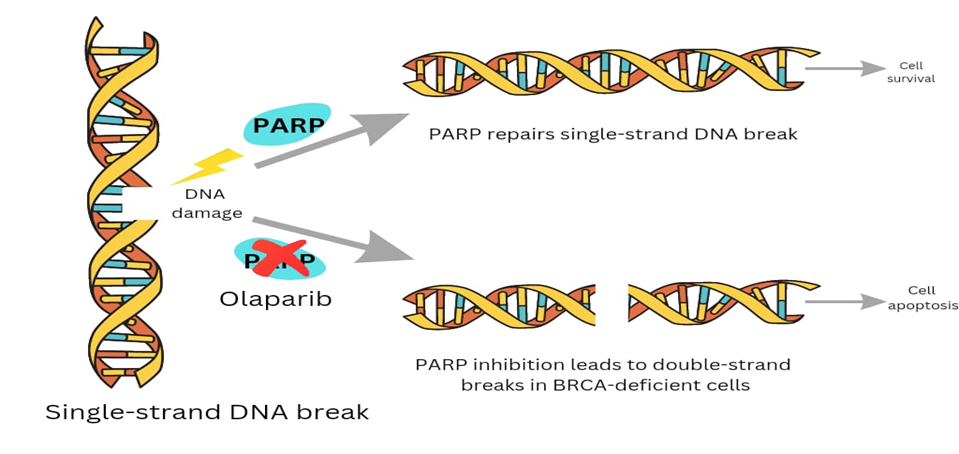
Figure 2: MOA of Olaparib
PHARMACOKINETIC
In oral administration, olaparib is rapidly absorbed.After administration of a single 300 mg dose of olaparib, the mean (CV%) Cmax was 5.4 μg/mL (32%) and AUC was 39.2 μg x h/mL (44%). The steady state Cmax and AUC following a dose of 300 mg twice daily was 7.6 μg/mL (35%) and 49.2 μg x h/mL (44%), respectively. Tmax is 1.5 hours. A high-fat and high-calorie meal may delay Tmax, but does not significantly alter the extent of olaparib absorption.
The mean (± standard deviation) apparent volume of distribution of olaparib is 158 ± 136 L following a single 300 mg dose.
The protein binding of olaparib is approximately 82% in vitro. In solutions of purified proteins, the olaparib fraction bound to albumin was approximately 56% and the fraction bound to alpha-1 acid glycoprotein was 29%.
Olaparib is metabolized by cytochrome P450 (CYP) 3A4/5 in vitro. Following an oral dose of radiolabeled olaparib to female patients, unchanged olaparib accounted for 70% of the circulating radioactivity in plasma. Olaparib undergoes oxidation reactions as well as subsequent glucuronide or sulfate conjugation. In humans, olaparib can also undergo hydrolysis, hydroxylation, and dehydrogenation. While up to 37 metabolites of olaparib were detected in plasma, urine, and feces, the majority of metabolites represent less than 1% of the total administered dose and they have not been fully characterized. The major circulating metabolites are a ring-opened piperazin-3-ol moiety and two mono-oxygenated metabolites. The pharmacodynamic activity of the metabolites is unknown.
Olaparib
A single dose of radiolabeled olaparib, 86% of the dosed radioactivity was recovered within a seven-day collection period, mostly in the form of metabolites. About 44% of the drug was excreted via the urine and 42% of the dose was excreted via the feces. Following an oral dose of radiolabeled olaparib to female patients, the unchanged drug accounted for 15% and 6% of the radioactivity in urine and feces, respectively.
Following a single oral dose in patients with cancer, the mean terminal half-life was 6.10 hours.
Following a single oral dose in patients with cancer, the mean apparent plasma clearance was 4.55 L/h.
PHARMACODYNAMICS
Olaparib is a cytotoxic and anti-tumour agent. Olaparib inhibits the growth of selective tumour cell lines in vitro and decreases tumour growth in mouse xenograft models of human cancer, both as monotherapy or following platinum-based chemotherapy. The drug exerts anti-tumour effects in cell lines and mouse tumour models with deficiencies in BRCA1/2, ATM, or other genes involved in the homologous recombination repair (HRR) of DNA damage and correlated with platinum response.
In preclinical models of cancer, olaparib demonstrated anti-tumour activity when used alone, in combination with chemotherapeutic agents, or radiotherapy.Olaparib can act as a chemosensitizer to potentiate the cytotoxicity of DNA-damaging chemotherapeutic agents such as alkylating agents and platinum-based drugs. It can also act as a radiosensitizer by preventing PARP-mediated DNA repair.4
PHYSICAL PROPERTY
The physical property of Olaparib are shown in Table 1:
Table 1: Physical Property of Olaparib
|
Physical Properties |
Olaparib |
|
Molecular formula |
C24H23FN4O3 |
|
Molecular weight |
434.5 g/mol |
|
State |
White to off-white crystalline solid |
|
Solubility |
Soluble in ethanol and dimethyl sulfoxide |
|
Log P |
1.96/2.724 |
TAXONOMICAL DATA
The taxonomical data of Olaparib are shown in Table 2:
Table 2: Taxonomical data of Olaparib
|
Taxonomy |
Olaparib |
|
Kingdom |
Organic compounds |
|
Super Class |
Organoheterocyclic compounds |
|
Sub Class |
Benzodiazines |
|
Direct Parent |
Phthalazinones |
|
Alternative Parents |
2-halobenzoic acids and derivatives / Benzamides / Benzoyl derivatives / Fluorobenzenes / Pyridazines and derivatives / Piperazines / Aryl fluorides / Cyclopropanecarboxylic acids and derivatives / Vinylogous halides / Tertiary carboxylic acid amides / Heteroaromatic compounds / Azacyclic compounds / Carbonyl compounds / Hydrocarbon derivatives / Organic oxides / Organofluorides / Organonitrogen compounds / Organopnictogen compounds |
|
Substituents |
1,4-diazinane / 2-halobenzoic acid or derivatives / Aromatic heteropolycyclic compound / Aryl fluoride / Aryl halide / Azacycle / Benzamide / Benzenoid / Benzoic acid or derivatives / Benzoyl / Carbonyl group / Carboxamide group / Carboxylic acid derivative / Cyclopropanecarboxylic acid or derivatives / Fluorobenzene / Halobenzene / Halobenzoic acid or derivatives / Heteroaromatic compound / Hydrocarbon derivative / Monocyclic benzene moiety / Organic nitrogen compound / Organic oxide / Organic oxygen compound / Organofluoride / Organohalogen compound / Organonitrogen compound / Organooxygen compound / Organopnictogen compound / Phthalazinone / Piperazine / Pyridazine / Tertiary carboxylic acid amide / Vinylogous halide |
|
Molecular Framework |
Aromatic heteropolycyclic compounds4 |
PHARMACEUTICAL ANALYSIS PROFILES FOR OLA PARIB
Various analytical methods for the estimation of Olaparib have been published in literature since 2014. Figure shows the annual papers published for Olaparib
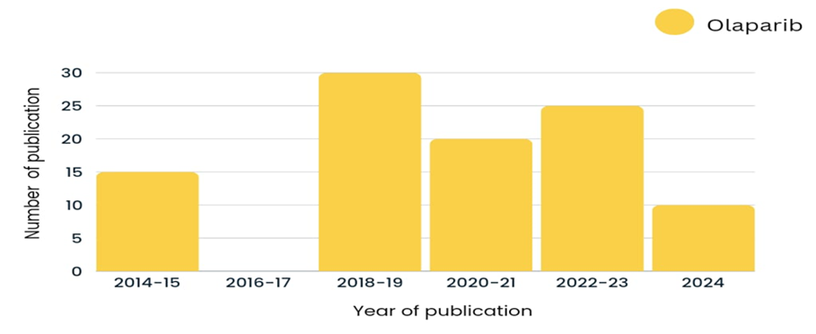
Figure 3: Year wise paper published of Olaparib
NON- COMPENDIAL ANALYTICAL METHODS
Non-compendial methods of analysis developed by various researches. These are spectroscopic, chromatographic techniques and hyphenated techniques represented graphically in figure
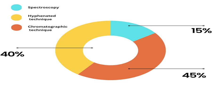
Figure 4: Non-compendial methods chart
ANALYTICAL ACCOUNT OF OLAPARIB
For the determination of Olaparib in bulk and pharmaceutical formulations, an exhaustive literature search found numerous analytical techniques such as UV/Visible Spectrophotometry, HPLC, HPTLC, LC-MS/MS, and bioanalytical approaches. Olaparib is measured as a single constituent in various dosage forms .Figure shows different analytical methods implemented for the estimation of Olaparib
Spectroscopy is the interaction of matter and electromagnetic radiation that, as a function of wavelength, provides a quantitative evaluation of the reflecting or transmitting properties of a material. These methods offer several advantages, including simplicity, cost-effectiveness, and reduced analysis time.They are the most widely used method by researchers and thosewho do not have access to advanced analytical tools. It was claimed that these methods were simple, quick, and relatively cost-effective.There are many spectrophotometric methods developed so far for estimation of Olaparib in various sample matrices. The maximum UV-visible wavelength range was between 400-200nm. The solvent used for the detection of Olaparib was found according to solubility. All of the UV-visible and spectrofluorometric methods of Olaparib are summarized in Table4.
Chromatography is defined as separation of the components of a sample, in which the components are distributed between two phases, one of which is stationary phase while other is mobile phase.The stationary phase may be solid or liquid supported on a solid or gel, and maybe packed in a column, spread as a layer or distributed as a film. The mobile phase may be gaseous or liquid.It is categorize as thin-layer (TLC) chromatography, gel permeation chromatography, affinity chromatography, high-pressure liquid chromatography (HPLC), ion-exchange chromatography and ultra-high-performance liquid chromatography (UHPLC) etc. All of the HPLC/UPLC methods of Olaparib are summarized in Table4.
In HPTLC the selection of mobile phase is based on adsorbent material used as stationary phase and physical and chemical properties of analyte. The mobile-phase systems are used based on their diverse selectivity properties are diethyl ether, methylene chloride, and chloroform combined individually or together with hexane as the strength-adjusting solvent for normal-phase TLC and methanol, acetonitrile, and tetrahydrofuran mixed with water for strength adjustment in reversed-phase TLC. All of the HPTLC methods of Olaparib are summarized in Table 4.
Hyphenated techniques are a combination of chromatographic techniques with spectral techniques. For the estimation of drugs in biological fluids such as plasma and urine, various advanced chromatographic techniques such as GC-MS, LC-MS/MS, UHPLC have been used. The LC-MS/MS techniques for TG is summarized in Table5.
Bio-analysis is a sub-discipline of analytical chemistry covering the quantitative measurement of xenobiotics(drugs and their metabolites, and biological molecules in unnatural locations or concentrations) and biotics(macromolecules, proteins, DNA, large molecule drugs,metabolites) in biological systems. The summary of the reported bioanalytical methods is shown in Table 6
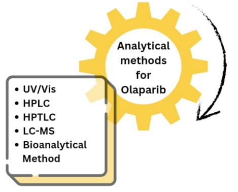
Fig. 5: Analytical methods of Olaparib
Table 4: Spectroscopic methods and Chromatographic methods (HPLC/TLC/UPLC) for the determination of Olaparib
|
Sr. No. |
Author and publication on year |
Method |
Column |
Mobile phase |
Detection wavelength (nm) |
Flow rate (ml/min) |
Retention time (min) |
Ref |
|
1 |
Robin dufour Et al (2018) |
Hplc for quantification of intracellular PARP inhibitor in cancer cells |
Nova-Pak® C18 column (150?×?3.9?mm, 4?μm) |
Acetonitrile and ultra-pure water in gradient mode |
254nm |
1.2?mL/min |
- |
5 |
|
2 |
C.M nijenhuis l. lucas Et al (2003) |
Hplc tandenmass spectrometry assay quantifying olaparib in human plasma |
C18 column |
Methanol, Ammonium acetate (>98%), water |
254nm |
- |
- |
6 |
|
3 |
Priyanka waje , Preeti kulkarni Et al (2021) |
Quantitative estimation of olaparib in tablet dosage form by RP- hplc method |
C18 column (250mm x 4.6 mm id 5µ) |
0.1% Trifluoroacetic buffer: Acetonitrile in the ratio of 60: 40 v/v |
276nm |
1.0 ml/min |
5.14 min |
7 |
|
4 |
Pressiat , Claire pharma Et al (2018) |
Simultaneous Quantification Method of Ruxolitinib, Vismodegib, Olaparib, and Pazopanib in Human Plasma Using Liquid Chromatography Coupled With Tandem Mass Spectrometry |
- |
10-mmol/L formate ammonium buffer containing 0.1%(vol/vol) formic acid (phase A) and acetonitrile with 0.1%(vol/vol) formic acid (phase B) |
254nm |
300 µL/min |
1.5–1.73 minutes. |
8 |
|
5 |
Shanthi DK puja Et al (2024) |
Stability indicating RP-HPLC method for the simultaneous estimation of olaparib and bevacizumab in pharmaceutical dosage form |
Inertsil ODS column |
|
258 nm |
|
Olaparib-2.336 min Bevacizumab -4.873 min. |
9 |
|
6 |
Takeo Yasu Et al (2023) |
HPLC- UV detection method for olaparib in patients with ovarian cancer |
Octadecylsilyl column(Capcell Pak C18 MG II; 250 mm × 4.6 mm; i.d., 5 μm) |
0.5% KH2PO4 (pH 4.5) and acetonitrile (71:29, v/v) |
218 nm |
0.8 mL/min |
13.6 min |
10
|
|
7
|
Dasameswaro kavitapu Et al (2019) |
New rapid stability indicating RP-UPLC method for the determination of olaparib, its related substances and degradation products in bulk drug and dosage form |
BEH C18 (2.1 mm × 100 mm, 1.7 micron) column |
Buffer (1.0ml Orthophosphoric acid in 1000ml water) and acetonitrile |
220 nm |
0.4mL/min |
15 min |
11
|
|
8 |
Ibrahim A Darwish Et al (2023) |
Development and comparative evaluation of two different label-free and sensitive fluorescence platforms for analysis of olaparib |
Nucleosil-CN HPLC column (250 mm length × 4.6 mm i.d., 5 µm particle diameter) |
Acetonitrile: water (25:75, v/v) |
360 nm for emission and 280 nm for excitation |
flow rate of 1.7 mL/min |
- |
12 |
|
9 |
Jeffrey Roth Et al (2014) |
A sensitive and robust ultra HPLC assay with tandem mass spectrometric detection for the quantitation of the PARP inhibitor olaparib (AZD2281) in human plasma |
BEH C18 column (2.1 × 50 mm, 1.7 µm) |
Acetonitrile, methanol, and ethyl acetate |
- |
1 ml/min |
- |
13 |
|
10 |
Revu Baby Nalanda Et al (2019) |
Olaparib Quantification in human plasma for clinical purposes using high-performance liquid chromatography with UV detector |
YMC C18 reverse phase column (250 × 4.6 mm i.d.; 5 μm) |
methanol-0.1 % orthophosphoric acid (60:40, v/v) |
220 nm |
1ml/min |
- |
14 |
|
11 |
Rajiv Tonk Et al (2021) |
Stability Indicating Assay Method For The Quantitative Determination Of Olaparib In Bulk And Pharmaceutical Dosage Form |
WATERS symmetry C18 (150 × 4.6 mm, 5µm) |
ammonium acetate buffer (pH adjusted to 3.5 by glacial acetic acid): methanol in the ratio of 50:50 v/v |
254 nm |
1.0 mL/min |
4.32 min |
15 |
Table 5: Hyphenated techniques for the determination of Olaparib
|
Sr. No |
Author |
Method |
Stationary Phase |
Mobile phase |
Description |
Ref |
|
1. |
Giovanni canil Et al (2023) |
LC-MS method of quantification of PARP inhibitors olaparib, rucaparib and niraparib in human plasma |
Cortecs-T3 column |
0.1% Formicacid, Acetonitrile, Ammonium formate,0.1%Ammonium hydroxide and methanol |
Wavelength – 254nm Range = 140-7000 (olaparib) = 100-5000(rucaparib) = 600-3000 (niraparib) Hematocrit range = 29-45% |
16 |
|
2. |
M.A.C. Bruin Et al (2020) |
LC-MS/MS assay for therapeutic drug monitoring of five PARP-inhibitors |
Reversed-phase UPLC BEH C18 column |
Acetonitrile and dilution of the supernatant with formic acid (0.1% v/v in water) |
Range -30–3000 ng/mL for niraparib, 100–10,000 ng/mL for olaparib, 50–5000 ng/mL for rucaparib, 0.5–50 ng/mL for talazoparib and 50–5000 for veliparib |
17 |
|
3.
|
Yuru Wei Et al (2024) |
Development and validation of a sensitive LC–MS/MS method for the assay of four PARP inhibitors in human plasma and its application in ovarian cancer patients |
Hypersil GOLD™ aQ C18 Polar Endcapped LC column (50 × 2.1 mm, 1.9 µm) |
50 % methanol with 0.1 % formic acid |
Range -10–2000 ng/mL, 25–5000 ng/mL, and 50–10,000 ng/mL for niraparib, olaparib and fluzoparib, and pamiparib |
18 |
|
4
|
Mohit Thummar Et al (2018) |
Validated stability indicating assay method of olaparib: LC-ESI-Q-TOF-MS/MS and NMR studies for characterization of its new hydrolytic and oxidative forced degradation products |
InertSustain C18 column (250 × 4.6 mm, 5 μm) |
10 mM ammonium acetate (pH 4.5) and acetonitrile (ACN) |
- |
19 |
|
5
|
Pramadvara Kallepalli Et al (2018) |
Separation, Identification and Quantification of process related Impurities and Stress Degradants of Olaparib by LC-ESI-Q-TOF-MS |
BDS Hypersil C8 column (250 mm x 4.6 mm, 5μm particle) |
- |
|
20 |
|
6
|
Roberta Ottria Et al (2019) |
Quantitative characterization of Olaparib in nanodelivery system and target cell compartments by LC-MS/MS |
Phenomenex Gemini C18 column |
Acetonitrile and ammonium formate 10 mM (0,1% formic acid) |
Range -0.1–10ng/mL LOQ- 1.54 ng/mL (liver), 2.87 ng/mL (kidney), and lower than 0.48 ng/mL (all matrices) |
21 |
|
7 |
Guosheng Su Et al (2020) |
Gender?dependent pharmacokinetics of olaparib in rats determined by ultra?high performance liquid chromatography/electrospray ionization tandem mass spectrometry |
BEH C18 column (50 × 2.1 mm, particle size 1.7 μm) |
Water containing 0.1% formic acid and acetonitrile |
Range -0.1–2000 ng/ml, correlation coefficient >0.9987. The lower LOQ-0.1 ng/ml. |
22 |
Table 6: Bio-analytical methods for the determination of Olaparib
|
Sr No. |
Author |
Method |
Description |
Reference |
|
1 |
Konrad lewandowski Et al (2024) |
Bioanalytical method validation for therapeutic drug monitoring of olaparib in patients with ovarian cancer |
Range – 100-20000ng/ml LOD –30 ng/ml R2 = 0.9993 |
23 |
CONCLUSION
This critical review comprehensively summarizes the reported analytical and bioanalytical methods developed for the estimation of olaparib in bulk drug, pharmaceutical dosage forms, and biological matrices. A wide range of analytical approaches—including UV–visible spectrophotometry, HPLC, HPTLC, UHPLC, and advanced hyphenated techniques such as LC–MS/MS—have been critically discussed with respect to their methodological parameters, sensitivity, selectivity, and applicability. Among these, LC–MS/MS and UHPLC-based methods have emerged as the most reliable techniques for therapeutic drug monitoring and pharmacokinetic studies due to their superior sensitivity and specificity.
Despite the availability of numerous analytical methods, significant variations exist in terms of method robustness, analysis time, sample preparation complexity, and environmental impact. Conventional UV and HPLC methods remain suitable for routine quality control; however, their limited sensitivity restricts their application in bioanalysis. Advanced hyphenated techniques have demonstrated enhanced performance, yet they require sophisticated instrumentation and skilled personnel, which may limit their widespread use.
Overall, this review provides a consolidated platform for researchers by highlighting the strengths and limitations of existing methods and identifying gaps that warrant further investigation. The compiled data can serve as a valuable reference for future analytical method development, validation, and regulatory submission related to olaparib.
FUTURE PERSPECTIVES AND EMERGING TRENDS
Future analytical research on olaparib is expected to shift toward high-throughput, eco-friendly, and clinically relevant methodologies. The integration of ultra-high-performance liquid chromatography (UHPLC) with tandem mass spectrometry (LC–MS/MS) is anticipated to dominate future bioanalytical applications, particularly for therapeutic drug monitoring and personalized medicine.
Emerging trends include the adoption of green analytical chemistry principles, focusing on reduced solvent consumption, safer mobile phases, and energy-efficient instrumentation. Additionally, micro-sampling techniques such as dried blood spots (DBS) and volumetric absorptive microsampling (VAMS) are gaining importance for minimizing sample volume and improving patient compliance.
The implementation of Quality by Design (QbD) and risk-based method development strategies, in line with recent ICH Q14 and Q2(R2) guidelines, is expected to enhance method robustness and regulatory acceptability. Furthermore, the application of automation, chemometric tools, and artificial intelligence-assisted optimization may significantly reduce method development time and improve analytical performance.
In conclusion, continued advancements in analytical technologies, combined with regulatory-driven method standardization, will play a crucial role in improving the accuracy, efficiency, and clinical relevance of olaparib analysis in both pharmaceutical and biological matrices
CREDIT AUTHORSHIP CONTRIBUTION STATEMENT:
Akanksha Vishwakarma: Conceptualization, investigation and writing – original draft and supervision.
Dakshaja Mahavarkar: Conceptualization and writing – review & editing.
Akshata Shelke: Methodology Writing – review & editing and supervision.
ABBREVIATIONS
SOURCE OF FUNDING
None.
CONFLICTS OF INTEREST
Authors have no conflicts of interest.
ACKNOWLEDGMENT
Authors are thankful to Oriental College of Pharmacy, Navi Mumbai-400705, Maharashtra, India for providing necessary facilities to gather the information about Olaparib.
REFERENCES


Akanksha Vishwakarma, Dakshaja Mahavarkar, Akshata Shelke, A Critical Review on Analytical Methods for FDA Approved Drugs: Olaparib, Int. J. of Pharm. Sci., 2026, Vol 4, Issue 1, 863-877. https://doi.org/10.5281/zenodo.18200051
 10.5281/zenodo.18200051