Anjuman-I-Islam Kalsekar Technical Campus/India.
Microneedle system is a type of new generation transdermal drug delivery systems (TDDS) which eliminate the main disadvantages of drug administration through the skin. This paper undertakes a review of the development of the microneedle technology which has been developed as a non-invasive procedure in drug delivery across the skin because it reduces the effectiveness of the first barrier to penetration, the stratum corneum. One improved characteristic of microneedles is that they span several wells yet their size makes them permeable to water and small molecule drugs therefore providing more efficient transport to deeper skin layers.Based on both the configuration and composition, there are numerous classifications of microneedles such as solid, hollow, coated, dissolvable and hydrogel-forming microneedles. Solid microneedles are mainly employed in rendering transient poration of the skin through which drugs can diffuse while hollow-needle delivery systems dispense controlled volumes of liquids into the skin. In dissolvable microneedles, which are composed of biodegradable materials, deliver drugs and then disintegrate. Hydrogel microneedles be exude interstitial fluid in order to form drug delivery.Techniques in the fabrication of microneedles include micromolding, laser ablation and atomized spraying for etching microneedles with specific dimensions and drug holding abilities. The advancement shown here has expanded the uses of the microneedle not limited to drug delivery but in vaccination, cosmetic dermatology, and diagnostic devices.This paper also considers the benefits of using microneedle systems including improved patient compliance, little or no pain, and better drug bioavailability especially for compounds with low oral absorbability or those which require duration release. Nevertheless, some problems are still pressing, for example, the problem of potentially incompleted medicament delivery to the deeper layers of the skin because of differences in the skin characteristics and certain probability of the irritation or allergic reactions. Overall, microneedle technology's proactive and personalized approach promises to significantly enhance drug delivery, surpassing the limitations of traditional methods. Further advancement could bring better management of a multitude of diseases and health problems affecting man.
Transdermal drug delivery systems (TDDS) are Constructed to produce therapeutically effective doses of medication through the skin. Traditional multi-dose medication methods often encounter issues such as poor bioavailability due to Pre-systemic metabolism, which TDDS effectively bypasses. The design goal for transdermal products is to optimize drug flux through the skin into the bloodstream while reducing drug retention and metabolism within the skin itself. TDDS are autonomous, individual dosage forms that, when applied to unbroken skin, deliver the drug at a controlled rate into the bloodstream. This method of administration is considered a suitable option for both localized and generalized drug delivery. Transdermal delivery not only allows for a steady and controlled release of medications but also supports continuous administration of drugs with short half-lives, avoiding the fluctuations in blood levels that can lead to side effects. Other innovative methods, like Prolonged release and Mucocutaneous systems, work alongside TDDS to enhance treatment effectiveness and safety (1).
Merits:
Transdermal drug delivery systems (TDDS) offer a convenient and safe method for administering medications.
Demerits:
Transdermal drug delivery systems (TDDS) have several limitations, including (1) :
Cutaneous Physiology
Skin is Most expansive tissue and it depth varies across different areas of the body, influenced by the depth of both the outer and inner layers. The most substantial skin thickness is located on the hairless areas of the hands and feet, where an extra layer called the stratum lucidum is found in the outermost skin layer. Conversely, areas without this layer are categorized as having thinner skin. The skin on the back is particularly thick, primarily due to a strong outer layer. However, despite its protective purpose, the skin is susceptible to a range of reddened and contagoius ailments. Furthermore, issues concerning wound recovery, changes in sensation, and aesthetic outcomes are significant in operative settings. A thorough grasp of the skin’s structure and function is crucial for effectively managing conditions in all medical fields (3).
Structure Of Skin (3)
1. Outermost Layer:
The outermost layer of the skin, called the epidermis, is made up of several layers and different types of cells that play crucial roles in its function..
Parts Of Outermost Layer
The epidermis consists of several layers, arranged from deepest to most superficial: Layer basale, Layer spinosum, Layer granulosum, Layer lucidum, layer corneum. The innermost layer of the epidermis, the basal layer, is connected to the underlying dermis by a specialized layer called the basement membrane. This connection is strengthened by structures called hemi desmosomes. This layer contains cube shape stem cell that are growing and continuously generating keratinizing cells, along with pigment producing cells. The spinous layer consists of 8 to 10 layers of irregular, facetted cells that feature , cellularprocess often referred to as spines. These projections connect with adjacent cell through Intercellular bridges. This layer contains antigen-presenting cells. The granular layer consists of 3 to 5 layers of Rhombic cells that contain Epidermal granule and Lipid-rich body.The Proteinrich granules hold Pre-keratin protein that aggregate and Interlink to form Cluster while the Lipid-rich body release glycolipids onto the cell membrane, acting as an adherent to ensure Intercellular adhesion. The clear layer, consisting of two to three layers of cells, is only present in thicker skin, like that on the palms and soles Eleidin, a derivative of keratohyalin, is found within this thin, translucent layer. The uppermost layer of the skin, the outermost layer of the epidermis, consists of 20 to 30 layers of tough, keratinized squamous cells. "The thickness of this layer varies, especially in areas with hardened skin Squamous cells in the stratum corneum secrete protective substances that contribute to the body's first line of defense."
Cell Of Outermost Layer
The epidermis contains several types of cells, including keratinizing cells, Pigment cells, dendritic cells, and Tactile cells. Keratinizing cells are the predominant in the outermost skin layer, originating from the bottom layer. They create Keratinous protein and Fatty substances that are essential for constructing the skin's waterproof barrier. Keratinizing cell also help control calcium levels by absorbing ultraviolet B radiation, which is necessary for activating vitamin D. Pigment-producing cell originate from specialized embryonic cells and are mainly responsible for producing melanin, the primary pigment in the skin. Found among the cells of the bottom layer of the epidermis, melanocytes increase pigment production in response to ultraviolet B radiation, shielding the skin from further UV damage and serving as a natural sunblock. Pigment is created by converting 4Hydroxyphenylalanine to a related compound using the enzyme monophenol oxidase. Pigment particles then move through lengthy extensions that link melanocytes to nearby keratinizing cell, transferring into their cytoplasm through a process called cytocrine transfer. In this process, keratinizing cell envelop the ends of these extensions. Epidermal dendritic cells are tree-like cells that act as the skin's initial defense mechanism and are crucial for foreign substance display.Specific staining methods can pinpoint these cells in the spiny layer. They originate from CD34-stem cells and are part of the Reticuloendothelial System (RES). Epidermal dendritic cells possess tennis racket-shaped organelles and exhibit Human leukocyte antigen (HLA) class I and II molecules. These cells capture foreign substances in the skin and transport them to the lymph nodes. Tactile cells are ovate, modified skin cells situated in the basal layer, just above the basement membrane. These cells act as sensory receptors for gentle touch and are primarily located in the palms, soles, and Mucus-lined tissues with the highest concentration in the fingertips. Tactile cells form connections with nearby skin cells through Maculae adherentes and contain intermediate keratin fibre. Their cell membranes interact with sensory nerve endings in the skin.
The middle layer, which is attached to the outer skin layer by the basal lamina, comprises two layers of supporting tissue: the superficial layer and the reticular layer, which merge without clear divisions. The superficial layer, situated above, is more slender and consists of aerolar tissue that interfaces with the epidermis. Conversely, the reticular layer is the more substantial, lower layer, composed of dense irregular connective tissue with collagen bundles and a reduced number of cells. The middle layer encompasses a variety of structures, including Sudoriferous glands, Pilosebaceous units, muscles, sensory nerves, and vasculature.
The innermost layer, also known as the subcutaneous fascia, is located below the middle layer . This layer contains ,Fat tissue clusters, sensory nerves, Vasculature, and a restricted number of skin structure, such as Pilosebaceous units.
The skin's diverse functions highlight its intricate nature and crucial role in maintaining general health and wellbeing. These roles are detailed below.
Host Defence: The skin serves as a critical component of both adaptive and non-specific immunity. In adaptive immunity,APCs stimulate T-cells mediated immunity, leading to the activation of diverse CD4+ T cells, including TH1, TH2, and TH17. Regarding non-sepcific immunity, the skin produces various polypeptides possessing Bacteriostatic Or Bactericidal and Mycostatic Or Mycotoxic capabilities. Skinassociated lymphatic tissue is a vital part of the immunuty, aiding in the infections, especially considering that even minor skin wounds can result in infections. Dendritic cells are essential to the adaptive host response, as they present foreign antigens encountered on the skin to T lymphocytes.
Homeostasis Control: The skin plays a vital role in regulating the body's temperature and hydration levels. It helps to maintain a stable internal environment by controlling thermal exchange with the external world, primarily through its Blood conduits and Sudoriferous glands. Moreover, the skin regulates the rate and extent of Desiccation and Absorption.
Humoral Regulation and Ducted Secretion: Skin cells play a vital role in vitamin D production when exposed to sunlight. They also have the necessary components to activate this vitamin, which is essential for their growth and development. The skin's external secretion functions helps to maintain body temperature and protect the skin through the production of sweat and oil.
Sensory Roles: The skin has specialized nerve units that allow us to feel things like touch, temperature (hot and cold), and pain. This helps us interact with our surroundings. These sensory abilities are essential for our motion, safety, and communication with the world.
Diagnostic Marker: The appearance of the skin, including its color, texture, flexibility, and firmness, can provide important clues about a person's general health. Examining the skin is a common part of a physical checkup.
Absorption Of Drugs Through Transdermal Route (4)
The transdermal method of delivery offers several benefits above traditional methods like oral or injectable treatments for various diseases and cosmetic applications. The skin serves as a reservoir, allowing for prolonged and sustained release of the delivered drug. This method can minimize hazardousness and local annoyance by utilizing multiple absorption sites and helps to avoid systemic adverse effects. But, the effectiveness of delivery by transdermal is often limited, as only a few drugs can be effectively administered at viable rates through this route. The stratum corneum acts as a significant barrier, hindering the penetration of most drugs. Fortunately, non-invasive techniques have been developed to enhance drug absorption through this barrier. Recently, the use of nano carriers has emerged as a promising alternative for expanding the selection of medication appropriate for transdermal administration. These nanocarriers can facilitate the delivery of both lipophilic and hydrophilic drugs across the stratum corneum, allowing for either localized or generelized effects to address various health conditions.
Drug Penetration Pathway Through the Skin (4)
Numerous studies have investigated how topical compounds penetrate the skin. For a medication to permeate the skin effectively, it must diffuse through the retained epidermis. Human skin contains sweat glands and hair follicles, which create additional pathways for drug absorption, constituting about 0.1% of the skin's total area. The stratum corneum regulates drug penetration, and two primary pathways are identified: transcellular and intercellular routes
The penetration of drugs through the skin can occur through two main pathways:
Lipid Pathway:
This pathway involves the diffusion of drugs through the lipid-rich stratum corneum, the outermost layer of the epidermis.
Lipophilic drugs, which have a high affinity for lipids, can easily penetrate this barrier.
The rate of penetration depends on the drug's lipophilicity and the thickness of the stratum corneum.
Transcellular Pathway:
This pathway involves the diffusion of drugs through the cells of the epidermis.
Hydrophilic drugs, which have a high affinity for water, can penetrate through the intercellular spaces between cells.
The rate of penetration depends on the drug's hydrophilicity and the tightness of the intercellular junctions.
Medicine Characteristics and Cutaneous Insertion (4)
Cutaneous drug delivery is influenced by the physical and chemical characteristics of the medication and the skin's condition. Critical factors impacting absorption include the daily intake, molecular size, lipid solubility , and fusion point. Ideally, the daily intake should be 20 milligrams or less, the molecular size should be below 500 Daltons, the Lipophilicity index should be between 1 and 3, and the fusion point should be below 200 degrees Celsius. Additionally, medications for skin-absorbed delivery must be non-irritating and non-allergenic. Biological membranes typically consist of a fat-loving part with a water-loving part. The movement of medications across biological membranes can be explained by Diffusion equation:
“J=P×CaqJ = P \times C_{aq}J=P×Caq”
Where,
J = Rate of drug movement through the membrane
P = Partition coefficient
C= Concentration of drug
The permeation coefficient is determined by:
“P=D×KhP = \frac{D \times K}{h}P=hD×K”
Where,
C= Diffusion coefficient of drug
K = Partition coefficient
H = Membrane thickness
The 'diffusion rate' can be estimated using the "Stokes-Einstein" equation :
“P=RT6πηrNP = \frac{RT}{6 \pi \eta r N}P=6πηrNRT”
Where,
This equation shows that a drug must be somewhat water-soluble while also being fat-soluble to pass through the membrane. The “Stokes-Einstein equation” also suggests that smaller molecules can permeate more easily than larger ones. Skin moisture is another key factor affecting drug absorption. Higher level of moisture generally improve the absorption of substance especially those that are water soluble. Moistening occurs through water diffusion from adjacent layers or accumulation from topical applications or occlusive vehicles. Other factors that affect skin absorbed delivery include the application location and method, specific skin area, application time, skin age, and the use of carrier that alter the skin's barrier function.
Transdermal Medication (5)
Transdermal medications are used to treat and control a number of illnesses, including migraines, motion sickness, hypertension, and discomfort. This exercise will go over the transdermal drug's indications, modes of action, and contraindications, highlighting the fact that, when used appropriately, these drugs can be very successful forms of treatment. The mechanism of action, possible side effects, and other crucial elements such as off-label applications, dosage, pharmacodynamics, pharmacokinetics, monitoring, and pertinent interactions will all be covered. The purpose of this information is to assist the interdisciplinary team in providing transdermal therapy to patients.
Objectives:
Since the 1970s , transdermal medications have been used to address various indications, including:
?Hypertension
When used as prescribed, transdermal administration has a long history of being safe and successful.
Mode Of Action (5)
The main obstacle to accomplishing successful transdermal drug delivery lies in getting active substances to diffuse through the skin's barrier, which consists of multiple layers. The outermost layer, known as the stratum corneum, is the thickest and is made up of many layers of corneocytes rich in keratin. Furthermore, when creating transdermal medications, two chemically unique sections of the stratum corneum must be taken into account. The keratin filaments have a lipid matrix positioned in between them and an aqueous layer at their outer surface. For transdermal drugs to function well, they must be able to diffuse through both of these regions.
Recent advancements have been made in developing methods to enhance drug delivery through the transdermal route. These methods include (5):
?Microneedles: These tiny, painless needles, it contains medication and might be solid or hollow, penetrate the stratum corneum without causing discomfort. This technique allows for the delivery of compounds with higher molecular weights.
?Iontophoresis: This method uses an electrical driving force to move charged particles across the stratum corneum through electrophoresis. A continuous low current voltage promotes the dispersion of chemicals, with the delivery rate being managed by the patient or a microprocessor.
?Thermal Infiltration: Heat therapy cause the skin to become permeable, which facilitates the easier passage of molecules through the stratum corneum.
?Electropermeabilization: Using this method, a strong electrical voltage is applied to stratum corneum, creating tiny holes that allow molecules to diffuse.
?Conventional Enhancers: These are substance that are used topically to change the active drug’s thermodynamics characteristics or to make the stratum corneum more permeable.
?Sonography: Sound waves are used to break up the stratum corneum, which increases its permeability.
Administration (5)
The administration of transdermal patches should begin with a thorough physical examination of the patient and consideration of any existing comorbidities. Here is a general overview of the steps for administering a transdermal patch:
?Clean the Skin: Disinfect and clean the area of skin where the patch will be applied.
?Apply the Patch: Position the patch where it is needed.
?Follow-Up : Make an appointment for follow-up to monitor for any unfavorable skin response.
?Adherence to Guidelines: For subsequent applications, be sure that the transdermal patch instruction are followed correctly.
While the administration process may vary based on the specific medication delivered via the patch, for all transdermal patches, typically follow these instructions:
Unfavorable Impact (5)
The main negative consequences associated with transdermal medication administration are skin responses. Transdermal patches are a popular way to apply active ingredients, but they can irritate the skin and cause symptoms like burning, itching, and redness in the vicinity. Reactions to allergens can also occur with all types of patches because of the active components that were utilized. Irritant contact dermatitis and allergic contact dermatitis are the two most commonly reported skin responses. These conditions are usually brought on by the medication, the patch, or ingredients such as adhesives and excipients. Furthermore, applying more than one patch when only one is required or leaving a patch on for longer than advised might lead to overdosing on medicine, which may result in an overdose.
Contraindication (5)
It is not advised for people with the following disorders to receive medication transdermally:
Microfabricated Microneedle
Microneedle technology denotes a remarkable advancement in controlled release technology. This innovative approach employs tiny needles, crafted from substances such as polysiloxane, metal, or glass, to penetrate the skin and create microscopic channels. These tiny channels help drugs pass through the top layer of the skin and enter deeper tissues. There, they are absorbed into the bloodstream for delivery throughout the body (6). One of the biggest advantages of microneedle technique is its ability to solve the problems that come with traditional skin patch drug delivery.. The skin's natural protection role often hinders the absorption of drugs applied topically. Microneedles, however, can effectively penetrate this barrier, enabling the efficient and successful transport of complex molecules, including proteins, vaccines, and peptides. This breakthrough has opened up new possibilities for drug administration, particularly for patients who struggle with swallowing or require alternative methods (7). Microneedle technology can be customized to fit the specific needs of different medicines and patients. The size, shape, and contents of microneedles can be changed to improve drug delivery for various purposes. Microneedles can also be covered with drugs or used to carry medicines through tiny channels, making them even more useful. These improvements have led to the use of microneedles in delivering drugs and vaccines, cosmetics, and diagnosing diseases (8). Microneedle technology can employ a single microneedle, a row of microneedles, or a whole array of microneedles with precise dimensions. Depending on the production method, microneedles can be shaped as cones or square pyramids. These variations allow for flexibility in design and application (9). Extraneous factors like skin problems, the type of drug, and weather can affect how well microneedles deliver drugs. Humidity and temperature can change how quickly the drug is released, with dry conditions slowing it down and wet conditions interfering. Too much sweat can stop the patch from sticking to the skin, which can prevent the drug from being delivered. The pH of the skin also plays a role, with extreme pH levels reducing drug absorption. Skin lipids can create a barrier, but removing them can improve drug absorption. Raising skin temperature can enhance drug delivery by increasing diffusion and dilating blood vessels (10). One of the significant merits of microneedle technology is the potential for lower dosages compared to oral ingestion. By bypassing the digestive system and first-pass metabolism, microneedles can reduce the amount of drug that is lost before reaching the bloodstream, allowing for more efficient and effective drug delivery. This can lead to improved therapeutic outcomes and potentially fewer side effects (11).
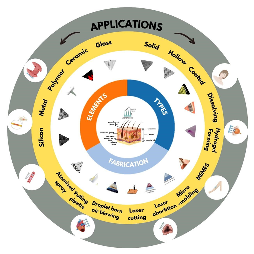
Element Of Microneedles
The composition and constituents of the patch determine the type of material used to make microneedles, which can range from metal to polymer. The strength of these substances needs to be sufficient to pierce flesh. Nondissolving materials are used to create inert, nontoxic, and powerful enough microneedles that can pierce skin without triggering an immune reaction. But coated or dissolving materials-based microneedles need to be biocompatible and dissolve in water. In the body, they need to likewise breakdown or disintegrate without harming someone. During production, storage, and delivery, it is critical that the materials used to make microneedles are compatible with the medications they contain. The characteristics of the various materials used to create microneedles are described (12).
Silicon: Silicon and materials derived from it are robust, stable, and simple to work with. These materials are commonly used in microneedle-based drug delivery systems. However, silicon may have negative effects on the body and microneedles made of silicon are brittle when placed into the skin. Researchers have attempted to coat silicon microneedles in gold or optimize their etching process in order to increase their mechanical strength and biocompatibility. Researchers have developed thin, tapered silicon microneedles that are simpler to implant into the skin without breaking by refining the etching process. Furthermore, the hardness of silicon microneedles can be increased by refining their size and structure. Silicon microneedles typically have a length of 150–1700 micrometers with a needle pitch of 260–2000 micrometers (13).
Metal: Numerous metals and their alloys, including stainless steel, titanium, palladium, nickel, tungsten, gold, and silver, have been used to create microneedles due to their strength and durability. Compared to glass and silicon microneedles, metallic microneedles are easier to make and less expensive. Two common methods for fabricating metallic microneedles are laser cutting and electrode position. Stainless steel and titanium are popular choices for metallic microneedles because they are safe for the body and strong. However, these metals can corrode and cause allergic reactions over time. To avoid these problems, microneedles made from these metals are often coated with other materials. Palladium and nickel are used to create hollow metallic microneedles through a process called electrode position. Palladium is safe for the body, but nickel can cause skin irritation or toxicity. To prevent this, nickel-based microneedles are often coated with a layer of biocompatible material. Silver and gold are also used as coatings on microneedles to improve their compatibility or for antibacterial or biosensor purposes. Recently, researchers have developed a new method for fabricating metallic microneedles using thermoplastic drawing of metallic glasses. This method is simpler and more convenient than traditional methods (14).
Polymer: Polymer microneedles present a viable substitute for conventional medication delivery techniques. Polymers are typically more affordable, biocompatible, and biodegradable than metal or silicon. Additional advantages for medication delivery come from their capacity to expand or dissolve in the body. Although polymers can be less strong than other materials, scientists are looking into ways to strengthen them mechanically by combining and adding other components. The design of the microneedle, the manufacturing method, and the polymer selection all provide a significant role in how effective polymer microneedles are. A vast variety of medications, from tiny compounds to macromolecules like biotherapeutics, can be delivered using polymers. When compared to typical injections, microneedles provide a more comfortable and efficient means of administering medication, particularly for biotherapeutics that are challenging to administer via other means (15).Microneedles have been produced from a diverse range of polymers, including “polylactideco-glycolide acid (PLGA), poly-L-lactic acid (PLA), polycaprolactone (PCL), poly-glycolic acid (PGA), fibroin, sodium alginate, chitosan, polyvinyl alcohol (PVA), hyaluronic acids (HA), polyvinyl pyrrolidone (PVP), polyvinyl alcohol (PVA), carboxymethyl cellulose (CMC), and others.”(16).
Ceramic: Micro molding, a technique that involves pouring ceramic slurry into tiny molds, is used to create ceramic microneedles. For large-scale production, this approach works well because it is scalable and inexpensive. For microneedles, alumina is the most widely utilized ceramic substance. In tension, alumina is brittle but strong under compression. It is applicable to medication delivery by drug-coated microneedle arrays. For microneedles, other biocompatible ceramics with strong mechanical qualities, such as calcium phosphate dehydrate and calcium sulfate dihydrate, can be utilized. Microneedle fabrication can also be done with Ormocer®, an organic-ceramic hybrid material. Its composition consists of silicon alkoxides and organic monomers, forming a three-dimensional network (17).
Glass: Glass microneedles are tiny needles made from glass. They are cheap, safe for the body, easy to sterilize, and can be made in various shapes. Glass microneedles are transparent, which allows for easy visualization of fluid flow. They can be used for dermal interstitial fluid extraction and intradermal insulin infusion. However, glass microneedles are difficult to produce using chemical methods and can be brittle. Borosilicate glass is a type of glass often used for microneedles. It is biocompatible and has been used in cortical implants. However, silica glass may cause granulomas in the skin. For pharmaceutical applications, borosilicate glass is generally preferred over silica glass (18).
Types Of Microneedle
Microneedle transdermal systems are a contemporary drug delivery technique that penetrates the skin using minuscule, needle-like devices. Microneedle systems can be categorized as solid, hollow, coated, dissolvable/dissolving, or hydrogel forming, depending on their composition and intended use. For the purpose of administering medications in a regulated and focused way, each kind has special benefits.
Solid microneedles are typically easier to manufacture, have superior mechanical properties, and offer sharper points, allowing for more efficient penetration of the skin barrier. These needles are designed to create microscopic channels in the skin, allowing for more efficient drug absorption into the deeper layers. Compared to hollow microneedles, solid microneedles are generally easier to manufacture, possess superior mechanical properties, and offer sharper tips, making them more effective in penetrating the skin barrier. Moreover, they can be fabricated from a wider range of materials, including silicon, metals, and polymers, providing versatility in their design and application. (10) (18). While solid microneedles offer several advantages, it's important to consider their potential limitations. One key challenge is maintaining prolonged drug release, as the skin's natural repair mechanisms can lead to the closure of the microscopic channels created by the needles. This can limit the duration of drug exposure and reduce the overall therapeutic efficacy. To address this issue, researchers are exploring strategies to prolong the permeability of the skin barrier and enhance the sustained release of drugs from solid microneedle systems. (19).
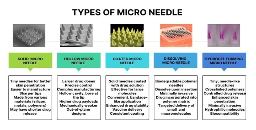
Hollow Microneedle
Hollow microneedles inject liquids through the skin. Hollow microneedles offer the benefit of larger drug doses and providing more precise control over drug administration, hollow microneedles are generally more complex to manufacture and mechanically weaker than solid microneedles, making them more susceptible to breakage or deformation. New advancements in microneedle technology have led to focus on addressing the limitations of hollow microneedles. Researchers have developed innovative designs, such as out-of-plane hollow microneedles, to mitigate clogging issues and improve drug delivery efficiency. Additionally, micromachining techniques have been working to constructed hollow microneedles with greater precision and consistency (20).Hollow microneedles can be constructed from a range of materials including ceramics, metals, silicon, and glass. Each needle typically contains a hollow space and a passage at the tip, allowing to deliver small volumes of drugs dermally. This design enables hollow microneedles to deliver higher drug payloads compared to solid, coated, or dissolving microneedle arrays (21).
Coated Microneedle
Coated microneedles are a type of method that employ solid needles cover in drug solution. Coated microneedle generally carry a smaller quantity of medication compare to other microneedle type , depending on the coating layer thickness. The effectiveness of coated microneedle relies on the ability to consistently apply a control medication layer onto the needle. These microneedles are particularly effective for delivering large molecules like “proteins and DNA” into the skin in a least invasive method (10). They offer a convenient, bandage-like application and can enhance drug stability, even at room temperature. Coated microneedles are also well-suited for delivering vaccines to the skin, as they can release antigens in the skin to target specific immune cells, leading to a stronger immune response (22). Coated microneedles can be made using different coating methods. Spray coating is one method, but it wastes a lot of drug. Inkjet printing is another method that is more precise but takes a long time for large patches. Dip coating is the most common method, but it has limitations in drug loading and large-scale production. Overall, precise drug dosing, consistent drug content, and efficient large-scale production remain challenges in the manufacturing of coated microneedles (23).
Dissolving Microneedle
Dissolving microneedles are a specialized method of drug delivery where needles made from biodegradable polymers are used. These microneedles dissolve when inserted into the skin., releasing the embedded drug payload straight intothe dermal tissue. This method providesa minimally Less invasive alternative to conventional injections, eliminating the associated pain and risk of infection (24). Dissolving microneedles can be fabricated using different water-soluble polymers, including polyvinylpyrrolidone (PVP), hyaluronic acid (HA), maltose, dextran, albumin, and chondroitin sulfate. The desired drug can be includedinto the polymer matrix during the manufacturing process, ensuring its controlled release upon dissolution. This allows for the targeted delivery of both small molecules and macromolecules, such as DNA and proteins, to the skin. Micromolding is a commonly employed technique for the fabrication of dissolving microneedles. This method involves the use of molds to shape the polymer material into the desired needle configuration (25).
Hydrogel Forming Microneedle
Hydrogel-forming microneedle belong to the category of drug delivery systemstiny, needle-like structures to penetrate the skin andand promote the delivery of therapeutic agents. These microneedles are composed of crosslinked polymers that absorb water and swell upon insertion into the skin, forming temporary conduits for drug diffusion. While Hydrogen-forming microneedle offer several advantages, including controlled drug release, improved skin absorption, and minimal invasiveness, they are primarily designed for the delivery of hydrophilic molecules. Hydrophobic drugs, which are often characterized by their low solubility in aqueous environments, present significant challenges for delivery via Hydrogen-forming microneedle. To overcome these challenges, various strategies have been explored, including the incorporation of solubilizing excipients and the modification of the hydrogel matrix to enhance its affinity for hydrophobic molecules. One promising approach is the use of solid dispersion (SD) techniques. SD involves dispersing hydrophobic drugs within hydrophilic carriers, increasing their surface area and promoting dissolution in aqueous environments. Commonly used hydrophilic carriers for SD include polyvinylpyrrolidone (PVP), polyethylene glycols (PEG), and polymethacrylates. These carriers possess favorable properties, such as biocompatibility, low melting points, and established safety profiles, making them suitable for use in drug delivery systems (26).
Fabrication Technique
Microneedle drug delivery systems can be fabricated utilizing multiple approaches, including microelectromechanical systems (MEMS), micromolding, laser ablation, laser cutting, droplet-born air blowing, pulling pipettes, and atomized spraying.
Microelectromechanical System (MEMS) (27)
Micro/nanostructures (MNs) are fabricated using a three-step process involving deposition, patterning, and etching.
Deposition
Deposition is the initial step in the fabrication process where a thin film is created on a substrate. This can be done using either chemical vapor deposition (CVD) or physical vapor deposition (PVD). In CVD, a chemical reaction on the substrate surface forms the film. In PVD, atoms are transferred directly from a source to the substrate through the gas phase.
Patterning
Patterning is a crucial step in the fabrication process where a desired pattern is transferred onto a photosensitivecoated substrate. This is typically achieved using lithography, such as photolithography, ion beam lithography, or X-ray lithography. Photolithography is a particularly important technique in this process. It involves exposing a substrate coated with a photoresist to UV light through a mask. The exposed photoresist undergoes a chemical reaction, making it either more or less soluble in a developer. This allows for the selective removal of the photoresist, leaving behind the desired pattern. Molds for microneedles can also be created using a similar process. A rigid silicone mold is first made with the desired pattern. Then, a negative mold is created using polydimethylsiloxane (PDMS). This negative mold can be used to replicate the pattern onto other materials.
Etching
Etching is the final step in the manufacturing process where unwanted material is removed to create the desired pattern or structure. Wet and dry are the primary types of etching. Wet etching involves immersing the reactant in a liquid chemical to remove unwanted material. Etching can be uniform in all directions (isotropic) or uneven (anisotropic).Dry etching employs a vapor phase or plasma etcher to remove material. Reactive ion etching (RIE) and ion-beam milling (IBM) are two common types of dry etching.RIE uses a reactive gas that interacts with the substrate to remove material. The level of uniformity can be controlled by adjusting the gas pressure and electric field. IBM physically removes material by bombarding it with inert ions. Although RIE can fabricate structures, it etches slowly and struggles to maintain a tall, narrow profile. Deep reactive ion etching (DRIE), or the Bosch process, is a specialized technique used for creating hollow microneedles with deep structures. Combining wet and dry etching techniques can often produce the best results, especially for creating well-defined and sharp microneedle tips.
Micromolding
Micromolding is a versatile technique used to fabricate microneedles, which are tiny needles designed for diverse uses, such as medication administration and diagnosis. This method involves creating a negative mold with cavities that correspond to the desired shape of the microneedles. The Matrix is then filled with an appropriate substance, which is subsequently hardened to form the completed microneedles. Micromolding offers several advantages. It is a straightforward, affordable, and highly repeatable process, making it suitable for large-scale production. Additionally, micromolding allows for the utilisation of a vast array of substances, encompassing "organic and artificial polymers, ceramics, hydrogels, and more". This versatility enables the creation of microneedles with diverse properties and functionalities. Furthermore, micromolding can be used to produce intricate microneedle configurations, such as multiregional, core-shell, stacked, swelling, and suction cup designs. These structures can have intricate release kinetics and functions, making them suitable for specific applications. For instance, multiregional microneedles can deliver different drugs to distinct layers of the skin, while swelling microneedles can expand upon insertion to increase drug delivery efficiency. However, micromolding also has limitations. One of the primary challenges is mold-filling. Surface tension can make it challenging to fill materials into microscale molds against gravity. Various techniques, such as vacuum, centrifugation, imprinting, spinning coating, atomized spray, and infiltration, have been developed to address this issue. Micromolding faces challenges in fabricating complex adhesive structures with barbs and hollow features. Moreover, Unmolding stress can impair the resulting microneedles, especially when working with delicate and brittle materials like hydrogel. Managing the extent of insertion, medication content, and physical properties of the polymer are also obstacles that require to be addressed Ultimately, micromolding is inappropriate for manufacturing materials such as metal, silicon, and glass. Despite these limitations, micromolding remains a valuable technique for manufacturing microneedles. Its simplicity, versatility, and reproducibility make it a popular choice for researchers and industries seeking to develop innovative microneedle-based technologies (28).
Laser Ablation
First, a mold insert is created using laser machining, where a femtosecond laser is employed to ablate the desired cavities. The laser parameters, such as wavelength, pulse repetition rate, and pulse energy, are optimized to ensure efficient material removal. A cross-hatching scanning strategy is used to form circular cavities with precise dimensions. Subsequently, the mold insert is employed in an injection molding process to duplicate the microneedle structure. The injection molding parameters, including mold temperature, injection temperature, injection rate, and holding pressure, are carefully adjusted to achieve high-quality microneedles. To characterize the fabricated microneedles, both the ablated cavities in the mold insert and the replicated structures in the injection-molded parts are analyzed using micro-CT and digital microscopy, respectively. This allows for the evaluation of the geometry and quality of the microneedles (29).
Laser Cutting
The initial stage of the technique entails fabricating a microneedle matrix utilizing a CO2 laser cutter. A polymethylmethacrylate (PMMA) sheet serves as the substrate, and the laser is used to engrave the desired microneedle patterns. By carefully controlling the laser power, speed, and scanning parameters, it is possible to achieve precise control over the depth and shape of the engraved grooves. To ensure accurate dimensions and consistent cone formation of the microneedles, the engraving process is monitored in real-time using a camera integrated into the laser cutter. The dimensions are further verified using a high-resolution 3D digital panoramic microscope. Once the microneedle mold is fabricated, the next step is to prepare the polymeric solutions that will be used to create the microneedle .A variety of hydrophilic materials, including gelatin, kollicoat IR, polyvinyl alcohol, and poloxamer 338, are dispersed in distilled water and warmed to ensure complete dissolution A variety of hydrophilic materials, including gelatin, kollicoat IR, polyvinyl alcohol, and poloxamer 338, are dissolved in distilled water and heated to ensure complete dissolution. The selected model drugs, are then incorporated into the polymeric solutions. The resulting mixtures are carefully stirred to ensure homogeneity and prevent phase separation. The medicated solutions are then poured into the microneedle matrix and allowed to dehydrated in a vacuum oven .During the drying process, the solutions infiltrate the engraved grooves, forming the microneedle structures. After drying, the microneedle patches are gently detached from matrix and stored in a Desiccation cabinet under phosphorous pentoxide to maintain a low humidity environment. This storage condition helps to prevent degradation of the patches and ensure their stability over time (30).
Droplet-Born Air Blowing Method
This novel technique presents a distinctive method of producing polymeric microneedles without the necessity for traditional silicon mold. Through the use of air blowing, polymer droplets are converted into microneedle structures, 'offering a delicate' fabrication 'procedure that eschews severe conditions like UV irradiation or heat. In the droplet-born air blowing technique, polymer solution drops are organized in a grid pattern on two plates. These plates are then joined together and moved at a regulated speed. When the plates reach their maximum distance, - the extended polymer is solidified using air blowing, resulting in the formation of microneedles. The recent inclusion of 'a cyclic contact and drying procedure' (CCDP process) has further improved this method, permitting the production of dissolvable microneedle patches. These patches offer the advantage of simple microneedle separation from the backing film. One of the principal advantages of this approach is the capability to accurately regulate droplet dimensions and concentration, enabling controlled medication loading without any drug wastage. The entire procedure generally takes approximately 10 minutes and has been effectively applied to produce drug-loaded microneedles (31).
Pulling Pipettes
In this method, microneedles fabricated using a micropipette puller and beveler. The microneedles were made from fire-polished borosilicate glass pipettes and were cleaned using chromic acid, followed by water and acetone rinses. The microneedle geometries were characterized using bright-field microscopy and image analysis. The effective tip opening radius was determined to be between 22 and 48 µm, with a tip bevel angle of 35 to 38 degrees. The oval shape of the tip opening was considered when calculating the effective radius (32,33).
Atomized Spraying Method
The atomised spray-filling method addresses the limitations associated with large-scale manufacturing of dissolving microneedles with desired shape and physical properties. It also reduces the difficulties presented by interfacial tension and thickness during matrix filling. Dissolving microneedles can be produced from a variety of materials, including sugars (fructose, and raffinose) and polymers (PVA, PVP, CMC, HPMC, and sodium alginate).The procedure involves connecting a orifice to an air vent and a fluid prepration generating an atomized mist.. The spray is then directed onto the PDMS mold, and the solution is allowed to dry. The atomised sprayfilling method can also be used to fabricate layered microneedles. By controlling the spraying parameters and the deposition of multiple layers, it is possible to create microneedles with distinct structures and properties (34).
ADVANTAGES
DISADVANTAGES
Microneedles, while offering several advantages, also have some potential disadvantages (37) :
Applications
Microneedles have developed as a useful tool for a variety of medicinal applications, with substantial benefits over previous approaches. Here's an overview of their main applications (13 , 38 , 39):
Microneedles are a Favorable vaccine administration method that offers several advantages over traditional injections.They provide a less invasive approach, stimulate a stronger immune response, reduce needle phobia, and facilitate efficient mass vaccination campaigns.
Microneedle-based insulin delivery offers improved glycemic control, a less invasive alternative to injections, and ensures proper biological effects.
Microneedle-based parathyroid hormone delivery offers a targeted approach for localized treatment and has Revealed favorable results in clinical trials for Enhancing bone health.
Microneedle-based anesthetic delivery, such as lidocaine offers a more comfortable and effective alternative to traditional injections, reducing pain and improving the overall patient experience.
Microneedle-based cosmetic procedures Promote collagen growth, refine skin texture, and minimize wrinkles and acne scars, and offer a minimally invasive approach to skin rejuvenation.
Microneedle patches have shown potential for treating opioid dependence and alcohol addiction.By delivering naltrexone, a medication that blocks the effects of opioids, microneedles can help individuals overcome addiction.
Microneedle-based disease diagnosis and monitoring systems offer continuous glucose monitoring for diabetes, detect biomarkers for various diseases, and extract interstitial fluid for analysis.
Microneedle patches loaded with ferric pyrophosphate can effectively treat iron deficiency anemia by delivering iron directly through the skin.
Microneedle masks fabricated using CLIP techniques can deliver proteins such as serum albumin and ovalbumin, offering a non-invasive approach for protein therapy.
Microneedle patches containing sodium hyaluronate and 5-aminolevulinic acid can be used to treat subcutaneous tumors through photodynamic therapy.
Microneedle biosensors are emerging as a valuable tool for continuous monitoring of various biomarkers. These innovative devices integrate sensing elements into microneedles, allowing for non-invasive and realtime measurement of glucose, lactate, alcohol, beta-lactam, and other substances. By providing continuous data, microneedle biosensors can aid in disease diagnosis, health monitoring, and treatment management.
Microneedles have proven to be valuable tools for various medical procedures beyond drug delivery. They can be used for fluid extraction, nerve stimulation, and recording, offering innovative solutions for healthcare. For fluid extraction, microneedle patches, hydrogel microneedles, and paper-based methods can effectively collect interstitial fluid, a valuable source of biomarkers for health monitoring. In nerve stimulation, microneedle electrodes can be used for transcutaneous electrical nerve stimulation (TENS) to manage pain. Additionally, microneedle sensor arrays can detect opioid drugs and organophosphate nerve agents, enabling rapid human body induction. For electrophysiological recordings, microneedle electrodes can be used to record electrical signals from the nervous system, providing insights into neural activity.
Microneedles are emerging as a promising technology for cancer therapy, offering efficient delivery of various therapeutic agents. By directly delivering anticancer drugs, proteins, and vaccines to tumor sites, microneedles can enhance bioavailability, reduce side effects, and enable targeted treatment. This minimally invasive approach holds great potential for improving outcomes in cancer patients, especially when combined with other therapies.
Microneedles provide a more comfortable alternative to intravitreal injections for ocular drug delivery, offering the likelihood of improved outcomes and fewer adverse reactions.
Overview On Microneedle
Research Article on Microneedle Patches:
|
Sr. No. |
Drug |
Method |
Citation |
|
1. |
Valsartan |
------ |
(40) |
|
2. |
Carvedilol |
------ |
(41) |
|
3. |
Lornoxicam |
------ |
(42) |
|
4. |
Insulin |
------ |
(43) |
|
5. |
Butorphanol tartrate |
Solvent casting method |
(44) |
|
6. |
Insulin |
Solvent casting method |
(45) |
|
7. |
Amphotericin B |
Probe sonication |
(46) |
|
8. |
Lidocaine |
Centrifugal lithography |
(47) |
|
9. |
Amphotericin B |
Solvent casting |
(48) |
|
10. |
Insulin |
Solvent casting |
(49) |
|
11. |
Vitamin B12 |
Solvent casting |
(50) |
|
12. |
Gentamicin |
Solvent evaporation |
(51) |
|
13. |
Dihydro- ergotamine mesylate |
Solvent casting |
(52) |
|
14. |
Influenza virus |
------ |
(53) |
|
15. |
Protein, Peptide and Antibody |
------ |
(54) |
|
16. |
Sodium nitroprusside and sodium thiosulfate |
Centrifugation casting method |
(55) |
|
17. |
Ibuprofen sodium |
Centrifugation casting method |
(56) |
|
18. |
Losartan potassium |
Film formation |
(57) |
|
19. |
Amlodipine besylate |
------- |
(58) |
|
20. |
Ferric pyrophosphate (FPP) |
Mold casting method |
(24) |
|
21. |
Meloxicam |
------ |
(59) |
|
22. |
Simvastatin |
Solvent casting method. |
(60) |
|
23. |
Propranolol hydrochloride |
Casting method |
(25) |
|
24. |
Etravirine |
Solvent Evaporation-Antisolvent Precipitation Method |
(61) |
|
25. |
Hyaluronic acid |
------ |
(62) |
|
26. |
Peptide, Collagen |
------ |
(63) |
|
27. |
Niacinamide |
------ |
(64) |
|
28. |
Hyaluronic Acid serum |
------ |
(65) |
|
29. |
Salicylic Acid, Hyaluronic Acid, Epiallocaine Gallet. |
(66) |
|
|
30. |
Collagen |
------ |
(67) |
|
31. |
Sodium Hyluronate –90% Peptide – 2% Vitamin E – 2% Collagen – 4% |
------ |
(68) |
|
32. |
Calcipotriol, Betamethasone dipropionate. |
------- |
(69) |
|
33. |
Adenosine |
------- |
(70) |
|
34. |
Tacrolimus |
------- |
(71) |
|
35. |
Doxazosin mesylate
|
------- |
(72) |
|
36. |
Itraconazole |
------- |
(73) |
|
37. |
Progesterone |
------- |
(74) |
|
38. |
Ibuprofen sodium |
------- |
(75) |
|
39. |
Primaquine |
------ |
(76) |
|
40. |
Sodium nitroprusside and sodium thiosulfate. |
Centrifugation casting method. |
(77) |
|
41. |
Recombinant human growth hormone |
------- |
(78) |
|
42. |
Acyclovir |
Laser-engineered |
(79) |
|
43. |
Besifloxacin |
Micromolding |
(80) |
|
44. |
Meloxicam |
------- |
(81) |
|
45. |
Ferrous sulfate nanoparticles (FS NPs) |
-------
|
(82) |
|
46. |
Minoxidil |
------- |
(83) |
|
47. |
Amoxicillin Sodium |
------- |
(84) |
|
48. |
Levodopa and Carbidopa |
Two-layer dissolving MAP casting. |
(85) |
|
49. |
Nimodipine |
------- |
(86) |
|
50. |
Olanzapine |
Nano-precipitation method - casting method |
(87) |
Marketed Microneedle Patches:
|
Sr. No. |
Drug |
Manufacturer |
Product Name |
Indication |
Citation |
|
1. |
Zolmitriptan |
Zosano pharma |
Qtrypta |
Migraine treatment, Cluster headache treatment |
(88) |
|
2. |
Salicylic Acid |
The derma co |
Micro tip Salicylic Acid |
Acne treatment, hyperpigmentation, psoriasis, callus removal, skin rejuvenation |
(89) |
|
3. |
Teriparatide acetate |
Nemaura Pharma |
Teriparatide micro patch |
Osteoporosis treatment, osteopenia treatment |
(90) |
|
4. |
Influenza vaccine |
Sanofi Pasteur |
Intanza |
Seasonal influenza prevention, pandemic influenza preparedness |
(91) |
|
5. |
Hyaluronic acid |
Dermatude |
Microneedle patch |
Hydration, anti-aging, wound healing, skin rejuvenation, acne treatment |
(65) |
|
6. |
Antibodies |
Vaxxas |
Microneedle patch for antibodies |
Infectious diseases, autoimmune diseases, cancer treatment, neurological disorders |
(92) |
|
7. |
Peptides |
Neutrogena |
Rapid wrinkle repair microneedle patch |
Anti-aging, skin rejuvenation, hair growth promotion, acne treatment, pain management, wound healing, inflammation reduction |
(93) |
|
8. |
Proteins |
Bioactive |
Protein microneedle patch |
Anti-aging, skin rejuvenation |
(94) |
|
9. |
Collagen |
Dermatude |
Microneedle patch |
Anti-aging, skin rejuvenation, hair growth promotion, acne treatment, pain management, wound healing, inflammation reduction Wound healing, burn care |
(95) |
|
10. |
Glutathione |
MDPI |
Microneedle patch for skin whitening |
Skin rejuvenation, wrinkle reduction, hyperpigmentation, skin brightening, detoxification |
(96) |
|
11. |
Niacinamide |
Dermatude |
Mesopure microneedle patch |
Anti-aging, skin brightening, hydration, acne treatment, skin elasticity improvement, hyperpigmentation, rosacea treatment |
(97) |
|
12. |
Sodium Hyaluronate |
Lumen |
Sodium Hyaluronate 3% microneedle patch |
Facial rejuvenation, lip augmentation, skin tightening, scar treatment, burn care, hydration, anti-aging |
(98) |
|
13. |
Tranexamic acid |
Skin republic |
Tranexamic acid micro- needle patch |
Hyperpigmentation, acne treatment, scar treatment, facial rejuvenation, skin brightening |
(99) |
|
14. |
Glycyrrhiza Glabra |
Dr. CYJ |
Glycyrrhiza Glabra microneedle patch |
Anti-inflammatory, anti-aging, hydration, skin brightening, wound healing, eczema, dermatitis treatment |
(100) |
|
15. |
Niacinamide |
Avareele |
Microneedle patch |
Multi Dart spot leach microneedle patch |
(64) |
|
16. |
Hyaluronic Acid serum |
China Pvt. Ltd. |
Microneedle eye patch |
Hyaluronic Acid microneedle eyepatch |
(65) |
|
17. |
Salicylic Acid, Hyaluronic Acid, Epiallocaine Gallet. |
BBOLD |
Dissolvable Microneedle acne patch |
Unique formulation to fight acne from the inside. |
(66) |
|
18. |
Collagen |
GharSaapos + Ayurved Science |
Skin microneedle infusion system |
Stimulates collagen whilst infusing active ingredients into skin. |
(67) |
|
19. |
Sodium Hyluronate – 90% Peptide – 2% Vitamin E – 2% Collagen – 4% |
Frownie Patches |
Dissolvable forehead microneedle patches |
Brighten dry and dull skin. |
(68) |
|
20. |
Calcipotriol, Betamethasone dipropionate. |
Nissna Co. Ltd. |
Microneedle patch |
Dissolving microneedle patches for cosmetics |
(69) |
Patented Microneedle Patch:
|
Sr. No. |
Drug |
Patent No. |
Country |
Date |
Citation |
|
1. |
Colchicine |
CN113133991B |
China |
2023-06-13 |
(101) |
|
2. |
Fluconazole and doxycycline |
CN116270666A |
China |
2023-06-23 |
(102) |
|
3. |
Liraglutide |
CN116350751A |
China |
2023-06-30 |
(103) |
|
4. |
Timolol maleate |
CN116421874A |
China |
2023-07-14 |
(104) |
|
5. |
1) Vitamin C or derivatives 2) Glutathione 3) Tocopherol or derivatives 4) Alpha lipoic acid. |
KR102563820B1 |
Korea |
2023-08-07 |
(105) |
|
6. |
Itraconazole |
CN116617150A |
China |
2023-08-22 |
(106) |
|
7. |
Taxol |
CN116687833A |
China |
2023-09-05 |
(107) |
|
8. |
Hyaluronic acid |
CN116725939A |
China |
2023-09-12 |
(108) |
|
9. |
Paeonol |
CN116763715A |
China |
2023-09-19 |
(109) |
|
10. |
Levosimendan |
CN116440062B |
China |
2023-09-26 |
(110) |
|
11. |
Interferon |
US20230330010A1 |
US |
2023-10-19 |
(111) |
|
12. |
Ginsenoside Rg3 |
CN116763716A |
China |
2023-10 -24 |
(112) |
|
13. |
Progesterone lipsosome |
CN117018422A |
China |
2023-11-10 |
(113) |
|
14. |
Sparfloxacin and manganese based nano drug |
CN117258131A |
China |
2023-12-22 |
(114) |
|
15. |
Levothyroxine sodium |
CN117503682A |
China |
2024-02-06 |
(115) |
|
16. |
Dutasteride |
CN117598988A |
China |
2024-02-27 |
(116) |
|
17. |
Metformin |
KR20240117782A |
Korea |
2024-08-02 |
(117) |
|
18. |
Lidocaine Hydrochloride, |
CN118615229A |
China |
2024-09-10 |
(118) |
|
19. |
Minoxidil |
WO2024187056A2 |
US |
2024-09-12 |
(119) |
|
20. |
Oligonucleotide |
WO2024186627A1 |
US |
2024-09-12 |
(120) |
CONCLUSION
Microneedles provide an highly efficient approach, to absorbing medication. They represent an advancement in drug delivery systems (TDDS). Constructed from materials, like silicon s, metal, polymers, glass and ceramics. These tiny synthetic devices form microchannels in the skin to aid in the absorption of drugs without penetrating. Microneedles come in various types, including solid, coated, hollow, and dissolvable, each offering distinct advantages depending on the desired treatment and drug properties. Various methods have been developed to create microneedles such, as Microelectromechanical Systems (MEMS) Micromolding techniques like Laser Ablation and Laser Cutting. Atomized Spraying Method and Droplet Born Air Blowing method are utilized along, with Pulling Pipettes in the fabrication process.Despite their constraints, in administering medication and the possibility of causing irritation microneedles have a diverse array of uses. As research progresses further into the field of fabrication techniques and the development of structures become more dynamic, in nature; they play a role, in enhancing the healthcare system. After examining 50 research papers on patches it is evident that there have been strides, in enhancing the delivery of drugs through the skin, for better release efficacy. The commercial bioavailability of the microneedle patch is showcased through the development of 20 marketed products and 20 patented works. Therefore the future prospects of microneedle patches, in healthcare seem thanks to research and advancements, in fabrication techniques that enhance their capabilities.
REFERENCES


Kadam Snehal, Kasu Daniya, Momin Ishrat, Shaikh Shama*, Khan Sharina, Poonam Patil, Microneedle Technology: A Revolutionary Approach to Drug Delivery, Int. J. of Pharm. Sci., 2025, Vol 3, Issue 4, 106-139 https://doi.org/10.5281/zenodo.15119727
 10.5281/zenodo.15119727
