
We use cookies to ensure our website works properly and to personalise your experience. Cookies policy
Department of Pharmaceutics, Channabasweshwar Pharmacy College (Degree), Latur-413512, Maharashtra, India.
Nanogel are nanoscale, crosslinked polymeric structures with the ability to absorb water and swell without disintegration. Their unique features, such as small particle size, high water content, and responsiveness to environmental stimuli like pH and temperature make them highly suitable for biomedical use, particularly in targeted and sustained drug delivery. This review explores their diverse synthesis methods, structural variations, and classifications based on responsive behavior and polymer types. Nanogels have found applications in several medical fields, including transdermal systems, oncology, vaccine formulation, insulin regulation, and treatment of inflammatory and autoimmune disorders. Their advantages include enhanced bioavailability, reduced side effects, and improved therapeutic performance. Despite these benefits, certain limitations such as high production costs and safety concerns remain. Additionally, this review discusses fabrication techniques like emulsion solvent diffusion and nanoprecipitation, and elaborates evaluation criteria such as pH measurement, particle size analysis, drug content determination, and Spreadability. Overall, nanogels represent an evolving and promising tool in advanced drug delivery systems.
Nanogels are nanoscale particles comprising crosslinked polymer networks that swell in suitable solvents. Initially termed “Nanogel”, they were developed for polynucleotide delivery using crosslinked polyethyleneimine (PEI) and polyethylene glycol (PEG) networks(1). Nanogels are highly crosslinked gel systems, 20-200 nm in size, formed from ionic or non-ionic monomers through polymerization. Nanogels are 3D hydrophilic networks that absorb fluids without structural changes, enabling ligand incorporation for targeted delivery, stimulus-responsive release, or composite material development(2). Nanogels, formed from natural, synthetic, or hybrid polymer, can be tailored for properties like size, charge, porosity, and degradability. Advances in synthesis enable diverse morphologies, including core-shell structures with high structural integrity. Their hydrophilic nature, biocompatibility, and stimulus-responsive behavior make them ideal for biomedical applications, protecting cargo and enabling controlled release at target sites. Recent innovations in nanocarriers, such as nanogels, liposomes, and nanoparticles, have transformed anticancer therapies, offering effective alternatives to traditional chemotherapy and radiation(3). Nanogels, with properties such as adjustable size, high surface area, and water content, enable controlled and sustained drug delivery. Their three-dimensional structure allows the entrapment of drugs, polymers, and liquid phases, while porous networks accommodate micro-and macromolecules. Acting as carriers, nanogel absorb active compounds through interactions like hydrogen bonding and hydrophobic forces. These properties enhance therapeutic effectiveness, particularly in topical applications, by prolonging skin exposure time.
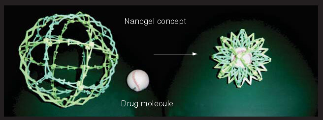
Fig no.1. Nanogel
Nanogels can deliver both hydrophilic and hydrophobic drugs and are influenced by polymer network functional groups, crosslinking density, and agents. Polymeric micellar nanogels exhibit slow dissociation rates and extended drug retention. They are suitable for various administration routes, with drug release driven by pH, thermosensitivity, volume transition, and other mechanisms. Their nanoscale size and 3D structure enable targeted drug delivery and bioconjugation for improved efficacy in biomedical applications(4). Nanogels can be made from natural, synthetic, or hybrid polymers, crosslinked either covalently or through non-covalent interactions like hydrogen bonds, electrostatic, and hydrophobic forces. Their high water absorption capacity is due to hydrophilic functional groups (e.g., –OH, –CONH–, –CONH2–, –SO3H) along the polymer chains. Nanogel are generally described as crosslinked polymer chains up to 100 nm, through accepted sizes range from 1 to 1000 nm, with typical dimensions extending up to 200 nm(5). Nanogels delivery hydrophobic and hydrophilic drugs, charged solutes, and diagnostic agents, with functionality shaped by polymer network properties and crosslinking. Designed as polymeric micellar systems, they offer gradual dissociation, reduced critical micelle concentrations, and prolonged drug retention. Administered via oral, pulmonary, nasal, parenteral, and intraocular routes, their release mechanisms respond to pH, temperature, and volume changes(6). Nanogels offer tuneable properties like swelling, degradation, and chemical functionality. Beyond drug delivery, they are used for quantum dots, dyes, and diagnostic agents. Their versatility, targeted delivery capabilities, and tuneable physicochemical properties make them valuable. Clinical studies highlight their potential in gene therapy, enabling gene delivery for silencing. Nanogel structure and functionality can be adjusted through solvent quality and polymer branching(7).
Advantages of Nanogel(8)(9)(7)
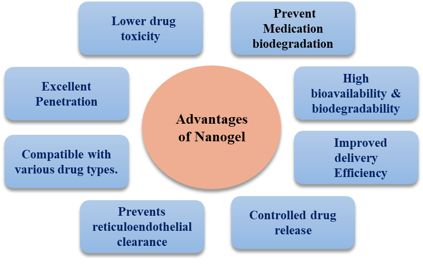
Fig no. 2. Advantages of Nanogel
Disadvantages of Nanogel(10)(11)(12)(4)
Applications of Nanogel (4,13–17)
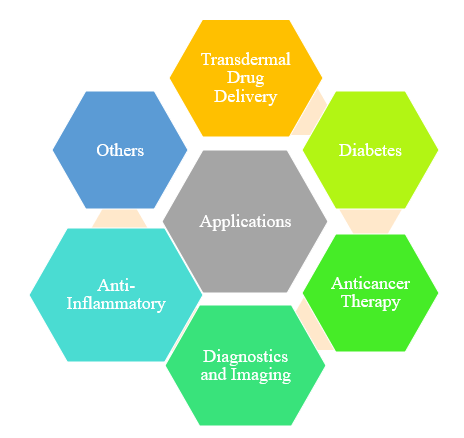
Fig.no.3 Application of Nanogel
Nanogel in Transdermal drug delivery:
Nanogels enhance transdermal drug delivery by passing the first-pass effect, improving drug efficacy, ensuring steady plasma levels, and increasing patient compliance. They facilitate better drug penetration, as seen with acelofenac, which was formulated into a nanogel using the emulsion solvent diffusion method. This approach improved its stability and permeability while reducing gastrointestinal side effects.
Nanogel-Based Insulin Delivery:
MIT and Boston Children’s Hospital have developed a glucose-responsive nanogel that stabilizes blood sugar for up to 10 days with a single injection. The nanogel, composed of dextran nanoparticles. Glucose oxidase enzymes, and insulin, releases insulin in response to rising glucose levels. The system is biocompatible and naturally degrades in the body.
Nanogel in Anticancer Therapy:
Nanogels can aid in tumour removal by filling the surgical void and providing protection. They can also be loaded with therapeutic agents to prevent tumour recurrence. Nanogels, including those made from polyethyleneimine and PEG/ Pluronic, show reduced toxicity in cancer treatments by enhancing drug delivery. Doxorubicin-loaded nanogels, pH-responsive glycol chitosan nanogels, and self-quenching pullulan-folate systems improve therapeutic efficacy while minimizing side effects. Polyplex nanogel with polyethyleneimine and PEG enhance drug activity, while heparin-Pluronic nanogels deliver RNAs enzymes. Additionally, cholesterol-bearing pullulan nanogels support tumour immunotherapy, and hyaluronic acid-loaded doxorubicin nanogels offer targeted delivery. Thermosensitive and quantum dot hybrid nanogels enable drug delivery and bioimaging. These nanogels, with an average size of ~100 nm, have enhanced circulation and tumour targeting capabilities.
Diagnostic and Imaging:
Diagnostic imaging provides non-invasive visualization of the body internal structures, aiding in the early detection and accurate diagnosis of medical conditions, thereby improving patient outcomes through timely intervention.
Anti-inflammatory:
Nanogels are effective topical delivery systems for NSAIDSs in dermatology, addressing the challenge of limited contact time by retaining water the gel matrix. This allows for the treatment of conditions like allergic contact dermatitis and psoriatic plaques. A nanogel formulation using poly(lactide-co-glycolic acid) and chitosan, modified with oleic acid, successfully delivers two anti-inflammatory drugs, ketoprofen. The system effectively permeates deeper skin layers, offering potential for treating various inflammatory disorders.
Vaccine Delivery:
Nanogels offer a promising approach to vaccine development by enhancing immune response while improving safety. Unlike live attenuated vaccines, they mitigate risks and avoid inflammation caused by traditional adjuvants like aluminium salts. Designed for targeted delivery, nanogels can modulate immune responses, making them suitable for preventing and treating infections, cancer, allergies, and autoimmune diseases.
Nanogel-Based Therapy for Autoimmune Diseases:
Nanogels offer a promising approach for autoimmune disease treatment by enhancing immunosuppressive therapy. A nanogel-based systems was developed for the controlled release of mycophenolic acid (MPA), utilizing UV-induced photopolymerization of PEG oligomers. This method improves drug delivery efficiency, optimizing therapeutic outcomes.
Nanogel for Targeted Drug Delivery:
Nanogels enable precise drug delivery by optimizing drug concentration at targeted sites while minimizing systemic loss. This is crucial for diseases like cancer, enhancing efficacy and reducing adverse effects. Researchers have developed nanogels for curcumin delivery in colon cancer, utilizing advanced formulations with potential applications in precision medicine.
Nanogels for Controlled Drug Delivery:
Nanogels tuneable properties enable precise, adaptable drug delivery. A promising nanogel has been developed for the controlled, on-demand release of camptothecin at tumour sites, enhancing targeted cancer therapy.
Properties of Nanogel (14,16,18–23)
Degradability and Biocompatibility of Nanogels:
Nanogels, composed of natural or synthetic polymers, are biocompatible and biodegradable, preventing long-term tissue accumulation. They are derived from materials like chitosan, ethyl cellulose, methylcellulose, and polysaccharide-based polymers such as dextran, pullulan, and dextrin. Polysaccharides, characterized by glycosidic linkages, offer stability, hydrophilicity, non-toxicity, and biodegradability, making them ideal for biomedical applications.
Swelling Properties in Aqueous Media:
Nanogels, due to their small and soft structures, swell in aqueous environments, a key factor in drug delivery. Swelling depends on the polymers chemical composition, cross-linking degree, and charge density in polyelectrolyte gels. Environmental factors such as pH, ionic strength, and charge density in polyelectrolyte gels. Environmental factors such as pH, ionic strength, and temperature also influence swelling. It occurs when an osmotic imbalance exists between medium ions and the polymer networks, enabling controlled responsiveness.
Higher Drug Loading Capacity:
Nanogels drug loading capacity is determined by functional group in their polymeric structure, influencing drug encapsulation, release, and targeting. Theses groups enable hydrogen bonding and van der Waals interactions, enhancing drug retention. Additionally, their presence at the drug/protein interface further increases loading efficiency.
Permeability and Particle Size:
Nano delivery systems leverage precise modifications in particle size, surface charge, and hydrophobicity to enhance permeability. While nanoparticles can diffuse through tissues and compromised endothelium, crossing the blood-brain barrier (BBB) remains a challenge. To overcome this nanogels are designed with a 20-200 nm diameter, enabling BBB penetration while minimizing rapid clearance for sustained therapeutic action.
Colloidal Stability:
The colloidal stability in nanoparticles is crucial to prevent aggregation and ensure efficacy. This is achieved by increasing the zeta potential (≥ ±30 mV) for electrostatic stabilization or incorporating PEG for steric and hydration effects. Polymeric nanogels offer superior stability over surfactant micelles, with lower critical micelle concentrations, reduced dissociation rates, prolonged drug retention, and excellent dispersion stability due to their high-water content.
Structure Of Nanogel:
Nanogel structure is defined by polymer composition, crosslinking degree, and charge density in polyelectrolyte gels. Environmental factors like pH, ionic strength, and ion composition influence polyelectrolyte gel behaviour, while temperature regulates the swelling of thermos responsive gels.
Non-immunologic response:
Nanoparticles in circulation are rapidly cleared by the Mononuclear Phagocyte System through opsonization and phagocytosis. To evade immune recognition, strategies focus on reducing protein binding, such as using hydrophilic polymers to create a protective shield that delays opsonin attachment and prolongs systemic retention.
Classification of Nanogels (10,14,20–22,24)
Different Types of Nanogel according to Their Reaction to Stimuli
Temperature-responsive nanogels exhibit a phenomenon called shrinkage-swelling behaviour, in which they alter size in reaction to environmental temperature fluctuations. This special quality masks it possible to titrate the release of medications in a controlled manner. The stimulation-induced reduction in particle size promoted intracellular absorption and it easier for the particles to accumulate in the disease-related microenvironment. Positive on therapeutic outcomes could result from these effects.
The ionizing groups in the design are principally responsible for the pH-dependent swelling-shrinking behaviour of nanogel; however, the pH value can modify the ionic behaviour. The pH ranges of different environments, such as lysosomes (pH 4.5 to 5.0), endosomes (pH 5.0 to 6.5), and cancer tissue (pH 6.5 to 6.5), as well as how these bodily environments change to physiological pH 7.4, have been identified. This information can be used to optimize the preparation of the nanogel for that particular pH.
The use of ultrasound-sensitive nanogel US-based drug delivery in transdermal administration for the treatment of conditions affecting the central nervous system has grown significantly. Acoustic waves capacity to deeply permeate tissue and the benefits that comes with it have inspired their usage in anticancer therapy. When administered, perfluoro hexanes evaporation from a liquid to a gaseous state was shown to aid in the medications release.
A magnetic field may be used to aid in the induction of hyperthermia. Moreover, magnetic targeting under a strong magnetic field is another application for these nanoparticles. In one study, temperature-sensitive nanogel and magnetic nanoparticles were combined to create hybrid nanogel that contained the medication. The dynamic process of shrinking and swelling, which is guaranteed by the action of an alternating magnetic field, facilitates the release of stimuli-responsive medications from nanogels.
Dual or multi-stimuli responsive nanogel have attracted a lot of attention because of their improved capacity to reliably maintain controlled drug release. Significantly progress has been made in examining combinations that show temperature and pH sensitivity.
The only processes that can be controlled by adding Di thioester molecules to a polymer are those that involves reversible addition-fragmentation chain transfer (RAFT). Through changes to the polymer’s length, structure, and properties, such as poly (N-vinyl caprolactam), RAFT technology can alter the micelle structure of an amphiphilic polymer.
Crosslinking between molecules brought on by radiation can produce atoms and radicals in a polymer that, when water molecules break down, generates nanogel.
A ligand can reach the precise receptors on the cells or subcellular structures it was intended to influence through active targeting. Additionally, to increase the effectiveness of medicinal nanoparticles, biological materials such as proteins, small molecules, peptides, and polysaccharides, biological materials such as proteins, peptides, and polysaccharides were included.
Nanogel Types Polymer-based:
Using polysaccharides such chitosan, sodium alginate, sodium hyaluronate, chondroitin, and cyclodextrin, nanogels containing oppositely charged polymers are created by intermolecular electrostatic interaction that produces ionic complexes. Nanogels having this characteristic can be used to detect and account for hydrophobic groups. Amphiphilic polymers are also used to create nanogels in aquatic systems.
A possible alternative for creating chitosan-based nanogels using oil-in-water lipid emulsion technology is chitosan, which can enhance emulsion stability and prevent coalescence through steric and electrostatic mechanisms. Importantly, utilizing chitosan eliminates the requirement for proteins or surfactants, Chemical crosslinking of chitosan nanogels could result in extremely stable matrices. Certain chitosan nanogel was produced by reactions with bi-functional agents, such as dialdehyde, glutaraldehyde, di-isocyanate, and di-epoxy compounds. Genipin is used as a crosslinking agent in the reverse microemulsion process to create chitosan nanogels for use in biomedical applications.
Pullulan-based nanogels that have been treated with cholesterol to add functional groups may be a useful tool. Distinct investigations have attached distinct functional groups to the pullulan backbone. These consist of urocanic acid, acryloyl groups, and tricarboxylate. The size and density of the pullulan-based hydrogel nanoparticles were associated with the quantity of cholesterol that replaced the hydrophobic moiety. The nanogels were made from self-associating hydrophilic polymers and are very durable and efficient in reducing the negative effects of proteins by, among other things, preventing aggregation and shielding them from enzyme degradation.
The animal-derived polysaccharides HA possesses advantageous properties that make it appropriate for creating nanogels. These characteristics which may be related to its unique glucuronic acid and N-acetylglucosamine composition, include biocompatibility, mucoadhesion, and non-immunogenicity. Its also important to remember that HA has a high level of bioactivity, mostly due to its distinct binding to particular cell receptors. The most well-known of these receptors is CD44 (Cell Surface Adhesion Receptor 44).
Alginate is a polysaccharide that contains alternating blocks of alpha guluronic and beta mannuronic acid residues. Alginates highly reactive groups (OH, COOH) make it chemically susceptible to oxidation, amidation, esterification, and sulfation. Chemical modifications can regulate solubility and lipophilicity, increasing the range of potential applications. Alginate nanogel can shield the proteins during oral administration, despite their well-known instability in the stomach acidic environment.
Cyclodextrins are cyclic oligosaccharides comprising D-glucopyranose units that are biologically compatible and connected by α

The biological source of gum acacia, often known as Arabic gum, is Acacia niloticais. It was used to make microparticles and nanoparticles. For this use, the polysaccharides main benefits are its high-water solubility, biocompatible, and low cost. To create nanogels, gum acacia was mixed with polysaccharides like chitosan and alginate or proteins like gelatine. Because Arabic gum is biocompatible and biodegradable, it possesses a variety of binding sites with negative charges that can interact with polycationic polymers like chitosan, unlike chemical agents.
Nanogels for a variety of applications have been made using gelatine, a collagen hydrolysate. There are further advantageous to using gelatine in the manufacture of nanogels. It has been demonstrated that this kind of biopolymer is extremely biocompatible. Second, it can be readily altered due to its several functionalities (COOH and NH2 groups). Third, the NGs might enter the cells through the porous endothelial connections of the tumour cells. Gelatine-based nanogels have been produced using a variety of methods, including inverse micro emulsion polymerization and precipitation polymerization. Among the crosslinkers that have been used for this purpose include aldehydes, genipin, carbodiimide/N-hydroxy succinimide, and enzymatic crosslinkers like transglutaminase. Nanogels based on uncross-linked gelatine were discovered to be unstable and to agglomerate with time. By oxidizing gum Arabic to gum Arabic aldehyde and crosslinking with gelatine, nanogels were created. Gelatine-based nanogels can develop triple helical structures at lower temperatures when the crosslinking density is decreased, which gives the nanogels thermoresponsive properties.
The isoelectric point of soy protein is around 4.8. A number of synthetic polymers that are frequently employed to create nanoparticles have poor stability, inadequate dose characteristics, and inadequate protein encapsulation. A more extensive purification of the original product is necessary due to the pyridines toxicity and the challenge of eliminating its remains. The particle size of the nanogel, which varies from 200 to 900 nm, is determined by the protein-polymer ratio. Protein-loaded nanogels are retained in rat stifle joints for over 14 days after a single intra-articular injection. The polyhydroxyethylmethacrylate-pyridine reactions modified catalyst has made it easier to achieve precise control over nanogel sizes, which are limited to a small range of 145 to 160 nm. Enabling the creation of vaccines using immunostimulatory self-assembly nanogel. The delivery of protein antigens to dendritic cells by these nanogel effectively primed ovalbumin-specific CD8+ T lymphocytes.
Techniques for Preparing Nanogel(25–30)
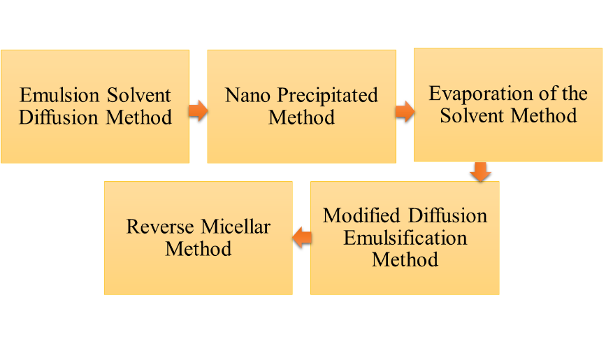
Fig No.4. Techniques Nanogel Preparation
Emulsion Solvent Diffusion Method for Nanogel Preparation:
In this method, the drug is dissolved in an organic solution, while a polymer and gelling agent are dissolved in water to form the drug phase. This drug phase is then added drop by drop into the aqueous phase, which has been homogenized for 30 minutes at 6000 rpm. During this process, an oil/water emulsion is formed as the homogenizer breaks the emulsion into tiny droplets. To convert this emulsion into a nanogel, triethanolamine is added, and the mixture is continuously stirred for an hour at 8000 rpm to complete the nanogel formation.
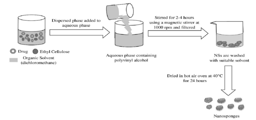
Fig No.5 Emulsion Solvent Diffusion Method
Nano Precipitation Method:
In this technique, the organic phase, comprising both the drug and polymer, interacts with the surfactant-containing aqueous phase, leading to the precipitation of the polymer. Following the elimination of excess solvent, polymeric nanoparticles remain as the final product. After moistening the particles, a gelling agent and the required quantity of nanoparticle dispersion are incorporated. Triethanolamine is then used to adjust and stabilize the pH.
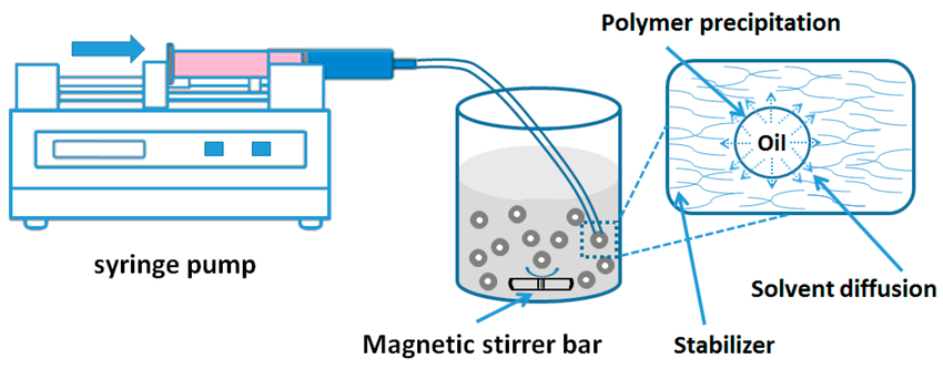
Fig No. 6 Nano Precipitation Method
Solvent Evaporation Method:
Over a two-hour period, the drug-polymer mixture is introduced into the aqueous phase while continuously stirred at 1000 rpm using magnetic stirrer. The resulting nano sponges undergo filtration and are subsequently dried in a hot air oven at 40? for 24 hours. Once dried, they are carefully stored in vials. For uniform dispersion, the polymer should be soaked in water for two hours before initiating gel formation. Following this, the polymer is agitated at 6000 rpm. A pH-adjusting agent is used to regulate the pH, after which the aqueous dispersion is blended with the optimized nanosponges suspension and permeation enhancers.
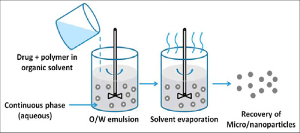
Fig no. 7 Solvent Evaporation Method
Reverse Micellar Method:
In this method, the polymer, drug, and surfactant are dissolved in an organic solvent. A cross-linking agent is then introduced gradually over an extended period, typically overnight. Once the nanoparticles are purified, the solvent is evaporated, resulting in a dried bulk produced. The nanogel formulation is prepared by dissolving the gelling agent in water. When the nanoparticles are dispersed into this aqueous phase containing the gelling agent, a nanogel is formed. The pH of the formulation is then adjusted using a neutralizing agent.
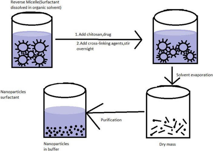
Fig no.8 Reverse Micellar Method
Modified Diffusion Emulsification Method:
In this approach, a solvent-containing polymer is blended with the drug in a carefully measured ratio. The organic phase is formed by continuously stirring the drug-polymer mixture in an aqueous phase at a speed of 5000 to 10000 rpm. Using a syringe with a needle, the organic phase is gradually introduced into the aqueous stabilizer solution at a controlled rate of 0.5 ml per minute. The mixture is then stirred for six minutes at a rotational speed of 10000 to 25000 rpm, followed by sonication for five to ten minutes to achieve uniform dispersion.
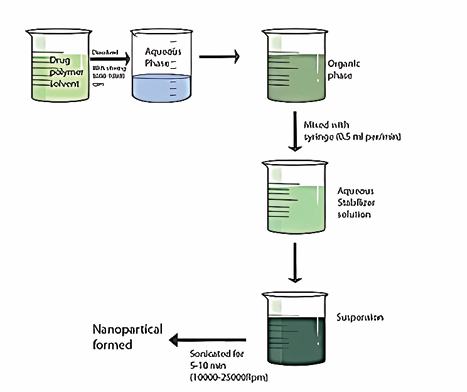
Fig No.9 Modified Diffusion Emulsification Method
Evaluation of Nanogel:(18,27,31–38)
Physical Characteristics of Nanogels:
The prepared Etoricoxib nanogel formulations were examined visually to assess their colour, clarity, and uniformity. After allowing the gels to settle in their containers, they were carefully observed for consistency and the presence of any visible aggregates.
pH Measurement:
The pH of the developed nanogels was assessed using a digital pH meter. Prior to use, the instrument was calibrated with standard buffer solutions of pH 4.0, 7.0, and 9.0. To prepare a 1% aqueous solution, a specific quantity of the nanogel was dissolved in 100 mL of distilled water. The glass electrode was then immersed in the solution to record the pH. Measurements were taken in triplicate, and the standard deviation was calculated. To minimize any risk of skin irritation, it is essential that the gel’s pH closely matches the natural pH of the skin.
Spreadability:
Spreadability is assessed to determine how efficiently the gel spreads when applied to the skin. A specific amount of gel was placed between two horizontal glass plates, each measuring 20 × 20 cm. A known weight was gently placed on the upper plate to facilitate spreading. After one minute, the diameter of the spread gel was measured in centimetres. Spreadability was then calculated using the formula:
S = (M × L) / T
Where:
S = Spreadability (gm.cm/s)
M = Weight applied on the upper plate (g)
L = Distance moved by the upper plate (cm)
T = Time taken (s)
Particle Size and Polydispersity Index (PDI):
The particle size of the developed nanogel was measured using photon correlation spectroscopy, which evaluates variations in light scattering caused by the Brownian motion of particles. A Malvern MasterSizer 2000 MS was used for the analysis. For measurement, 1 mL of the nanogel was diluted with 10 mL of distilled water to obtain a clear solution. The average droplet size, polydispersity index (PDI), and zeta potential were subsequently analysed using the Malvern Zetasizer.
Drug Content:
The amount of drug present in the formulation was quantified using UV spectrophotometry and high-performance liquid chromatography (HPLC) techniques.
Dynamic Light Scattering (DLS):
Dynamic light scattering is a technique used to evaluate the size distribution of nanoparticles suspended in liquids. It measures fluctuations in light scattering on a microsecond scale during various experimental conditions. This method helps assess the influence of cross-linking agents and the charge density of polymer chains on the hydrodynamic radius of the resulting nanogels. DLS is also effective in monitoring nanogel swelling behavior in different media. However, it's important to note that very small polymer particles may not always be accurately represented in DLS results. To gain a comprehensive understanding of particle characteristics, DLS is often complemented with other analytical techniques. In this study, both the average particle diameter and the polydispersity index were determined using DLS.
REFERENCES


Nivrutti Kotsulwar, Dr. Shivappa Nagoba*, Rachita Malshette, Maithili Kamble, Mayur Upade, Amrapali Rajput, Nanogel in Modern Drug Delivery: A Comprehensive Review of Synthesis, Properties, Applications, And Evaluation Techniques, Int. J. of Pharm. Sci., 2025, Vol 3, Issue 5, 4681-4695. https://doi.org/10.5281/zenodo.15542653
 10.5281/zenodo.15542653