View Article

Abstract

Diabetes mellitus is a long-term metabolic disease which hinders wound healing and raises the chance of developing chronic diabetic foot ulcers (DFUs), infections, and relapse. Chronic high blood glucose levels interfere with the normal process of wound repair by causing prolonged inflammation, oxidative stress, poor angiogenesis, peripheral neuropathy, and abnormal remodelling of the extracellular matrix. This review focuses on the possible benefits of medicinal plants, such as Rosmarinus officinalis, Punica granatum, Moringa oleifera, Curcuma longa, and Azadirachta indica, since these possess antioxidant, anti-inflammatory, antimicrobial, and possible glucose-regulating properties. The review also looks at conventional antidiabetic drugs, including metformin, DPP-4 inhibitors, pioglitazone, and GLP-1 receptor agonists, as these might help tissue repair by bringing about glycaemic control and by influencing the pathways related to inflammation, angiogenesis, and cellular regeneration. Even though promising results have been obtained, further clinical studies are still required to determine their safety, effectiveness, and value as additions to standard diabetic wound care.

Keywords

Diabetes mellitus, peripheral neuropathy, medicinal plants, Curcuma longa metformin oxidative stress, angiogenesis

Introduction

× Popup Image

Diabetes mellitus is a complex condition arising either from a failure to sense glucose or from insufficient insulin production, from autoimmune destruction of the β-cells in the case of type 1 diabetes, or from inadequate compensation for insulin resistance in the case of type 2 diabetes [5-7]. It is currently estimated that about 500 million people are affected by diabetes mellitus, with forecasts showing that the number will increase considerably in the future. In the United States alone, annual expenditures on medical bills and lost working days because of diabetes mellitus exceed $300 million [1,2].Moreover, one estimate suggests that between one in three and one in five people with diabetes mellitus will develop a chronic non-healing wound during their lives, for example a diabetic foot ulcer (DFU), which has a worrying recurrence rate of 40% within one year and 65% within five years [3,4].In the case of type 1 diabetes, the beta cells in body cease to produce insulin, so the patient have to depend completely on insulin injection in order to survive. Type 2 diabetes, which accounts for about 90%of all cases, is caused by the body becoming resistant to insulin. Although type 2 diabetes can generally be controlled by changes in diet, exercise, and the use of oral medications, both types of diabetes have similar symptoms consist of high blood sugar levels, a constant feeling of thirst, needing to go to the bathroom frequently, unexplained weight loss even though the person is hungry, fatigue, blurred vision, nausea, and sudden changes in mood [8].

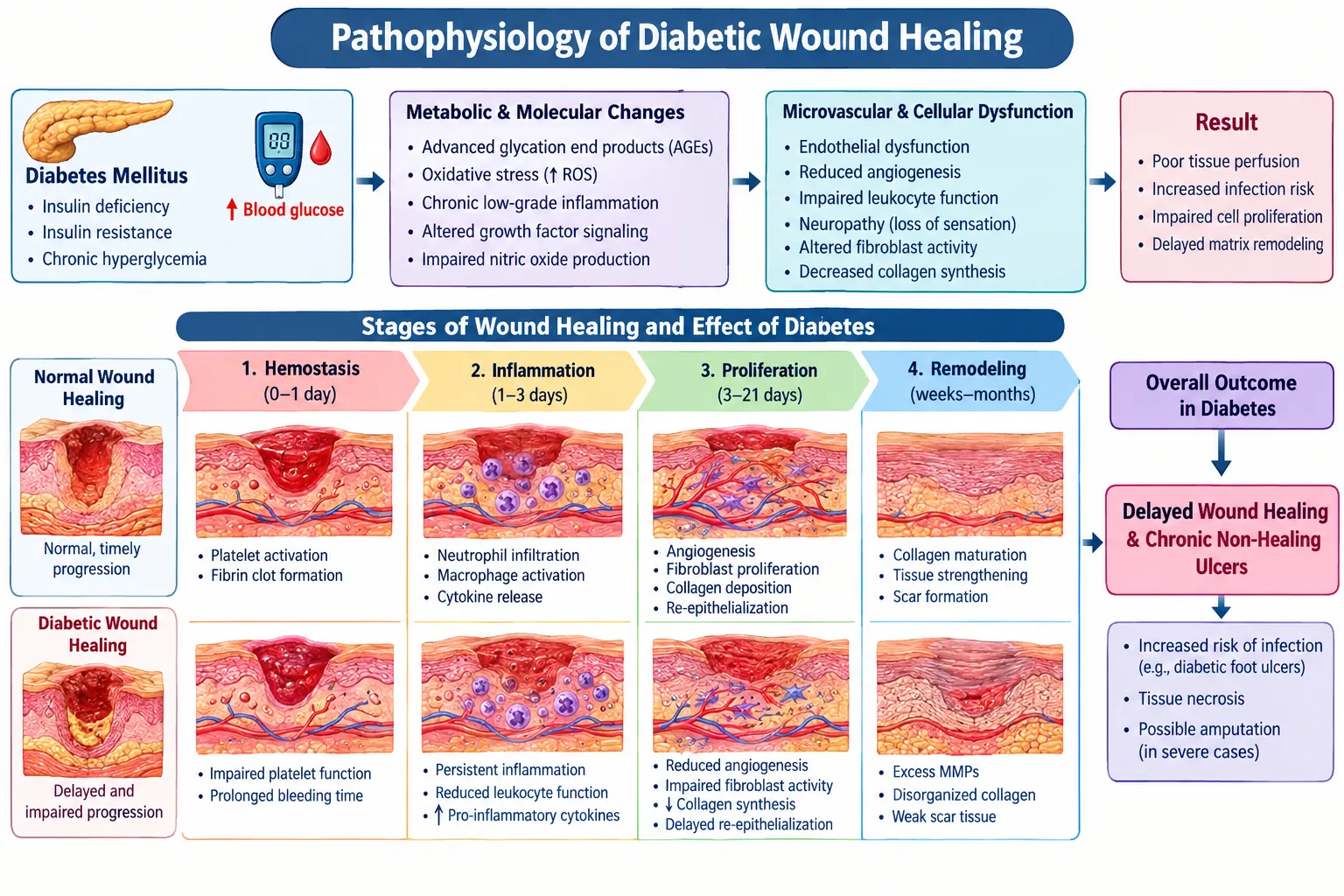
Normal Wound Healing Vs. Diabetic Wound Healing

Normal Wound Healing
figure

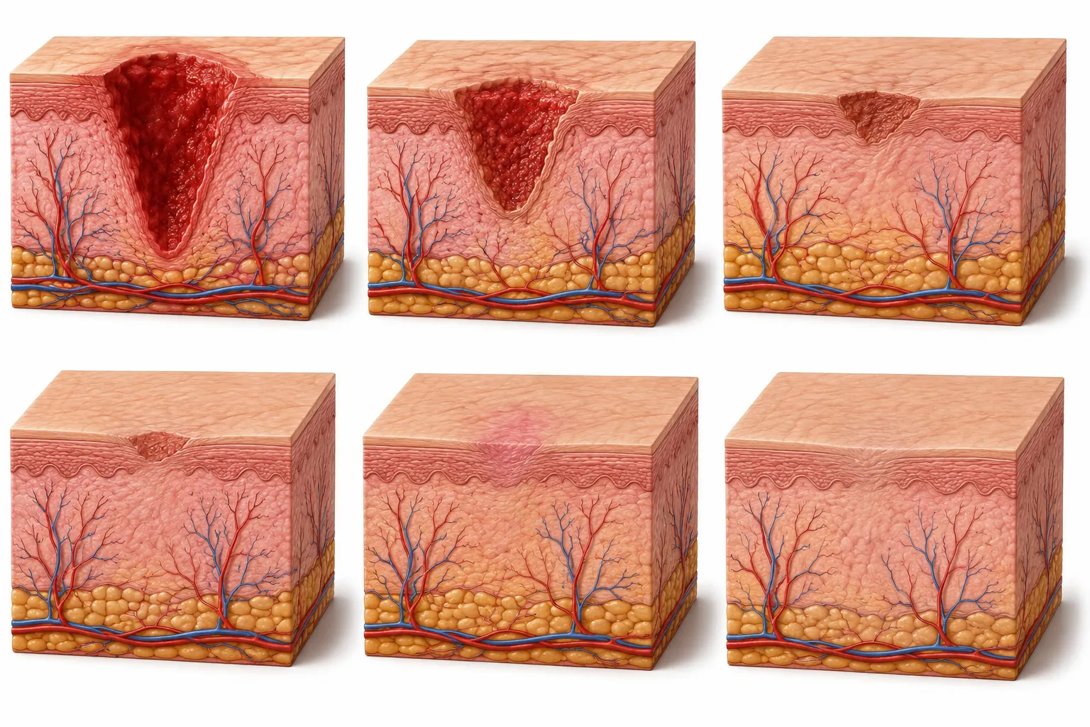
A wound is described as a disruption of the normal anatomical structure and function [9]. Healing a tissue refers to the process of restoring its anatomical continuity and function. For tissues that cannot regenerate, healing occurs through the deposition of connective tissue and results in scarring. An example of this is the replacement of parenchymal tissue with connective tissue in the heart following a myocardial infarction [10].

Normal wound healing Stages

Haemostasis

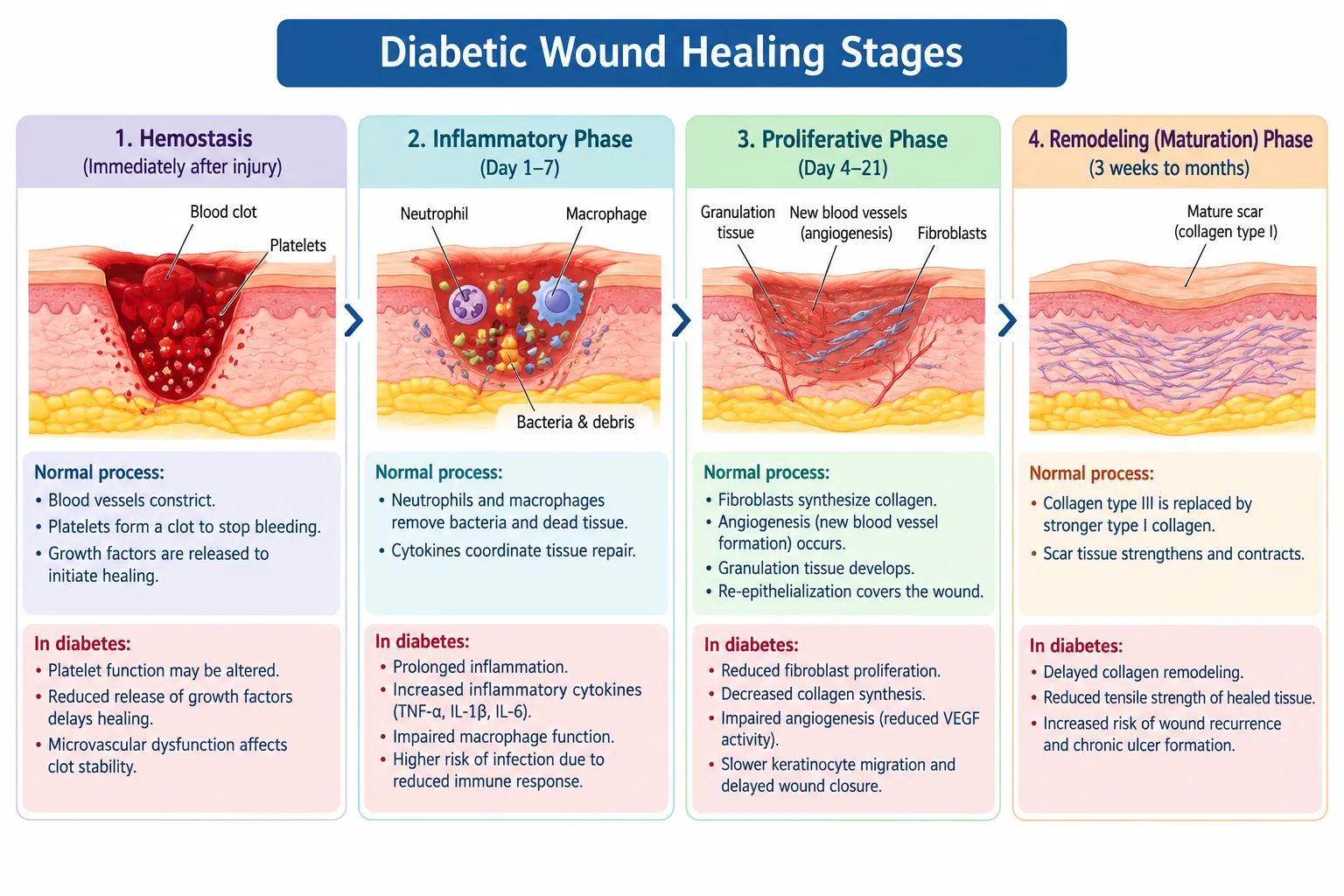
In the event of tissue injury, the body begins haemostasis, a response that stops blood from leaving a skin wound.[11]. To do this the body tightens the blood vessels near the injury, which reduces blood loss for a time. When collagen fibres and the inner lining of the vessel are exposed, platelets in the blood stick to the vessel wall. Release growth signals and other clotting substances. After this the platelets gather to create a platelet plug, which's essential to halt further bleeding from the damaged vessel. The platelet plug stops bleeding immediately [12]. The resulting fibrin clot acts as a shield that keeps bacteria out of the bloodstream. It also provides a framework, for immune cells fibroblasts and endothelial cells to move in and mend the injured tissue. The fibrin clot remains as a barrier while the repair process continues [13].

Inflammation

Inflammation is marked by the movement of neutrophils and macrophages which play a role in clearing away foreign substances, bacteria and damaged tissue to stop infection. During this stage the blood vessels that were initially narrowed begin to widen and become more permeable allowing neutrophils and macrophages to reach the area. The widening of blood vessels leads to redness, warmth and swelling at the site of the injury. The pain experienced is caused by the swelling and the presence of chemicals, such, as bacterial toxins at the wound. The presence of pain and swelling can reduce the function of the affected area. The five main signs of inflammation are swelling (tumor) redness (rubor) warmth (Calor) pain (dolor) and loss of function (functio lease) [14].

Proliferation

Angiogenesis is the process involving the formation of new blood vessels which may arise from existing blood vessels or from progenitor cells. It is triggered by growth factors such as growth factor-1 (FGF-1), FGF-2 and vascular endothelial growth factor (VEGF). These growth factors are emitted by platelets, macrophages, fibroblasts and keratinocytes [15,16,17]. Following this, the endothelial cells begin to move, to divide and arrange themselves in the fibrin-fibrinogen matrix. They produce buds of blood vessels. The buds then join one another to form blood vessels. This process goes on until the number of blood vessels in the wounded area is three times greater than in the surrounding tissue [18].

Maturation

The final stage of wound healing, in lude PDGF, TGF-β1, and EGF. At the same time, cells release inflammatory messengers such as IL-1[26]. Those signals pull in macrophages and neutrophils. After they arrive, they boost the inflammation around the injury [27,28]. Next, chemotactic signals push neutrophils into an active state. The neutrophils then gather at the wound, stick to nearby tissue, and release matrix parts outside the cells. Still, poor small blood flow can interfere with how neutrophils attach. Inflammation may stay on for too long. That leads to a late wound healing process overall [29,30].

Diabetic Wound Healing

Diabetic Wound Inflammation

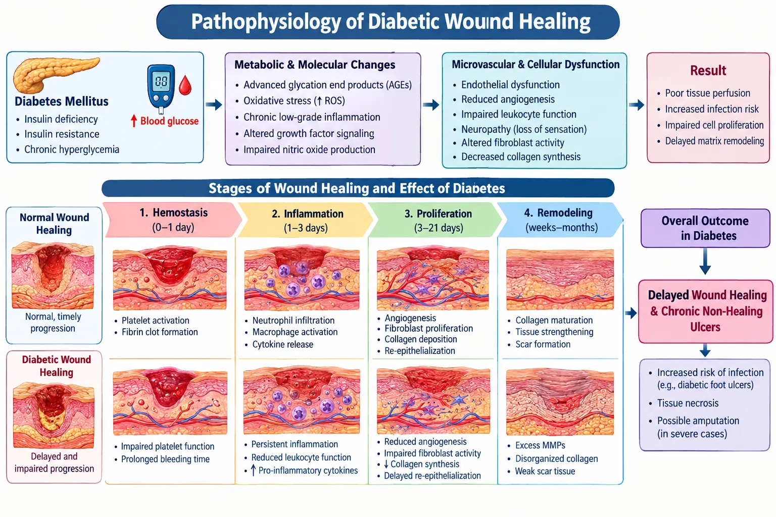
Compared to wounds in people without diabetes, wounds in individuals with diabetes take longer to go through the inflammatory stage of healing. This prolonged period of inflammation slows down the healing process and can result in the development of a chronic wound [19]. Neutrophils, a type of white blood cell, play a role in inflammation by releasing harmful enzymes, inflammatory substances, and free radicals, which increase oxidative stress. This oxidative stress causes more tissue damage and slows down the healing process in diabetic wounds. Neutrophils also create structures called neutrophil extracellular traps (NETs), which are used to trap harmful microorganisms. In diabetic wounds, the production of NETs is increased, which keeps the inflammatory response going and makes it harder for the wound to heal. Factor that contributes to extreme inflammation in diabetic healing is miRNA. While miRNA levels related to healing are similar in both diabetic and non-diabetic skin that is not injured, they behave differently once the skin is wounded. This suggests that miRNA also plays a role in the abnormal or dysregulated inflammation seen in diabetic wounds [20].

Angiogenesis in Diabetic wound healing

In wounds associated with diabetes, a lack of adequate angiogenesis significantly impacts poor healing, which results from several factors. To begin with, diabetic wounds exhibit a shortage of essential proangiogenic factors, likely due to a reduced presence of macrophages that generate them. Moreover, there is an increase in anti-angiogenic factors, while factors that promote capillary maturation are diminished. This lack of maturation factors slows down the healing process, heightening the chances of the wound becoming chronic or recurring [21]. Furthermore, changes in the blood vessels of people with diabetes and insufficient oxygen delivery impede the movement of white blood cells to the area of the wound, thus increasing the risk of infections [22].

Scarring of Diabetic Wound Healing

The normal healing process of wounds ends with maturation, remodelling, and the formation of a scar. During this stage, extra collagen breaks down, the wound tightens, and a scar forms [23]. The scar is made up of collagen and extracellular matrix produced by fibroblasts. New skin scars differ from the original tissue in several ways; they lack hair follicles and sebaceous glands, and their collagen fibers are tightly packed [24]. In individuals with diabetes, the healing process results in a scar that has reduced collagen production and structural alterations when compared to scars in healthy individuals. These changes cause the scar to have reduced contraction ability and higher collagen density, both of which reduce the scar's tensile strength and negatively affect wound healing. The inadequate contraction observed in diabetic wounds is partly due to fibroblasts that have become resistant to multiplication and have entered a state of senescence. In the absence of adequate contraction, diabetic wounds depend more heavily on granulation tissue and reepithelialization for healing, which may lead to a diminished ability of the scar matrix to withstand tensile forces and shear stress [25].
figure

Molecular and Cellular Pathophysiology of Diabetic Wounds

Dysregulated Innate Immune Responses in Diabetic Wound Healing

In typical wound healing, M1 macrophages are brought in during the initial inflammatory phase, where they aid in eliminating pathogens and damaged cells through proinflammatory functions. As the wound healing process moves into the proliferative and remodelling phases, macrophages gradually take on an M2-like phenotype. M2 macrophages facilitate tissue healing by releasing growth-promoting factors, aiding in ECM accumulation, and encouraging angiogenesis in injured regions. The usual shift from proinflammatory to reparative macrophage phenotype in diabetic wounds is greatly hindered. Continual buildup of these inflammatory and oxidative substances leads to harm to cells and tissues, undermines the function and stability of growth factors, and disrupts the formation of healthy granulation tissue. Collectively, these irregularities extend the inflammatory stage and significantly hinder the typical advancement of diabetes [31].

Impaired Angiogenesis and Microvascular

Restoring proper microvascular flow is essential for supplying oxygen and nutrients to the healing tissue. In typical hypoxic conditions after an injury, signals that promote angiogenesis, like vascular endothelial growth factor (VEGF)are increased. The movement of endothelial cells is impaired pro-angiogenic signalling is lacking, and anti-angiogenic elements (like pigment epithelium-derived factor) are abnormally heightened. Furthermore, shortages in vascular stabilization elements, like platelet-derived growth factor (PDGF), hinder the stabilization of newly formed capillaries, resulting in weak, non-operational microvascular. [32].

Matrix Remodelling in Diabetic Wounds

In wounds of people with diabetes the process of rebuilding tissue is not working properly. Lasting inflammation, high blood sugar levels, too much free radicals and low oxygen levels affect how skin cells work and how the structure of the skin is made. Wounds in people with diabetes often have collagen and it is not arranged the right way. Less activity from a protein called TGF-β, which usually helps skin cells make collagen has been seen in these wounds. These issues cause the tissue that helps wounds heal to not form [33]. A major problem in the way the skin structure is built in diabetes is much cutting and breaking down of the structure. Enzymes called MMPs are important for healing because they clean up damaged parts help cells move around and are part of rebuilding tissue. Normally these enzymes stop working once the inflammation goes down. In long-lasting wounds from diabetes the inflammation continues, blood sugar stays high oxygen is not enough and certain chemicals like TNF-α and IL-1β keep causing more of these enzymes to be made. More of MMP-1, MMP-2, MMP-8 and especially MMP-9 have been found in wounds on the feet of people, with diabetes [34].

Metabolic and Molecular change

High blood sugar and insulin resistance stop wound healing by breaking the normal function of cells and the signals inside the body. These issues alter how inflammation happens how new blood vessels grow how fibroblasts work how collagen is made and how tissues are rebuilt [35]. When high blood sugar stays for a time it triggers harmful chemical changes, in the body. These changes involve the polyol pathway, the protein kinase C pathway and the advanced glycation product pathway. A lot of sugar builds up glycation end products [36]. A key feature of diabetic wounds is extended and unregulated inflammation. In a typical wound, inflammatory cells eliminate damaged tissue and pathogens prior to the wound advancing into the proliferative phase. In diabetes, irregular activity of neutrophils and macrophages, an overabundance of inflammatory mediators, and continuous activation of inflammatory signalling pathways hinder the prompt resolution of inflammation. This extended inflammatory condition leads to ongoing tissue injury and hinders advancement toward regeneration [37].

Neuropathy

Aside from raising the likelihood of DFU development, every form of neuropathy (sensory, Motor and/or autonomic functions can distinctly affect the healing of DFUs. For instance, autonomic neuropathy reduces sweat gland function, resulting in dry and cracked skin, thus heightening the likelihood of itching and infection, which impedes wound healing [38]. Furthermore, due to dry skin and inadequate circulation, diabetic neuropathy is linked, for reasons that are not well understood. Accompanied by itching [39,40]. Simultaneously, motor neuropathy elevates pressure on the plantar. Surface of the foot, causing tissue ischemia and necrosis [18]. In general, neuropathic skin possesses a lower density of neurons and shows impaired skin healing [41].
figure

The Role of Medicinal Plants in Diabetic Wound Healing

Due to the ethnobotanical survey, numerous types of plants and herbs that aid in wound recovery Activities related to agriculture have been recognized in Africa and various other regions. Emerging nations. The application of medicinal herbs in Wound care and management includes cleaning and disinfection, debridement, along with ensuring a suitable environment treatment for the body's natural healing process [42]. It is presumed that components derived from medicinal plants exhibit lower toxicity and exhibit reduced side effects in comparison to conventional therapeutic substances; therefore, the heightened and revitalized fascination with the utilization and implementation of therapeutic flora during the process of healing wounds in those with diabetes and conditions that are not diabetic. Difficulty in the recovery of diabetic wound is recognized as a significant health concern. For healthcare providers worldwide, and this is associated due to the unclear cause in certain instances; consequently, a therapeutic method for treatment is the utilization of healing herbs especially in impoverished resource configurations [43].

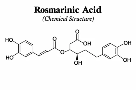
Rosmarinus officinalis

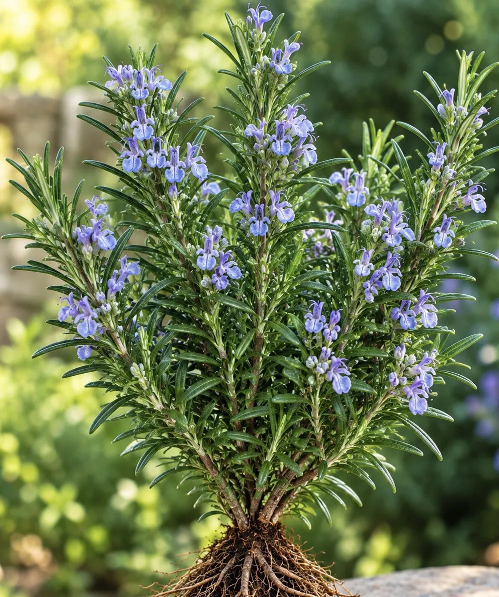
The scientific name of (Rosmarius officinalis) is Salvia rosmarinus ,its belong to family of mint in Lamiaceae , Rosmarinus officinalis was also known as rosemary, the Rosmarinus officinalis found in geographical region like Morocco, Spain, France, Algeria and some parts of India like Kerala in the region of Nilgiris Hills District [44].

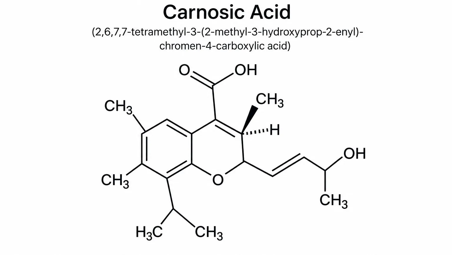
This plant is recognized for its antioxidant and antibacterial effects. In the animal model, Abul-Al-Basal [45] investigated the therapeutic capabilities of the extract of the plant on complete-thickness excision cut necrotic injuries in alloxan-induced diabetic BALB/c mice. For this research, 6-week-old male BALB/c mice weighing 18-20 g were utilized. Diabetes in the rodents was initiated by administering 0.2 ml of alloxan monohydrate within the peritoneal cavity. After the experiment, several assessments were conducted involving wound contraction, granulation tissue, and histopathology. Results indicated that on day 3, the moist weight of granulation tissue had significantly elevated in comparison to the untreated controls. Quantitative assessments of wound dimensions were commonly employed to evaluate the initial dimensions of a wound prior to and following debridement and advancement in wound closure. The rate of wound healing was measured by observing the reduction in wound size as a percentage on days 3, 6, 10, and 15 after injury. . The author concluded that the essential oil from the aerial parts of the plant had a more effective healing effect than the aqueous extract when applied topically on wounds of diabetic mice. This healing ability of the plant can be attributed to its strong antimicrobial, anti-inflammatory, and antioxidant properties [46,47].
figure

figure

figure

Application

Enhanced Tissue Regeneration: Promotes fibroblast proliferation, reepithelialization, and the regeneration of healthy granulation tissue [48].

Collagen Deposition & Angiogenesis: Stimulates collagen synthesis and new blood vessel formation, which are often impaired in chronic diabetic ulcers [49].

Punica granatum

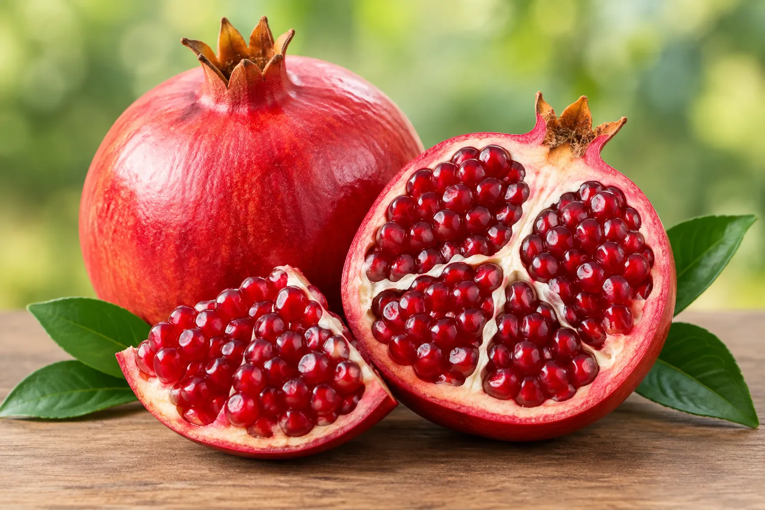
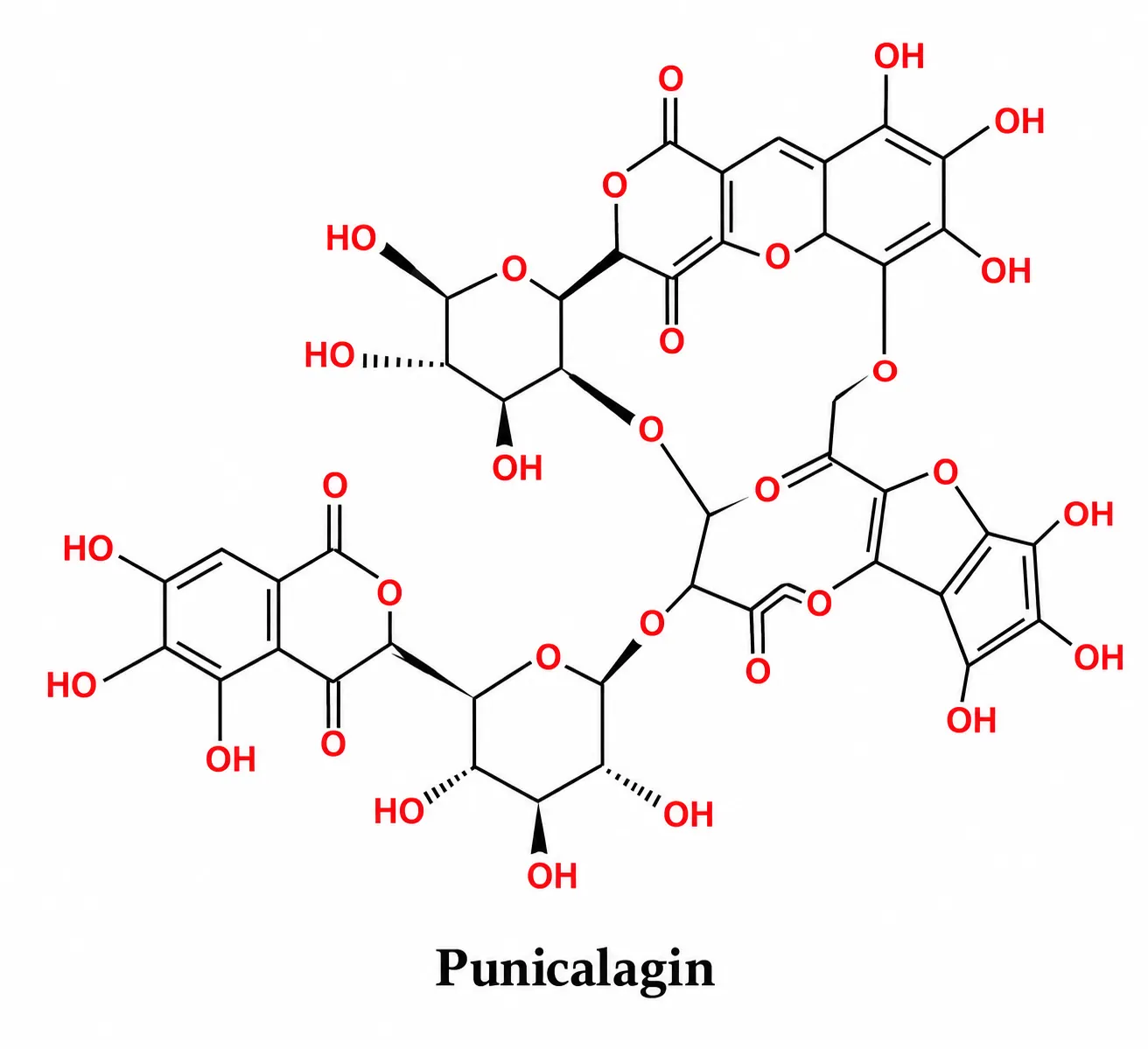
The pomegranate's scientific name is Punica granatum, and it is part of the Lythraceae family (previously grouped under Punicaceae); it includes both fresh and dried fruit [50].

Pomegranate extracts may be useful in preventing and treating type 2 diabetes, according to the findings. Later reports that addressed the therapeutic effects of pomegranate, in general [51], have indirectly discussed the connection between the pomegranate and type 2 diabetes. Moreover, a recent review by Medjakovic and Jungbauer [52] addressed the potential use of the pomegranate and its active compounds as therapy for metabolic syndrome. Hence, further research, particularly clinical, focusing on the effects of pomegranate needs to be performed to fully characterize the relationship between this fruit and type 2diabetes. In the study presented here, we sought to examine the short-term impact of fresh pomegranate juice on fasting serum glucose and insulin levels in individuals with type 2 diabetes. We proposed that drinking pomegranate juice might help lower insulin resistance and reduce blood glucose levels three hours after consumption. If our findings support this, pomegranate juice could be considered as a potential aid in managing type 2 diabetes. It could also provide dietitians with evidence-based nutritional information regarding the use of PJ in T2D patients [53].
figure

figure

figure

figure

Applicationa

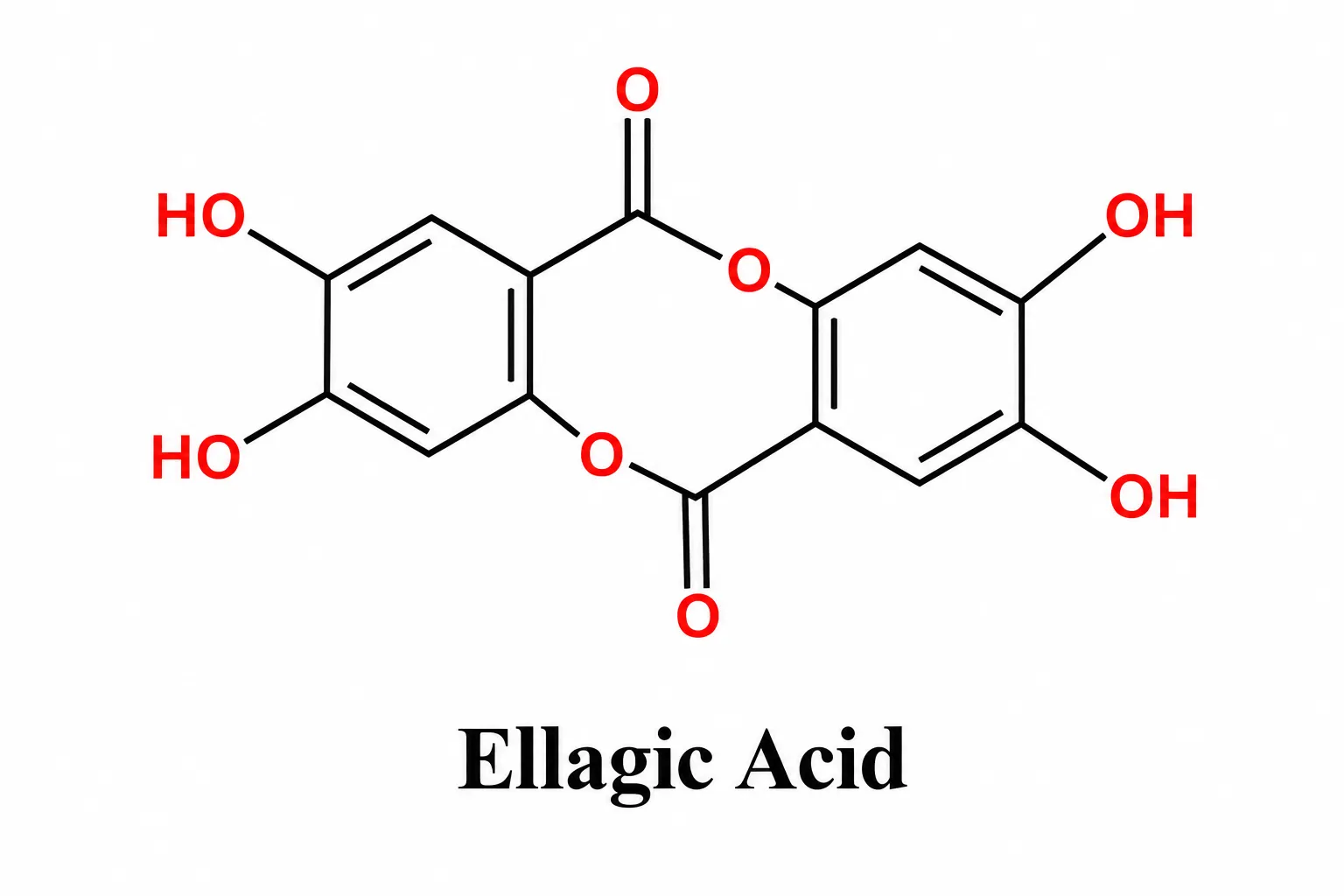
Blood sugar support: Certain compounds found in pomegranates, such as punicalagins and ellagic acid, may aid in slowing the absorption of carbohydrates and enhancing the body's response to insulin.Research from clinical studies suggests that these compounds could lead to lower levels of fasting blood glucose and improved resistance to insulin [54].

Moringa oleifera

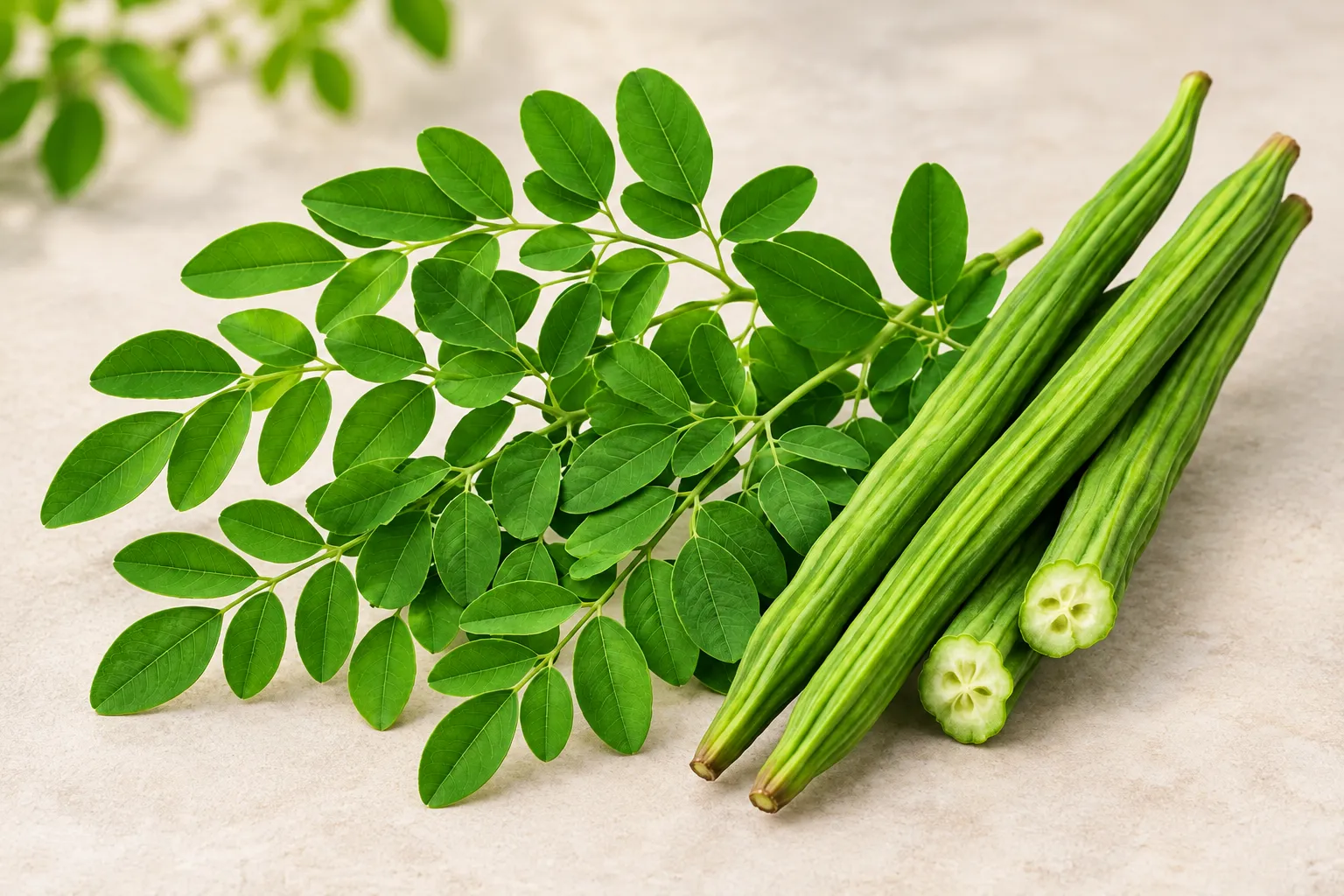
The scientific name of moringa oleifera is Moringa pterygosperma Gaertn and it belong to family of Moringaceae, The plant source consists of the dried or fresh leaves, seeds, roots, and bark.It is globally distributed across tropical and subtropical regions and the tree is indigenous to South Asia, specifically the sub-Himalayan foothills of northern India, Pakistan, Bangladesh, and Nepal [55].

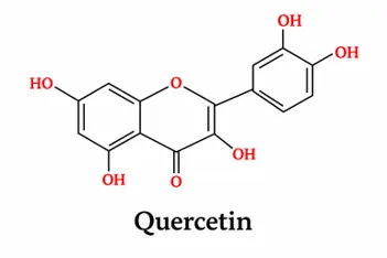
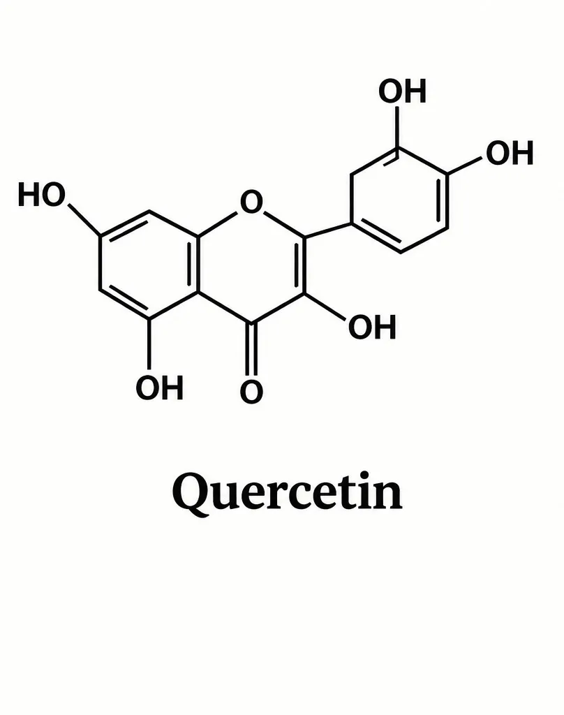
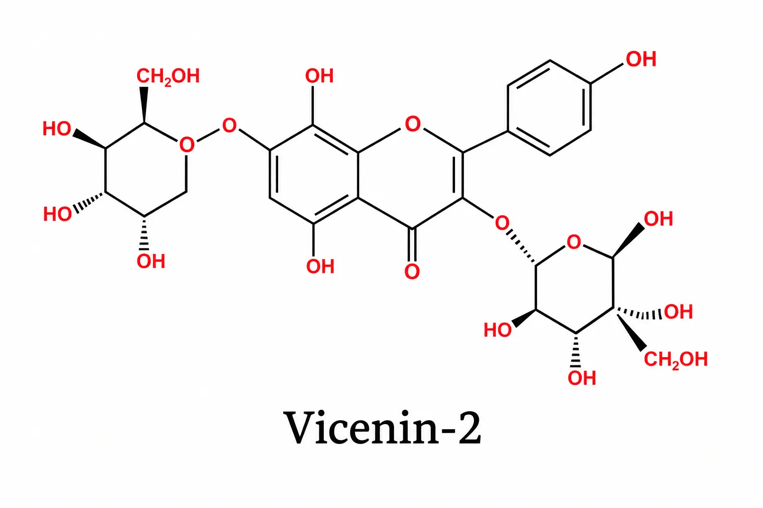
Morninga oleifera helps diabetic wounds heal faster. It does this by reducing stress decreasing inflammation, fighting infections and promoting tissue repair. The plant has compounds like quercetin and kaempferol. These compounds neutralize free radicals in the wound area. This helps the skin heal better and faster. It’s a way to support the body’s own healing process. This reduction in stress helps diabetic wounds close more effectively. Research indicates that the extract is effective against drug- bacteria such as Staphylococcus aureus and MRSA which are common causes of serious infections in people with diabetes. When applied to the skin Moringa oleifera promotes the growth of fibroblasts increases the formation of blood vessels and enhances collagen production. These processes help rebuild the skin structure and accelerate the formation of skin, over the wound [56]
figure

figure

figure

Application

Enzyme Inhibition: Enzyme inhibition delays the carbohydrates’ breakdown and the absorption of glucose. It acts by inhibiting enzymes, such as α-amylase and α-glucosidase [57].

Reduction of Oxidative Stress: The antioxidant power fights cell damage and reduces problems that come from diabetes [58].

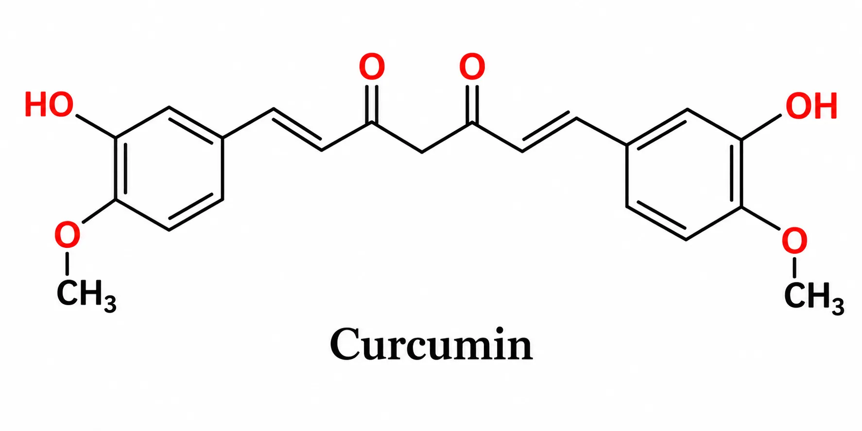
Curcumin

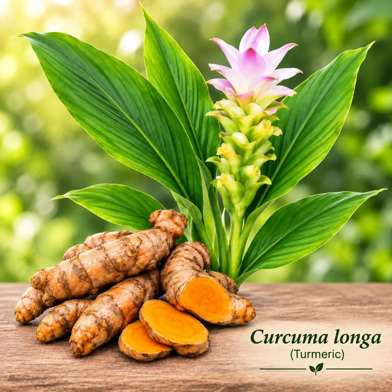
Curcumin scientific name is Curcuma longa is it also known as turmeric, is a plant of the ginger family, Zingiberaceae. It is mostly cultivated in Southwestern Asia. This plant is widely used in the cooking of many countries including Iran, Malaysia, India, China, Polynesia and Thailand. It is a spice that can change the taste, color and smell of food. In many recipes turmeric is mixed with other spices [59].

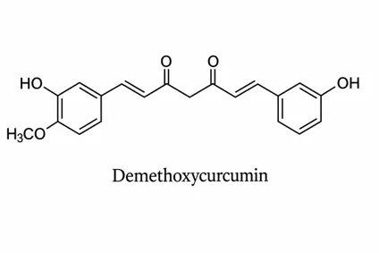
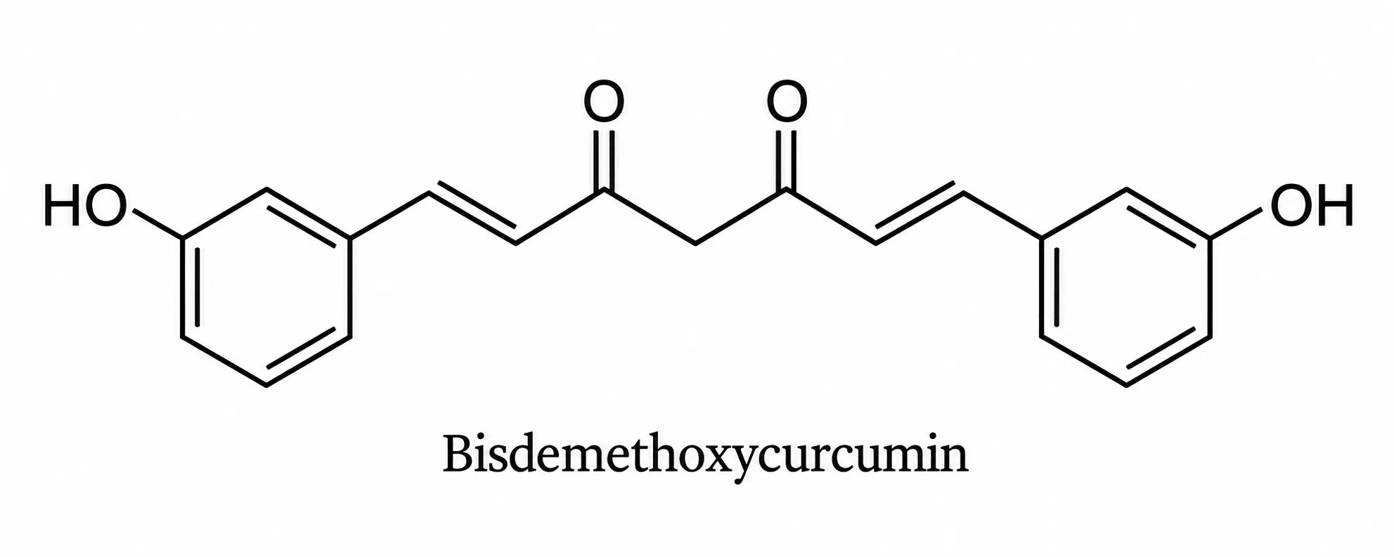
The main part of the plant is the rhizomes, and the most common active ingredients are the curcuminoids, which include curcumin, demethoxycurcumin, and bisdemethoxycurcumin[60,61,62].These curcuminoids are non-toxic polyphenolic compounds that have a wide variety of biological effects[63,64].They are known to produce significant immunosuppressants that help reduce the production of IL-2 and IL-12.This compound also inhibits the expression of several pro-inflammatory molecules, including iNOS (inducible nitric oxide synthase), COX-2 (cyclooxygenase-2), lipoxygenase-5, and various inflammatory cytokines such as TNF-α, IL-1, IL-6, and IL-8 [65-67].

Curcuminoids can regulate apoptosis and inhibit neurotoxic elements in macrophages and alveolar monocytes activated by lipopolysaccharides. Additionally, it hinders the phosphorylation and breakdown of IκBα (nuclear factor kappa light polypeptide gene enhancer in B-cells inhibitor, alpha) and triggers the γ receptor pathway stimulated by peroxisome proliferators, diminishing the inflammation profile caused by the NF-κB pathway [68-69].

Application

Diabetic Complications: Aids in preventing damage to blood vessels, nerve damage, and eye conditions such as retinopathy by reducing oxidative stress [70].

Bioavailability Limits: Natural curcumin has limited absorption by the body, prompting the development of advanced forms like nanocurcumin to improve its effectiveness [71].
figure

figure

figure

A

figure

Azadirachta indica

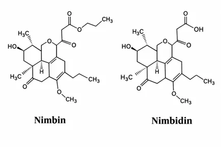
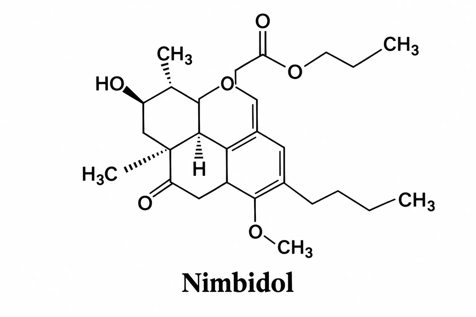
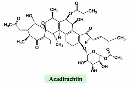
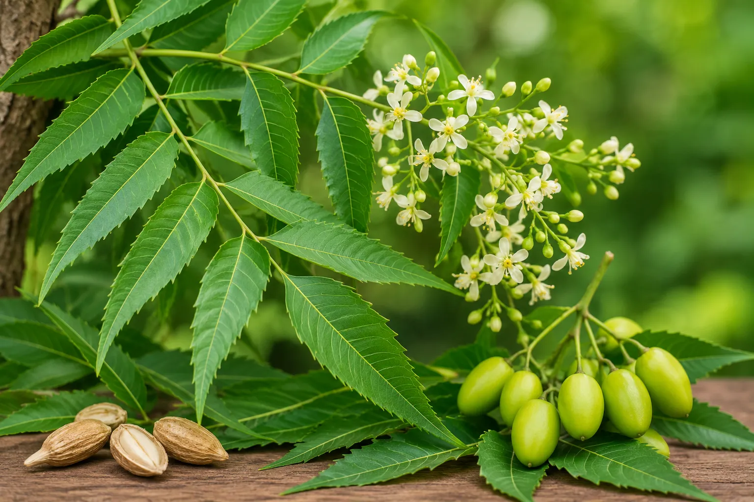
The neem tree, also known scientifically as Azadirachta indica and belonging to the Meliaceae family is an evergreen that grows widely in Asia. Many people use neem leaves and neem seeds in home remedies in the traditional system of Ayurveda. In Sanskrit the neem tree is called rishtha, which means "Reliever of Sickness ". It is also called Sarba-roga-nibarini. Several chemicals from neem such, as Azadirachtin, Nimbidiol, Gedunin and Azadiradione have shown the ability to lower blood sugar but these chemicals can be toxic. The bitter flavor of neem seed oil comes from a compound called Meliacin. Another compound, Tignic acid (5-methyl-2butanoic acid) taken from neem seeds gives neem seed oil its special smell. All these compounds are a class of products known as triterpenoids [72].The effect of neem leaf water extract on lowering blood sugar may happen because more insulin is released from the cells in the pancreas or because the body takes in and uses glucose better in parts of the body. Insulin made by the cells that are still working or new cells helps the extract fight diabetes brings back the bodys ability to respond to insulin lowers how much sugar, from food is not absorbed in the intestine or helps the body take in sugar through the tissues using a special transporter called GLUT-4. The neem leaf extract has chemicals called alkaloids that help increase the amount of insulin released by repairing the parts of the pancreas that make insulin [73].

Application

Alpha-Glucosidase Inhibition: Bioactive constituents in Neem act as inhibitors of enzymes. Alpha-Glucosidase Inhibition blocks the breakdown of carbohydrates into sugars in the digestive system thereby stopping rapid post-meal glucose surges [74]

Activates insulin-mimicking pathways. This improves the responsiveness of insulin receptors allowing muscle and fat cells to absorb glucose from the bloodstream more efficiently [75].

Neem contains phytochemicals, such, as flavonoids, triterpenoids, nimbin and nimbidin that affect pancreatic beta cells and increase insulin release [76].
figure

Active Constituent

figure

figure

figure

figure

Synthetic and Conventional Drugs of Diabetes Mellitus

1.Metformin

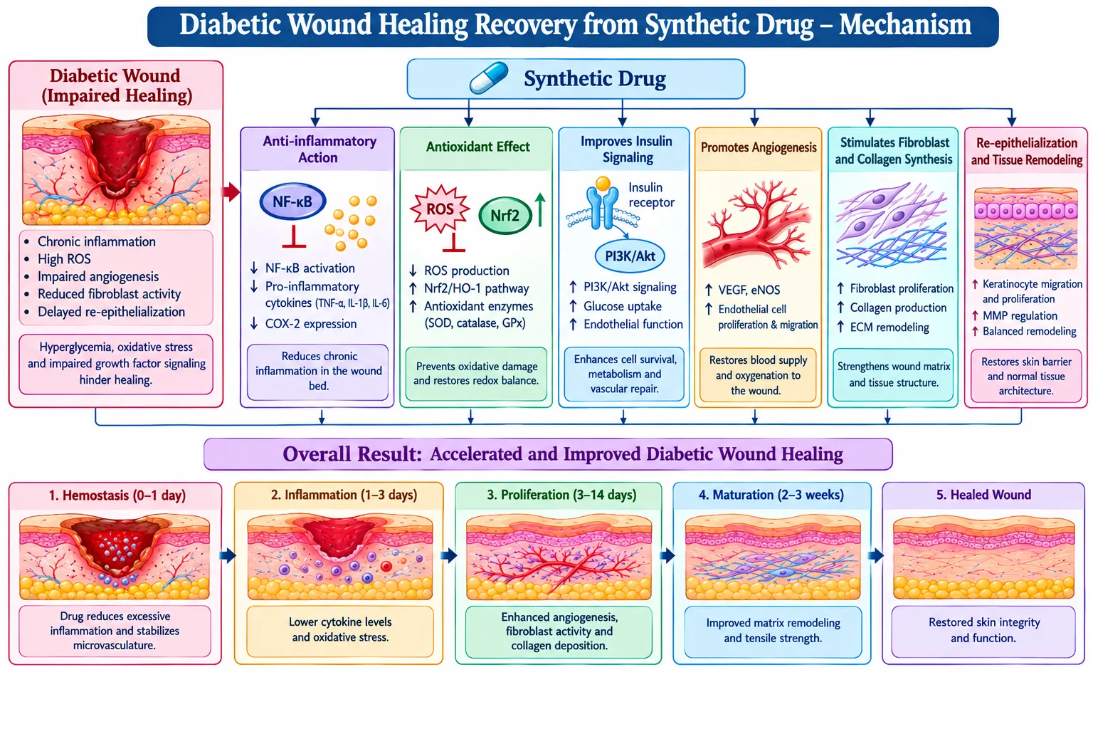
Clinical applications Metformin can help heal the wounds of diabetic patients by decreasing inflammation, preventing proliferation of cells, promoting angiogenesis and enhancing collagen synthesis when it is administered topically and systemically [80]. In terms of collagen synthesis, it increases the supply of collagen-I and collagen-III proteins and serves as a satisfactory foundation for new tissue formation. In terms of blood vessels generation, angiogenesis, it helps the living tissue develop new blood vessels, which carry oxygen and nutrients to the wound. In terms of inflammation decrease, it soothes the immune system and erases pro-inflammatory markers [77].

Mechanism

Activation of the AMPK pathway: Initiates cellular survival signals and diminishes cellular harm caused by high glucose or low oxygen levels in the wound area.Enhanced Angiogenesis: Boosts the quantity and activity of circulating endothelial progenitor cells (EPCs) and elevates Vascular Endothelial Growth Factor (VEGF) to form new blood vessels [78]. Enhances Collagen Production: Boosts levels of collagen types I and III, reinforcing the extracellular matrix for quicker healing.Anti-Inflammatory & Immunomodulatory Effects: Redirects macrophages to the pro-repair M2 phenotype while inhibiting detrimental pro-inflammatory cytokines and matrix-degrading enzymes (MMPs).

Decreased Apoptosis: Minimizes cell death occurrences in injured tissues by blocking pro-apoptotic indicators such as p53 [79].

2. DPP-4 Inhibitors

Inhibition of peptidase (DPP)-4 is a way to lower glucose levels, in the treatment of type 2 diabetes [83]. This approach was developed based on the idea that gut-derived glucagon-like peptide (GLP)-1 is an antidiabetic hormone. DPP-4 inhibition stops the inactivation of GLP-1, thereby elevating the circulating intact (active) GLP-1 levels beyond physiological levels that exhibit antidiabetic effects[84].

Mechanism

The function of DPP-4 inhibiting drugs like sitagliptin and linagliptin is to block the enzyme DPP-4. Normally, DPP-4 quickly degrades hormones that are released after a meal.These drugs stop the breakdown of the incretins. The incretins that are raised by DPP-4 inhibition are GLP-1 (glucagon-like peptide-1) and GIP (glucagon-like peptide-1) and GIP (glucose-dependent insulinotropic polypeptide) [80].

Reducing Blood Sugar: Lower levels of glucagon and increased levels of insulin inhibit the liver from producing more sugar and promote the body to utilize it, thus lowering blood sugar levels. Lowering of the Secretion of Glucagon: These drugs also result in the reduction of the glucagon secretions. It is a hormone responsible for the stimulation of the liver to start production of glucose and to add it to the bloodstream [81].

Releasing Insulin Mechanism: An incretion in the level of incretin causes the pancreas to release more insulin in the bloodstreams [82].

3.Pioglitazone

Pioglitazone accelerates the healing of wounds in diabetic patients by functioning as an agonist of PPAR-γ, thus reducing chronic inflammation, promoting tissue repair and stimulating the growth of new blood vessels [85].

Mechanism

Activation of PPAR-γ: Pioglitazone activates the PPAR-γ receptor it helps reduce long-term inflammation. This action of Pioglitazone helps manage conditions where inflammation plays a role. The PPAR-γ receptor Pioglitazone influences the body’s response to inflammation effectively. The result is an effect on chronic inflammation over time [86].

Pioglitazone also supports tissue repair. I notice that Pioglitazone encourages the formation of blood vessels, which are essential for wound healing. Another important effect of Pioglitazone [87].

MMP-9 is an enzyme that builds wounds and blocks the healing process. When pioglitazone reduces MMP-9 it allows the tissue to repair itself quickly. These actions together make pioglitazone a useful agent, in improving wound healing in patients [88].

4. GLP1As

Glucagon-like peptide-1 receptor agonists (GLP-1 RAs), which are commonly prescribed to control blood glucose and assist in weight reduction, have numerous other advantages for promoting tissue repair. For example, in the case of chronic wounds like diabetic foot ulcers, GLP-1 RAs do more than simply regulate blood glucose; they actually affect the wound directly by reducing inflammatory responses, increasing cell migration toward the wound, and enhancing new blood vessel formation [89].

Mechanism

Macrophage Polarization (Shift from M1 to M2)

Chronic diabetic wounds stay inflamed for a time mainly because of M1 macrophages. Macrophage Polarization is driven by M1 macrophages. GLP-1 receptor agonists interact with GLP-1 receptors on cells, and this lowers signals from NF-κB and TNF-α. GLP-1 receptor agonists change Macrophage Polarization from M1 type to M2 reparative type. M2 macrophages create anti-inflammatory cytokines such as IL-10 and the enzyme arginase-1. These substances help the healing process by encouraging the repair of damaged tissues [90].

Enhancement of Angiogenic Factors (VEGF and HIF-1α)

To fix oxygen and bad blood flow GLP-1 receptor agonists increase the activity of key factors that help blood vessels grow. These factors include Vascular Endothelial Growth Factor Receptor-2, called VEGFR-2 and Hypoxia-Inducible Factor-1 alpha, also called HIF-1α. At the time GLP-1 receptor agonists boost Sirtuin 1 also called SIRT1. SIRT1 protects cells that can become blood vessels and helps build functional small vessel networks. GLP-1 receptor agonists activate the phosphoinositide 3-kinase (PI3K) and protein kinase B (Akt) pathways at the wound edge. This helps skin cells called keratinocytes survive and move which is vital for skin renewal. Also GLP-1 receptor agonists activate the AMPK pathway, called Adenosine monophosphate-activated protein kinase. AMPK cuts down reactive oxygen species also called ROS and stops cell death that comes from advanced glycation end-products also called Ages [91].

Extracellular Matrix (ECM) Remodelling through MAPK: The change of fibroblasts, into myofibroblasts helps shrink the wound. GLP-1 receptor agonists control the Mitogen-Activated Protein Kinase, called MAPK and ERK1/2 pathways. These pathways manage enzymes called matrix metalloproteinases also called MMPs, which break down the matrix. They also encourage orderly collagen build-up of messy scar tissue [92].
figure

CONCLUSION

The repair of wounds in individuals suffering from diabetes is a difficult and gradual process involving chronic inflammation, oxidative stress, inadequate angiogenesis, altered cellular responses, neuropathy, and a defective remodelling of the extracellular matrix. Review of the available evidence shows that both synthetic drugs and medicinal plants can influence the various stages and molecular pathways taking place in this improper healing process. Synthetic drugs such as metformin, the DPP-4 inhibitors, pioglitazone, and the Glucagon-Like Peptide-1 Receptor Agonists (GLP-1 RAs) all have an impact on the various molecular pathways and cellular mechanisms associated with inflammation, the mobilisation of progenitor cells, angiogenesis, collagen synthesis, and tissue repair. In the same way, various medicinal plants such as Rosmarinus officinalis, Moringa oleifera, Curcuma longa, Punica granatum, and Azadirachta indica contain several phytoconstituents that have antioxidant, anti-inflammatory, antimicrobial, glucose-modulating, and tissue-repair properties. It is therefore impossible, based on this review, to assert that either synthetic medicines or natural medicines are generally quicker or better at healing diabetic wounds. The results show that the effectiveness of these medicines varies according to the drug or plant, the formulation, the mechanism of action, the properties of the wound, and the quality of the evidence. Even though medicinal plants show promise as potential complementary treatments and for future therapeutic approaches, synthetic drugs have more precise molecular targets. Yet further well-designed clinical studies are required to establish the effective doses, formulations, safety, and comparative efficacy in promoting wound healing before these methods can be routinely recommended for use in diabetic wounds.

REFERENCES

  1. Gao, D.; Zhang, Y.; Bowers, D.T.; Liu, W.; Ma, M. Functional hydrogels for diabetic wound management. APL Bioeng. 2021, 5, 031503. [CrossRef]
  2. Glover, K.; Stratakos, A.C.; Varadi, A.; Lamprou, D.A. 3D scaffolds in the treatment of diabetic foot ulcers: New trends vs.conventional approaches. Int. J. Pharm. 2021, 599, 120423. [CrossRef]
  3. Hajhosseini, B.; Gurtner, G.C.; Sen, C.K. Abstract 48. Plast. Reconstr. Surg. Glob. Open 2019, 7, 34–35. [CrossRef]
  4. Chang, M.; Nguyen, T.T. Strategy for Treatment of Infected Diabetic Foot Ulcers. Accounts Chem. Res. 2021, 54, 1080–1093.[CrossRef]
  5. Goboza, M., Meyer, S., Aboua, Y.G. and Oguntibeju, O.O. (2017) Diabetes mellitus: Economic and health burden, treatment and the therapeutical effectsof Hypoxis hemerocallidea plant. Med. Technol. SA,30 (2): 39-46.
  6. Kim, L.E., Lee, J.H., Kim, S.H. and Jung, Y. (2018) Skin regeneration with self-assembled peptide hydrogels conjugated with substance in a diabetic rat model. Tissue Eng., 24 (1-2): 1-15.
  7. WHO. (2018) Global Reports on Diabetes. World HealthOrganization, Geneva, Switzerland. Available from: http://www.who.int.org Last accessed on 12/12/2018
  8. 8.Matteucci, E. and Giampietro, O.: Oxidative stress in families of type 1 diabetic patients. Diabetes Care, 23, 1182–1186,2000.
  9. Lazarus GS, Cooper DM, Knighton DR, et al. Definitions and guildlines for assessment of wounds and evaluation of healing. Wound Repair Regen 1994; 2:165-70
  10. Robbins SL, Cotran RS. Pathologic Basis of Disease. 2nd ed, Saunders, Philadelphia, 1979; 55-106.
  11. Pradhan L, Andersen ND, LoGerfo FW, et al. Molecular targets for promoting wound healing in diabetes. Recent Patents on Endocrine, Metabolic & Immune Drug Discovery 2007;1:1-13
  12. Lorenz HP, Longaker MT. Wounds: biology, pathology, and management. In. Norton, JA, Bolinger RR, Chang AE, et al.(Edit). Essential practice of surgery: basic science and clinical evidence. Springer-Verlag, New York, 2002;77-88
  13. Mostafa G, Cathey L, Greene FL. Review of Surgery: Basic Science and Clinical Topics for ABSITE. Springer, New York 2005; 17-20
  14. Myers BA. Wound management: principles and practice.n Pearson Education, New Jersey, 2004; 1-32
  15. Robbins SL, Cotran RS. Pathologic Basis of Disease. 2nd ed,Saunders, Philadelphia, 1979; 55-106
  16. Mostafa G, Cathey L, Greene FL. Review of Surgery: Basic Science and Clinical Topics for ABSITE. Springer, New York 2005; 17-20
  17. Davidson JM, Dipietro L. The wound-healing process. In. Veves A, Guirini GM & LoGerfo FW (Edit). The Diabetic Foot. 2nd ed, Humana Press, Totowa, 2006; 59-82
  18. WHO. World Health Statistics, Geneva, World Health Organization, 2008; 30
  19. Zhao R, Liang H, Clarke E, Jackson C, Xue M. Inflammation in chronic wounds. Int J Mol Sci 2016;17 (12):E2085.
  20. Davis FM, Kimball A, Boniakowski A, Gallagher K. Dysfunctional wound healing in diabetic foot ulcers: new crossroads. Curr DiabRep 2018;18 (01):2
  21. Okonkwo UA, DiPietro LA. Diabetes and wound angiogenesis. Int J Mol Sci 2017;18 (07):141
  22. 22.Greenhalgh DG. Wound healing and diabetes mellitus. Clin Plastic Surgery 2003;30 (01):37–45
  23. Wallace HA, Basehore BM, Zito PM. Wound Healing Phases. In: StatPearls. StatPearls Publishing; 2021 Accessed April 25, 2021 at: http://www.ncbi.nlm.nih.gov/books/NBK470443/
  24. Sorg H, Tilkorn DJ, Hager S, Hauser J, Mirastschijski U. Skin wound healing: an update on the current knowledge and concepts. EurSurg Res 2017;58 (1-2):81–94 25 Berlanga-Acosta J, Schultz GS, López-Mola E, Guillen-Nieto García-Siverio M, Herrera-Martínez L. Glucose toxic effects on granulation tissue productive cells: the diabetics’ impaired healing. BioMed Res Int 2013;2013:256043
  25. 26.Barrientos, S.; Stojadinovic, O.; Golinko, M.S.; Brem, H.; TomicCanic, M. Growth factors and cytokines in wound healing. Wound Repair Regen., 2008, 16(5), 585-601. http://dx.doi.org/10.1111/j.1524-475X.2008.00410.x PMID:19128254
  26. Shah, J.M.; Omar, E.; Pai, D.R.; Sood, S. Cellular events and biomarkers of wound healing. Indian J. Plast. Surg., 2012, 45(2), 220-228.http://dx.doi.org/10.4103/0970-0358.101282 PMID: 23162220
  27. Gianino, E.; Miller, C.; Gilmore, J. Smart wound dressings for diabetic chronic wounds. Bioengineering (Basel), 2018, 5(3), 51.http://dx.doi.org/10.3390/bioengineering5030051 PMID: 29949930
  28. Dinh, T.; Veves, A. Microcirculation of the diabetic foot. Curr.Pharm. Des., 2005, 11(18), 2301-2309.http://dx.doi.org/10.2174/1381612054367328 PMID: 16022669
  29. Pereira, L.P. Focusing on metabolomic dysregulation and modulation of retinal metabolism to develop novel therapeutic strategies for diabetic retinopathy. 2015
  30. Grover, J. K., Yadav, S., & Vats, V. (2002). Medicinal plants of India with antidiabetic potential. Journal of Ethnopharmacology, 81 (1), 81-100.
  31. Boulton, A. J. M., Armstrong, D. G., Kirsner, R. S., et al. (2018). Diagnosis and Management of Diabetic Foot Complications. American Diabetes Association.
  32. Kyriakides TR, et al. The role of extracellular matrix in the pathophysiology of diabetic wounds. Matrix Biology Plus. 2020;6-7:100037. DOI: 10.1016/j.mbplus.2020.100037.
  33. Zhang W-Q, et al. Effect of matrix metalloproteinases on the healing of diabetic foot ulcer: A systematic review. Journal of Tissue Viability. 2023. DOI:10.1016/j.jtv.2022.12.001.
  34. 35.. Xiong Y, et al. Mechanisms and therapeutic opportunities in metabolic aberrations of diabetic wounds: a narrative review. Cell Death & Disease. 2025;16:341. doi:10.1038/s41419-025-07583-3.
  35. Porel P, Kaur M, Sharma V, Aran KR. Understanding molecular mechanism of diabetic wound healing: addressing recent advancements in therapeutic managements. Journal of Diabetes & Metabolic Disorders. 2025;24:76. doi:10.1007/s40200-025-01588-7.
  36. Jreije S, Fadel M Karam C, Ghadieh HE Ghanem A. Diabetes and delayed wound healing: molecular mechanisms and dermatological interventions. International Wound Journal. 2026;23 (6):e70972
  37. Deng, L.; Du, C.; Song, P.; Chen, T.; Rui, S.; Armstrong, D.G.; Deng, W. The Role of Oxidative Stress and Antioxidants in Diabetic Wound Healing. Oxidative Med. Cell. Longev. 2021, 2021, 1–11. [CrossRef]
  38. Hachisuka, J.; Chiang, M.C.; Ross, S.E. Itch and neuropathic itch. Pain 2018, 159, 603–609. [CrossRef]
  39. 40.Yamaoka, H.; Sasaki, H.; Yamasaki, H.; Ogawa, K.; Ohta, T.; Furuta, H.; Nishi, M.; Nanjo, K. Truncal Pruritus of Unknown Origin May Be a Symptom of Diabetic Polyneuropathy. Diabetes Care 2009, 33, 150–155. [CrossRef] [PubMed]
  40. Barker, A.R.; Rosson, G.; Dellon, A.L. Wound Healing in Denervated Tissue. Ann. Plast. Surg. 2006, 57, 339–342. [CrossRe
  41. Firdous, S.M. and Sautya, D. (2018) Medicinal plants with wound healing potential. Bangladesh J. Pharmacol., 13 (1): 41-52.
  42. Marrie, T.J. (1992) Bacteraemic pneumonia: A continuously evolving disease. J. Infect., 24 (3): 247-255
  43. National Center for Biotechnology Information (NCBI) - Rosemary Overview
  44. Abul-Al-Basal, M.A. (2010) Healing potential of Rosmarinus officinalis on the full-thickness excision cutaneous wounds in alloxan-induced diabetic BALB/c mice. J.Ethnopharmacol., 131 (2): 443-450.
  45. Firdous, S.M. and Sautya, D. (2018) Medicinal plants with wound healing potential. Bangladesh J. Pharmacology 13 (1): 41-52
  46. Takaki, I., Bersani-Amado, L.E., Vendruscolo, A. Sartoretto, S.M., Diniz, S.P. and Bersani-Amado, C.A. (2008) Anti-inflammatory and anti-nociceptive effects of Rosmarinus officinalis essential oil in experimental animal models. J. Med. Food, 11 (4): 741-746.
  47. Razieh Sanjari1, Mohsen Forozanfar ,Fatemeh Honarasa, Mohammad Amin Edalatmanesh1, Heydar Aqababa , Synthesis and Characterization of a Rosemary Extract-Based Nano-Ointment for Enhanced Diabetic Wound Healing in Rats
  48. 49.Oluwafemi O Oguntibeju Medicinal plants and their effects on diabetic wound healing
  49. Vandna Kalsi, Simranjeet Kaur. An Overview of Biological Activities of a Common Fruit Pomegranate- Punica q L.. Research Journal Pharmacy and Technology. 2025;18 (11):5582-8. doi: 10.52711/0974- 360X.2025.00805
  50. Jurenka JS. Therapeutic applications of pomegranate (Punica granatum L.): a review. Altern Med Rev 2008;13:128 44.
  51. Medjakovic S, Jungbauer A. Pomegranate: a fruit that ameliorates metabolic syndrome. Food Funct 2013;4:19 39.
  52. Makino-Wakagi Y, Yoshimura Y, Uzawa Y, Zaima N, Moriyama T, Kawamura Y. Ellagic acid in pomegranate suppresses resistin secretion by a novel regulatory mechanism involving the degradation of intracellular resistin protein in adipocytes.Biochem Biophys Res Commun 2012;417:880 5.
  53. Hossein Bahari a bi, Damoon Ashtary-Larky , Kian Goudarzi dE, Seyedeh Nooshan Mirmohammadali ex, Omid Asbaghi f9,Moslem Naderian jo& Ali Hosseini & &1, Kosar sadat Hosseini kolbadi, The effects of pomegranate consumption on glycemic indices in adults: A systematic review and meta-analysis.
  54. Ashutosh Pareek , Malvika Pant', Madan Mohan Gupta, Pushpa Kashania , Yashumati Ratant, Vivek Jain, Aaushi Pareek, Anil ies of Moringa oleifera Extracts: Focusing on Oxidative Stress and Inflammation as Main Therapeutic Targets
  55. 56.Abdullah A Al-Ghanayem Mohammed Sanad Alhussaini, Mohammed Asad 1, Babu Joseph, Moringa oleifera Leaf Extract Promotes Healing of Infected Wounds in Diabetic Rats: Evidence of Antimicrobial, Antioxidant and Proliferative Properties
  56. 57.Jamil Ahmad, Imran Khan, Renald BlundelL, Moringa oleifera and glycemic control:A review of current evidence and possible mechanisms.
  57. Fikile T. Mthiyane, Khanyisani Ziqubu ,Ndivhuwo Muvhulawa ,Bongani B.Nkambule ,Phiwayinkosi V. Dludla, Sinenhlanhla X. H. Mthembu N ,Nokulunga Hlengwa ,S,Sithandiwe E. Mazibuko-Mbeje A Review on the Antidiabetic Propert 49. Oluwafemi O Oguntibeju Medicinal plants and their effects on diabetic wound healing
  58. Kocaadam, B.; Şanlier, N Curumin, an active component of turmeric (Curcuma longa), and its effects on health. Crit. Rev. Food Sci. Nutr. 2017, 57, 2889-2895. [Google Scholar] [CrossRef]
  59. Almeida, H.H.; Barros, L.; Barreira, J.C.; Calhelha, R.C.; Heleno, S.A.; Sayer, C.; Miranda, C.G.; Leimann, EV.; Barreiro, M.F.; Ferreira, I.C. Bioactive evaluation and application of different formulations of the natural colorant curcumin (E100) in a hydrophilic matrix (yogurt). Food Chem. 2018, 261, 224-232. [Google Scholar [CrossRef] [Scilit]
  60. Kocaadam B, Sanlier N. Curcumin, an Active Component of Turmeric (Curcuma Longa), and its Effects on Health. Crit Rev Food Sci Nutr (2017) 57:2889-95. 10.1080/10408398.2015.1077195 [DOI D] [PubMed]
  61. Jena S, Ray A, Banerjee A, Sahoo A, Nasim N, Sahoo S, et al. Chemical Composition and Antioxidant Activity of Essential Oil from Leaves and Rhizomes of Cureuma Angustifolia Roxb.Natural Product Res (2017) 31:2188-91. 10.1080/14786419.2017.1278600 [DOI a] (PubMed) [Google Scholar z]
  62. Lobo R, Prabhu KS, Shirwaikar A, Shirwaikar A. Curcuma Zedoaria Rosc. (White Turmeric): A Review of its Chemical, Pharmacological and Ethnomedicinal Properties. J Pharm Pharmacology (2009) 61:13-21. 10.1211/jpp/61.01.0003
  63. 64.Itokawa H, Shi Q, Akiyama T, Morris-Natschke SL, Lee KH. Recent Advances in the Investigation of Curcuminoids. Chin Med 2008) 3:11. 10.1186/1749-8546-3-11 [DOI1
  64. Srivilai J, Phimnuan P, Jaisabai J, Luangtoomma N, Waranuch N Khorana N, et al. Curcuma Aeruginosa Roxb. Essential Oil Slows Hair-Growth and Lightens Skin in Axillae; a Randomised, Double Blinded Trial. Phytomed Int J phytother Phytopharmacology (2017) 25: 29-38.10.1016/j.phymed.2016:12.007 [DOI] [PubMed]
  65. Seo EJ, Fischer N, Efferth T. Phytochemicals as Inhibitors of NF-b for Treatment of Alzheimer's Disease. Pharmacol Res (2018) 129:262-73. 10:1016/j,phrs.2017.11.030 [DOI) [PubMed]
  66. Cunha Neto F Marton LT, de Marqui SV, Lima TA, Barbalho SM. Curcuminoids From Curcuma Longa: New Adjuvants for the Treatment of Crohn's Disease and Ulcerative Colitis? Crit Rev Food Sci Nutr (2019) 59:2136-43. 10.1080/10408398.2018.1456403 [DOI c] [PubMed]
  67. Baliga MS, Joseph N, Venkataranganna MV, Saxena A, Ponemone V, Fayad R. Curcumin, an Active Component of Turmeric in the Prevention and Treatment of Ulcerative Colitis: Preclinical and Clinical Observations. Food Funct 2012) 3:1109-17.10.1039/c2fo30097d [DOI] [PubMed) [Google Scholar
  68. 69.Hanai H, Sugimoto K. Curcumin has Bright Prospects for the Treatment of Inflammatory Bowel Disease. Curr Pharm Design (2009) 15:2087-94. 10.2174/138161209788489177 [DOIc] [PubMed
  69. Hatcher H, Planalp R, Cho J, Torti FM, Torti SV. Curcumin: From Ancient Medicine to Current Clinical Trials. Cell Mol Life Sci: CMS (2008) 65:1631-52. 10.1007/s00018-008-7452-4 [DOIa] [MC free article] [PubMed]
  70. Dong-wei Zhang, Min Fu, Si-Hua Gao, Jun-LiLiu Curcumin and Diabetes: A Systematic Review
  71. Susana Rivera-Mancía , Joyce Trujillo,_ José Pedraza ChaverriC Utility of curcumin for the treatment of diabetes mellitus: Evidence from preclinical and clinical studies
  72. Vishal Biswas, Yogita Dhuri*, Shamili Singh, Divyani Soni, Shruti Rathore, A Review On Anti-Diabetic Effect Of Neem (Azadirachta Indica) Leaves
  73. Amjad, L., & Shafighi, M. (2013). Evaluation of antioxidant activity, phenolic and flavonoid content in Punica granatum var. Isfahan Malas flowers. International Journal of Agriculture and Crop Sciences, 5 (10), 1133.
  74. Fatemeh Yarmohammadi 1,2, Soghra Mehri 3,2, Nahid Najafi 1,2, Sanaz Salar Amoli 14, Hossein Hosseinzadeh, The protective effect of Azadirachta indica (neem) against metabolic syndrome: A review
  75. Shashank M Patil, Prithvi S Shirahatti, Ramith Ramu Azadirachta indica A. Juss (neem) against diabetes mellitus: a critical review on its phytochemistry, pharmacology, and toxicology
  76. Pradipta Banerjee Preetha Bhadra Satya Prasad NandaRole of Phytochemicals in Human Physiological Disorders:
  77. Fatma Kubra Tombulturk*,*, Tugba Soydas?, Gönül Kanigur-Sultuybek Topical metformin accelerates wound healing by promoting collagen synthesis and inhibiting apoptosis in a diabetic wound model
  78. 79..Xue Han , Yulong Tao Yaping Deng , Jiawen Yu †, Yuannan Sun 1, Guojun Jiang Metformin accelerates wound healing in type 2 diabetic db/db mice
  79. Nancy A. Thornberry PhD a, Baptist Gallwitz MD (Professor, Doctor) Mechanism of action of inhibitors of dipeptidyl-peptidase-4 (DPP-4)
  80. 81.Chinyere Aloke , Oluwasola Abayomi Adelusi , Olalekan Olugbenga Onisuru , Emmanuel Amarachi Iwuchukwu , Ikechukwu Achilonu Dipeptidyl Peptidase 4 Inhibitors: Novel Therapeutic Agents in the Management of Type II Diabetes Mellitus
  81. Bilal Omar; Bo Ahrén Pleiotropic Mechanisms for the Glucose-Lowering Action of DPP-4 Inhibitors
  82. Ahrén B. Dipeptidyl peptidase-4 inhibitors: clinical data and clinical implications. Diabetes Care 2007;30:1344–1350 [PubMed]
  83. Ahrén B, Simonsson E, Larsson H, et al. Inhibition of dipeptidyl peptidase IV improves metabolic control over a 4-week study period in type 2 diabetes. Diabetes Care 2002;25:869–875 [PubMed]
  84. 85.Jawahar Natarajan, Bharat Kumar Reddy Sanapalli , Mehjabeen Bano , Sachin Kumar Singh , Monica Gulati , Veera Venkata Satyanarayana Reddy Karri Nanostructured Lipid Carriers of Pioglitazone Loade Collagen/Chitosan Composite Scaffold for Diabetic Wound Healing
  85. Dalia H. Abdelkader a, Samar M. Elshaer a, Eman A. Elkordy b, Naglaa I. Sarhan c, Ebtessam A. Essa a Pioglitazone repurposing via in-situ gelling system: An effective topical strategy for wound management
  86. Yiqiang Hu , Ranyang Tao , Lang Chen Yuan Xiong Hang Xue 1,2, Liangcong Hu , Chenchen Yan, Xudong Xie , Ze Lin , Adriana C Panayi 3, Bobin Mi , Guohui Liu Exosomes derived from pioglitazone-pretreated MSCs accelerate diabetic wound healing through enhancing angiogenesis
  87. 88.Jawahar Natarajan , Bharat Kumar Reddy Sanapalli , Mehjabeen Bano ,Sachin Kumar Singh , Monica Gulati , Veera Venkata Satyanarayana Reddy Karri Nanostructured Lipid Carriers of Pioglitazone Loade Collagen/Chitosan Composite Scaffold for Diabetic Wound Healing
  88. 89.Johanna Ghebrehiwet-Kuflom 1, Alisha Mehta 2, Pallas Lim 3, Sara Dahle 4, R Rivkah Isseroff 5 GLP-1 Receptor Agonists as Emerging Modulators of Inflammation and Angiogenesis in Chronic Cutaneous Wound Healing
  89. Xingqian Wu 1, Wenjie He 2, Xingrui Mu 3, Ye Liu 4, Junyu Deng 5, Yiqiu Liu 6, Xuqiang Nie Macrophage polarization in diabetic wound healing
  90. Johanna Ghebrehiwet-Kuflom 1, Alisha Mehta 2, Pallas Lim 3, Sara Dahle 4, R Rivkah Isseroff GLP-1 Receptor Agonists as Emerging Modulators of Inflammation and Angiogenesis in Chronic Cutaneous Wound Healing
  91. 92.Xin Liu; Hui Lin; Yuting Shao; Jiaqi Shen; Yanlong Bi Study on the Regulatory Mechanism of GLP-1 Receptor Agonist Liraglutide in Diabetic Corneal Epithelial Wound Healing.

Reference

  1. Gao, D.; Zhang, Y.; Bowers, D.T.; Liu, W.; Ma, M. Functional hydrogels for diabetic wound management. APL Bioeng. 2021, 5, 031503. [CrossRef]
  2. Glover, K.; Stratakos, A.C.; Varadi, A.; Lamprou, D.A. 3D scaffolds in the treatment of diabetic foot ulcers: New trends vs.conventional approaches. Int. J. Pharm. 2021, 599, 120423. [CrossRef]
  3. Hajhosseini, B.; Gurtner, G.C.; Sen, C.K. Abstract 48. Plast. Reconstr. Surg. Glob. Open 2019, 7, 34–35. [CrossRef]
  4. Chang, M.; Nguyen, T.T. Strategy for Treatment of Infected Diabetic Foot Ulcers. Accounts Chem. Res. 2021, 54, 1080–1093.[CrossRef]
  5. Goboza, M., Meyer, S., Aboua, Y.G. and Oguntibeju, O.O. (2017) Diabetes mellitus: Economic and health burden, treatment and the therapeutical effectsof Hypoxis hemerocallidea plant. Med. Technol. SA,30 (2): 39-46.
  6. Kim, L.E., Lee, J.H., Kim, S.H. and Jung, Y. (2018) Skin regeneration with self-assembled peptide hydrogels conjugated with substance in a diabetic rat model. Tissue Eng., 24 (1-2): 1-15.
  7. WHO. (2018) Global Reports on Diabetes. World HealthOrganization, Geneva, Switzerland. Available from: http://www.who.int.org Last accessed on 12/12/2018
  8. 8.Matteucci, E. and Giampietro, O.: Oxidative stress in families of type 1 diabetic patients. Diabetes Care, 23, 1182–1186,2000.
  9. Lazarus GS, Cooper DM, Knighton DR, et al. Definitions and guildlines for assessment of wounds and evaluation of healing. Wound Repair Regen 1994; 2:165-70
  10. Robbins SL, Cotran RS. Pathologic Basis of Disease. 2nd ed, Saunders, Philadelphia, 1979; 55-106.
  11. Pradhan L, Andersen ND, LoGerfo FW, et al. Molecular targets for promoting wound healing in diabetes. Recent Patents on Endocrine, Metabolic & Immune Drug Discovery 2007;1:1-13
  12. Lorenz HP, Longaker MT. Wounds: biology, pathology, and management. In. Norton, JA, Bolinger RR, Chang AE, et al.(Edit). Essential practice of surgery: basic science and clinical evidence. Springer-Verlag, New York, 2002;77-88
  13. Mostafa G, Cathey L, Greene FL. Review of Surgery: Basic Science and Clinical Topics for ABSITE. Springer, New York 2005; 17-20
  14. Myers BA. Wound management: principles and practice.n Pearson Education, New Jersey, 2004; 1-32
  15. Robbins SL, Cotran RS. Pathologic Basis of Disease. 2nd ed,Saunders, Philadelphia, 1979; 55-106
  16. Mostafa G, Cathey L, Greene FL. Review of Surgery: Basic Science and Clinical Topics for ABSITE. Springer, New York 2005; 17-20
  17. Davidson JM, Dipietro L. The wound-healing process. In. Veves A, Guirini GM & LoGerfo FW (Edit). The Diabetic Foot. 2nd ed, Humana Press, Totowa, 2006; 59-82
  18. WHO. World Health Statistics, Geneva, World Health Organization, 2008; 30
  19. Zhao R, Liang H, Clarke E, Jackson C, Xue M. Inflammation in chronic wounds. Int J Mol Sci 2016;17 (12):E2085.
  20. Davis FM, Kimball A, Boniakowski A, Gallagher K. Dysfunctional wound healing in diabetic foot ulcers: new crossroads. Curr DiabRep 2018;18 (01):2
  21. Okonkwo UA, DiPietro LA. Diabetes and wound angiogenesis. Int J Mol Sci 2017;18 (07):141
  22. 22.Greenhalgh DG. Wound healing and diabetes mellitus. Clin Plastic Surgery 2003;30 (01):37–45
  23. Wallace HA, Basehore BM, Zito PM. Wound Healing Phases. In: StatPearls. StatPearls Publishing; 2021 Accessed April 25, 2021 at: http://www.ncbi.nlm.nih.gov/books/NBK470443/
  24. Sorg H, Tilkorn DJ, Hager S, Hauser J, Mirastschijski U. Skin wound healing: an update on the current knowledge and concepts. EurSurg Res 2017;58 (1-2):81–94 25 Berlanga-Acosta J, Schultz GS, López-Mola E, Guillen-Nieto García-Siverio M, Herrera-Martínez L. Glucose toxic effects on granulation tissue productive cells: the diabetics’ impaired healing. BioMed Res Int 2013;2013:256043
  25. 26.Barrientos, S.; Stojadinovic, O.; Golinko, M.S.; Brem, H.; TomicCanic, M. Growth factors and cytokines in wound healing. Wound Repair Regen., 2008, 16(5), 585-601. http://dx.doi.org/10.1111/j.1524-475X.2008.00410.x PMID:19128254
  26. Shah, J.M.; Omar, E.; Pai, D.R.; Sood, S. Cellular events and biomarkers of wound healing. Indian J. Plast. Surg., 2012, 45(2), 220-228.http://dx.doi.org/10.4103/0970-0358.101282 PMID: 23162220
  27. Gianino, E.; Miller, C.; Gilmore, J. Smart wound dressings for diabetic chronic wounds. Bioengineering (Basel), 2018, 5(3), 51.http://dx.doi.org/10.3390/bioengineering5030051 PMID: 29949930
  28. Dinh, T.; Veves, A. Microcirculation of the diabetic foot. Curr.Pharm. Des., 2005, 11(18), 2301-2309.http://dx.doi.org/10.2174/1381612054367328 PMID: 16022669
  29. Pereira, L.P. Focusing on metabolomic dysregulation and modulation of retinal metabolism to develop novel therapeutic strategies for diabetic retinopathy. 2015
  30. Grover, J. K., Yadav, S., & Vats, V. (2002). Medicinal plants of India with antidiabetic potential. Journal of Ethnopharmacology, 81 (1), 81-100.
  31. Boulton, A. J. M., Armstrong, D. G., Kirsner, R. S., et al. (2018). Diagnosis and Management of Diabetic Foot Complications. American Diabetes Association.
  32. Kyriakides TR, et al. The role of extracellular matrix in the pathophysiology of diabetic wounds. Matrix Biology Plus. 2020;6-7:100037. DOI: 10.1016/j.mbplus.2020.100037.
  33. Zhang W-Q, et al. Effect of matrix metalloproteinases on the healing of diabetic foot ulcer: A systematic review. Journal of Tissue Viability. 2023. DOI:10.1016/j.jtv.2022.12.001.
  34. 35.. Xiong Y, et al. Mechanisms and therapeutic opportunities in metabolic aberrations of diabetic wounds: a narrative review. Cell Death & Disease. 2025;16:341. doi:10.1038/s41419-025-07583-3.
  35. Porel P, Kaur M, Sharma V, Aran KR. Understanding molecular mechanism of diabetic wound healing: addressing recent advancements in therapeutic managements. Journal of Diabetes & Metabolic Disorders. 2025;24:76. doi:10.1007/s40200-025-01588-7.
  36. Jreije S, Fadel M Karam C, Ghadieh HE Ghanem A. Diabetes and delayed wound healing: molecular mechanisms and dermatological interventions. International Wound Journal. 2026;23 (6):e70972
  37. Deng, L.; Du, C.; Song, P.; Chen, T.; Rui, S.; Armstrong, D.G.; Deng, W. The Role of Oxidative Stress and Antioxidants in Diabetic Wound Healing. Oxidative Med. Cell. Longev. 2021, 2021, 1–11. [CrossRef]
  38. Hachisuka, J.; Chiang, M.C.; Ross, S.E. Itch and neuropathic itch. Pain 2018, 159, 603–609. [CrossRef]
  39. 40.Yamaoka, H.; Sasaki, H.; Yamasaki, H.; Ogawa, K.; Ohta, T.; Furuta, H.; Nishi, M.; Nanjo, K. Truncal Pruritus of Unknown Origin May Be a Symptom of Diabetic Polyneuropathy. Diabetes Care 2009, 33, 150–155. [CrossRef] [PubMed]
  40. Barker, A.R.; Rosson, G.; Dellon, A.L. Wound Healing in Denervated Tissue. Ann. Plast. Surg. 2006, 57, 339–342. [CrossRe
  41. Firdous, S.M. and Sautya, D. (2018) Medicinal plants with wound healing potential. Bangladesh J. Pharmacol., 13 (1): 41-52.
  42. Marrie, T.J. (1992) Bacteraemic pneumonia: A continuously evolving disease. J. Infect., 24 (3): 247-255
  43. National Center for Biotechnology Information (NCBI) - Rosemary Overview
  44. Abul-Al-Basal, M.A. (2010) Healing potential of Rosmarinus officinalis on the full-thickness excision cutaneous wounds in alloxan-induced diabetic BALB/c mice. J.Ethnopharmacol., 131 (2): 443-450.
  45. Firdous, S.M. and Sautya, D. (2018) Medicinal plants with wound healing potential. Bangladesh J. Pharmacology 13 (1): 41-52
  46. Takaki, I., Bersani-Amado, L.E., Vendruscolo, A. Sartoretto, S.M., Diniz, S.P. and Bersani-Amado, C.A. (2008) Anti-inflammatory and anti-nociceptive effects of Rosmarinus officinalis essential oil in experimental animal models. J. Med. Food, 11 (4): 741-746.
  47. Razieh Sanjari1, Mohsen Forozanfar ,Fatemeh Honarasa, Mohammad Amin Edalatmanesh1, Heydar Aqababa , Synthesis and Characterization of a Rosemary Extract-Based Nano-Ointment for Enhanced Diabetic Wound Healing in Rats
  48. 49.Oluwafemi O Oguntibeju Medicinal plants and their effects on diabetic wound healing
  49. Vandna Kalsi, Simranjeet Kaur. An Overview of Biological Activities of a Common Fruit Pomegranate- Punica q L.. Research Journal Pharmacy and Technology. 2025;18 (11):5582-8. doi: 10.52711/0974- 360X.2025.00805
  50. Jurenka JS. Therapeutic applications of pomegranate (Punica granatum L.): a review. Altern Med Rev 2008;13:128 44.
  51. Medjakovic S, Jungbauer A. Pomegranate: a fruit that ameliorates metabolic syndrome. Food Funct 2013;4:19 39.
  52. Makino-Wakagi Y, Yoshimura Y, Uzawa Y, Zaima N, Moriyama T, Kawamura Y. Ellagic acid in pomegranate suppresses resistin secretion by a novel regulatory mechanism involving the degradation of intracellular resistin protein in adipocytes.Biochem Biophys Res Commun 2012;417:880 5.
  53. Hossein Bahari a bi, Damoon Ashtary-Larky , Kian Goudarzi dE, Seyedeh Nooshan Mirmohammadali ex, Omid Asbaghi f9,Moslem Naderian jo& Ali Hosseini & &1, Kosar sadat Hosseini kolbadi, The effects of pomegranate consumption on glycemic indices in adults: A systematic review and meta-analysis.
  54. Ashutosh Pareek , Malvika Pant', Madan Mohan Gupta, Pushpa Kashania , Yashumati Ratant, Vivek Jain, Aaushi Pareek, Anil ies of Moringa oleifera Extracts: Focusing on Oxidative Stress and Inflammation as Main Therapeutic Targets
  55. 56.Abdullah A Al-Ghanayem Mohammed Sanad Alhussaini, Mohammed Asad 1, Babu Joseph, Moringa oleifera Leaf Extract Promotes Healing of Infected Wounds in Diabetic Rats: Evidence of Antimicrobial, Antioxidant and Proliferative Properties
  56. 57.Jamil Ahmad, Imran Khan, Renald BlundelL, Moringa oleifera and glycemic control:A review of current evidence and possible mechanisms.
  57. Fikile T. Mthiyane, Khanyisani Ziqubu ,Ndivhuwo Muvhulawa ,Bongani B.Nkambule ,Phiwayinkosi V. Dludla, Sinenhlanhla X. H. Mthembu N ,Nokulunga Hlengwa ,S,Sithandiwe E. Mazibuko-Mbeje A Review on the Antidiabetic Propert 49. Oluwafemi O Oguntibeju Medicinal plants and their effects on diabetic wound healing
  58. Kocaadam, B.; ?anlier, N Curumin, an active component of turmeric (Curcuma longa), and its effects on health. Crit. Rev. Food Sci. Nutr. 2017, 57, 2889-2895. [Google Scholar] [CrossRef]
  59. Almeida, H.H.; Barros, L.; Barreira, J.C.; Calhelha, R.C.; Heleno, S.A.; Sayer, C.; Miranda, C.G.; Leimann, EV.; Barreiro, M.F.; Ferreira, I.C. Bioactive evaluation and application of different formulations of the natural colorant curcumin (E100) in a hydrophilic matrix (yogurt). Food Chem. 2018, 261, 224-232. [Google Scholar [CrossRef] [Scilit]
  60. Kocaadam B, Sanlier N. Curcumin, an Active Component of Turmeric (Curcuma Longa), and its Effects on Health. Crit Rev Food Sci Nutr (2017) 57:2889-95. 10.1080/10408398.2015.1077195 [DOI D] [PubMed]
  61. Jena S, Ray A, Banerjee A, Sahoo A, Nasim N, Sahoo S, et al. Chemical Composition and Antioxidant Activity of Essential Oil from Leaves and Rhizomes of Cureuma Angustifolia Roxb.Natural Product Res (2017) 31:2188-91. 10.1080/14786419.2017.1278600 [DOI a] (PubMed) [Google Scholar z]
  62. Lobo R, Prabhu KS, Shirwaikar A, Shirwaikar A. Curcuma Zedoaria Rosc. (White Turmeric): A Review of its Chemical, Pharmacological and Ethnomedicinal Properties. J Pharm Pharmacology (2009) 61:13-21. 10.1211/jpp/61.01.0003
  63. 64.Itokawa H, Shi Q, Akiyama T, Morris-Natschke SL, Lee KH. Recent Advances in the Investigation of Curcuminoids. Chin Med 2008) 3:11. 10.1186/1749-8546-3-11 [DOI1
  64. Srivilai J, Phimnuan P, Jaisabai J, Luangtoomma N, Waranuch N Khorana N, et al. Curcuma Aeruginosa Roxb. Essential Oil Slows Hair-Growth and Lightens Skin in Axillae; a Randomised, Double Blinded Trial. Phytomed Int J phytother Phytopharmacology (2017) 25: 29-38.10.1016/j.phymed.2016:12.007 [DOI] [PubMed]
  65. Seo EJ, Fischer N, Efferth T. Phytochemicals as Inhibitors of NF-b for Treatment of Alzheimer's Disease. Pharmacol Res (2018) 129:262-73. 10:1016/j,phrs.2017.11.030 [DOI) [PubMed]
  66. Cunha Neto F Marton LT, de Marqui SV, Lima TA, Barbalho SM. Curcuminoids From Curcuma Longa: New Adjuvants for the Treatment of Crohn's Disease and Ulcerative Colitis? Crit Rev Food Sci Nutr (2019) 59:2136-43. 10.1080/10408398.2018.1456403 [DOI c] [PubMed]
  67. Baliga MS, Joseph N, Venkataranganna MV, Saxena A, Ponemone V, Fayad R. Curcumin, an Active Component of Turmeric in the Prevention and Treatment of Ulcerative Colitis: Preclinical and Clinical Observations. Food Funct 2012) 3:1109-17.10.1039/c2fo30097d [DOI] [PubMed) [Google Scholar
  68. 69.Hanai H, Sugimoto K. Curcumin has Bright Prospects for the Treatment of Inflammatory Bowel Disease. Curr Pharm Design (2009) 15:2087-94. 10.2174/138161209788489177 [DOIc] [PubMed
  69. Hatcher H, Planalp R, Cho J, Torti FM, Torti SV. Curcumin: From Ancient Medicine to Current Clinical Trials. Cell Mol Life Sci: CMS (2008) 65:1631-52. 10.1007/s00018-008-7452-4 [DOIa] [MC free article] [PubMed]
  70. Dong-wei Zhang, Min Fu, Si-Hua Gao, Jun-LiLiu Curcumin and Diabetes: A Systematic Review
  71. Susana Rivera-Mancía , Joyce Trujillo,_ José Pedraza ChaverriC Utility of curcumin for the treatment of diabetes mellitus: Evidence from preclinical and clinical studies
  72. Vishal Biswas, Yogita Dhuri*, Shamili Singh, Divyani Soni, Shruti Rathore, A Review On Anti-Diabetic Effect Of Neem (Azadirachta Indica) Leaves
  73. Amjad, L., & Shafighi, M. (2013). Evaluation of antioxidant activity, phenolic and flavonoid content in Punica granatum var. Isfahan Malas flowers. International Journal of Agriculture and Crop Sciences, 5 (10), 1133.
  74. Fatemeh Yarmohammadi 1,2, Soghra Mehri 3,2, Nahid Najafi 1,2, Sanaz Salar Amoli 14, Hossein Hosseinzadeh, The protective effect of Azadirachta indica (neem) against metabolic syndrome: A review
  75. Shashank M Patil, Prithvi S Shirahatti, Ramith Ramu Azadirachta indica A. Juss (neem) against diabetes mellitus: a critical review on its phytochemistry, pharmacology, and toxicology
  76. Pradipta Banerjee Preetha Bhadra Satya Prasad NandaRole of Phytochemicals in Human Physiological Disorders:
  77. Fatma Kubra Tombulturk*,*, Tugba Soydas?, Gönül Kanigur-Sultuybek Topical metformin accelerates wound healing by promoting collagen synthesis and inhibiting apoptosis in a diabetic wound model
  78. 79..Xue Han , Yulong Tao Yaping Deng , Jiawen Yu †, Yuannan Sun 1, Guojun Jiang Metformin accelerates wound healing in type 2 diabetic db/db mice
  79. Nancy A. Thornberry PhD a, Baptist Gallwitz MD (Professor, Doctor) Mechanism of action of inhibitors of dipeptidyl-peptidase-4 (DPP-4)
  80. 81.Chinyere Aloke , Oluwasola Abayomi Adelusi , Olalekan Olugbenga Onisuru , Emmanuel Amarachi Iwuchukwu , Ikechukwu Achilonu Dipeptidyl Peptidase 4 Inhibitors: Novel Therapeutic Agents in the Management of Type II Diabetes Mellitus
  81. Bilal Omar; Bo Ahrén Pleiotropic Mechanisms for the Glucose-Lowering Action of DPP-4 Inhibitors
  82. Ahrén?B. Dipeptidyl peptidase-4 inhibitors: clinical data and clinical implications. Diabetes Care?2007;30:1344–1350 [PubMed]
  83. Ahrén?B, Simonsson?E, Larsson?H, et al. Inhibition of dipeptidyl peptidase IV improves metabolic control over a 4-week study period in type 2 diabetes. Diabetes Care?2002;25:869–875 [PubMed]
  84. 85.Jawahar Natarajan, Bharat Kumar Reddy Sanapalli , Mehjabeen Bano , Sachin Kumar Singh , Monica Gulati , Veera Venkata Satyanarayana Reddy Karri Nanostructured Lipid Carriers of Pioglitazone Loade Collagen/Chitosan Composite Scaffold for Diabetic Wound Healing
  85. Dalia H. Abdelkader a, Samar M. Elshaer a, Eman A. Elkordy b, Naglaa I. Sarhan c, Ebtessam A. Essa a Pioglitazone repurposing via in-situ gelling system: An effective topical strategy for wound management
  86. Yiqiang Hu , Ranyang Tao , Lang Chen Yuan Xiong Hang Xue 1,2, Liangcong Hu , Chenchen Yan, Xudong Xie , Ze Lin , Adriana C Panayi 3, Bobin Mi , Guohui Liu Exosomes derived from pioglitazone-pretreated MSCs accelerate diabetic wound healing through enhancing angiogenesis
  87. 88.Jawahar Natarajan , Bharat Kumar Reddy Sanapalli , Mehjabeen Bano ,Sachin Kumar Singh , Monica Gulati , Veera Venkata Satyanarayana Reddy Karri Nanostructured Lipid Carriers of Pioglitazone Loade Collagen/Chitosan Composite Scaffold for Diabetic Wound Healing
  88. 89.Johanna Ghebrehiwet-Kuflom 1, Alisha Mehta 2, Pallas Lim 3, Sara Dahle 4, R Rivkah Isseroff 5 GLP-1 Receptor Agonists as Emerging Modulators of Inflammation and Angiogenesis in Chronic Cutaneous Wound Healing
  89. Xingqian Wu 1, Wenjie He 2, Xingrui Mu 3, Ye Liu 4, Junyu Deng 5, Yiqiu Liu 6, Xuqiang Nie Macrophage polarization in diabetic wound healing
  90. Johanna Ghebrehiwet-Kuflom 1, Alisha Mehta 2, Pallas Lim 3, Sara Dahle 4, R Rivkah Isseroff GLP-1 Receptor Agonists as Emerging Modulators of Inflammation and Angiogenesis in Chronic Cutaneous Wound Healing
  91. 92.Xin Liu; Hui Lin; Yuting Shao; Jiaqi Shen; Yanlong Bi Study on the Regulatory Mechanism of GLP-1 Receptor Agonist Liraglutide in Diabetic Corneal Epithelial Wound Healing.

Photo
Akshada Wagh
Corresponding author

PRES’s college of pharmacy (for women) chincholi,Nashik

Photo
Roshan Sanap
Co-author

PRES’s college of pharmacy (for women) chincholi,Nashik

Photo
shrilekha Bodke
Co-author

PRES’s college of pharmacy (for women) chincholi,Nashik

Photo
Rutuja Sanap
Co-author

PRES’s college of pharmacy (for women) chincholi,Nashik

Akshada Wagh, Roshan Sanap, Srilekha Bodhke, Rutuja Sanap, A Comprehensive Review of Diabetic Wound Healing: Mechanism, Medicinal Plant and Therapeutic Approaches , Int. J. of Pharm. Sci., 2026, Vol 4, Issue 11, 632-648. https://doi.org/10.5281/zenodo.23938685

More related articles
Pharmacognostical and Antimicrobial Activity of Me...
Rituraj Singh Songar, Saurabh Sharma, S. C. Mahajan, Mughisa Nago...
Semaglutide in Cardiovascular Disease: Integrating...
Akif Ahamad Baig, Kothapalli Akanksha, Muhammed Sumayya Qatun, Ye...
Regulating AI-Driven Software as a Medical Device ...
Shraddha Jagtap, Nikita Thorat, Atharva Vikhe...
Novel Topical Formulations for Hyperpigmentation: Recent Advances and Therapeuti...
Christina Viju, Shrutika Patil, Shreeya Shelar, Saniya Nadhavale, Nadim Khan, Anushka More...
Lipid Nanoparticles for Oral Delivery of Poorly Water-Soluble Drug...
Archana Rautela, Kriti Dabral, Gulfsha Parveen, Anisha Arya...
Microspicule-Assisted Skin Delivery: Mechanisms, Formulation Strategies and Ther...
Sonal Bangar, Tripthi Hinger, Soni Suryavanshi, Shrutika Patil, Shravani Shingate, Khan Zoya Almas...
Related Articles
Quality Control and Quality Assurance of Topical Gel Formulations: Critical Qual...
Mantasha Khan, Shelake Poonam, Rohit Khairnar, Khope Sanjana, Jadhav Sanjana, Jadhav Pratiksha...
Bacteriophages Therapy as an Emerging Alternative for Multidrug-resistance Klebs...
Todkar Ganesh, Shredatta Shetkar, Ramprasad Khandare, Nikita Kale, Anvirappa Katte, Amarja Bhoge, Dn...
Formulation of Natural Antimicrobial Wound-Healing Hydrogels Using Biopolymer-Ba...
Ganesh Todkar, Shredatta Shetkar, Nikita Kale, Dnyaneshwari Marlapalle, Anvirappa Katte, Amarja Bhog...
Pharmacognostical and Antimicrobial Activity of Methanolic Extract of Neolamarck...
Rituraj Singh Songar, Saurabh Sharma, S. C. Mahajan, Mughisa Nagori...
More related articles
Pharmacognostical and Antimicrobial Activity of Methanolic Extract of Neolamarck...
Rituraj Singh Songar, Saurabh Sharma, S. C. Mahajan, Mughisa Nagori...
Semaglutide in Cardiovascular Disease: Integrating Mechanisms with Evidence from...
Akif Ahamad Baig, Kothapalli Akanksha, Muhammed Sumayya Qatun, Yenduri Harikrishna Vignesh...
Pharmacognostical and Antimicrobial Activity of Methanolic Extract of Neolamarck...
Rituraj Singh Songar, Saurabh Sharma, S. C. Mahajan, Mughisa Nagori...
Semaglutide in Cardiovascular Disease: Integrating Mechanisms with Evidence from...
Akif Ahamad Baig, Kothapalli Akanksha, Muhammed Sumayya Qatun, Yenduri Harikrishna Vignesh...