

We use cookies to ensure our website works properly and to personalise your experience. Cookies policy
1 Department of Pharmaceutical chemistry, Arunai College of Pharmacy, Tiruvannamalai – 606603,Tamil Nadu
2 M.Pharm, Ph.D.,Principal and Professor,Arunai College of Pharmacy,Tiruvannamalai – 606603,Tamil Nadu
Background: Inflammation is a complex biological response associated with tissue injury and several chronic diseases. Natural products are considered promising sources of anti-inflammatory agents due to their diverse phytoconstituents. Objectives: The present study was undertaken to phytochemical screening and assessment of the in vitro anti-inflammatory activity of ethanol and n-hexane extracts of Mangifera indica L. flowers using the protein denaturation assay. Methods: Dried flowers of Mangifera indica L. were extracted separately with ethanol and n-hexane. Both extracts were subjected to preliminary phytochemical screening using standard qualitative methods. The in vitro anti-inflammatory activity was evaluated by the inhibition of heat-induced protein denaturation at concentrations ranging from 100–500 µg/mL. Aspirin (100 µg/mL) was used as the reference standard. All experiments were performed in triplicate, and the results were expressed as Mean ± SEM. Results: The ethanol extract exhibited a concentration-dependent inhibition of protein denaturation, with percentage inhibition increasing from 71.86% at 100 µg/mL to 91.21% at 500 µg/mL. In contrast, the n-hexane extract showed negative inhibition at lower concentrations and only 3.23% and 35.60% inhibition at 400 and 500 µg/mL, respectively. Aspirin showed 99.56% inhibition at 100 µg/mL. The superior anti-inflammatory activity of the ethanol extract suggests that polar phytoconstituents extracted by ethanol may be responsible for the observed biological activity. Conclusion: The findings indicate that the ethanol extract of Mangifera indica L. flowers possesses significant in vitro anti-inflammatory activity compared with the n-hexane extract. The observed activity is likely associated with the presence of polar phytoconstituents in the ethanol extract.
Inflammation is the local response in living tissue of mammal caused by injury or infection due to causative agent such as physical, chemical, infective and immunological agent. It is one of innate defence mechanism in the body. Inflammation is an important for immune response by the host which is enabled by the removal of harmful stimuli along with healing of damaged tissue. In higher organisms it plays a protective role in the host for renewing cellular homeostasis in injured or damage condition.
The commonly used drug for the treatment of inflammatory conditions are NSAID, which is having a various adverse drug reactions like irritation of GIT, which causes gastric ulcer. The in-vitro anti-inflammatory activity of plant extracts was determined by protein denaturation of egg albumin.
Medicinal plants are accepted to be an essential source of new chemical substances with potential therapeutic effects. In recent years, the use of herbal medicines and natural products has expanded because of minimal cost and lesser side effects. In Ayurvedic medicine, many of natural plant compounds are used to inhibit inflammatory pathways for centuries with low side effects.
Mangifera indica L. (Mango) belong to the family of Anacardiaceae is used in this research work to evaluate the Anti-inflammatory activity. The plant grown in many parts of the world, particularly in tropical countries. It is the national fruit of India. It is also having several important medicinal uses throughout the world. The previous researchers have found some pharmacological properties on Mangifera indica L. but the current research was conducted to evaluate the possible in-vitro anti-inflammatory activity against protein denaturation of egg albumin method.
PLANT PROFILE:
1. Synonym:
Mangifera indica L.
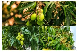
Fig.1: Mangifera indica L.
2. Common Names:
Aam, Manga, Ma, Mango, Maavu, Mangoo
3. Taxonomical classification:
4. Morphological characters:
5. Chemical Constituents:
6. Pharmacological activities:
7. Part used:
Flowers
MATERIALS AND METHOD:
Plant material:
The flowers of Mangifera indica L. were collected from vandavasi, Tiruvannamalai district in Tamil Nadu. which was authenticated by Dr. J. Sureshkumar, M.Sc, M.Phil, Ph.D, PGDCA.,
Chemicals:
Ethanol, n-Hexane, Tween 80 were obtained from GREAT SCIENTIFIC INDUSTRIES Seriyenthal, Tiruvannamalai, Tamil Nadu.
Methods of Extraction:
Mangifera indica L. (Flower) were collected and shade dried at room temperature for 3 weeks. The dried flowers were powdered and passed through sieve no: 22# and 42#.
Requirements:
Plant: Dried flower powder (Mangifera indica L.)
Solvent: Ethanol, n-Hexane
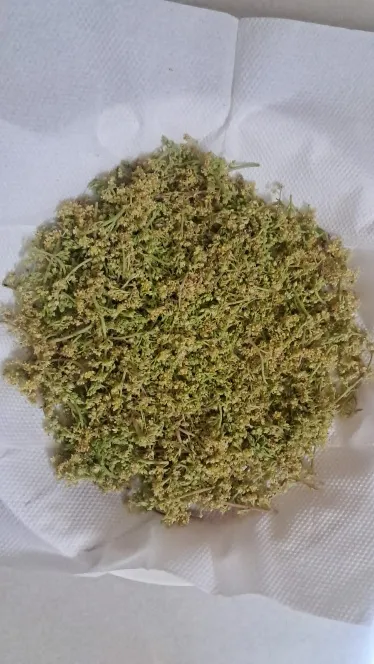
Fig.2: Mangifera indica L. Flower
Extraction process of Mangifera indica L.:
Soxhlet Process:
35 g of dried coarse powder was packed into a cellulose thimble, which was placed in the chamber of a Soxhlet apparatus. The extracting solvent in the flask was heated, and its vapours were condensed in the condenser. The condensed solvent dripped into the thimble containing the crude drug, allowing it to come into contact with the sample. When the liquid level in the chamber rose to the tip of the siphon tube, the liquid contents of the chamber were siphoned back into the flask. This process was continuously carried out until a drop of the solvent from the siphon tube left no residue.
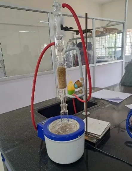
Fig.3: Soxhlet process.
PHYTOCHEMICAL SCREENING:
All the Mangifera indica L. (Ethanol and n-Hexane) extracts were analysed for preliminary phytochemical screening for identification of various phytoconstituents.
TEST FOR ALKALOIDS
HAGER’S TEST
To 1mL of the extract, add 3mL of Hager’s reagent (saturated aqueous solution of picric acid), yellow coloured precipitate indicates the presence of alkaloids.
TEST FOR CARBOHYDRATES
FEHLING’S TEST
1mL of the extract, add equal quantities of Fehling’s solution A and B. Upon heating formation of the brick red precipitate indicates the presence of sugar.
TEST FOR PROTEIN AND AMINO ACIDS
MILLION’S TEST
1mL of the test solution is made acidic with sulphuric acid and add million’s reagent (Mercuric nitrate in nitric acid) and boil this solution. A yellow precipitate is formed indicates the presence of protein.
TEST FOR FLAVONOIDS
LEAD ACETATE
1mL of plant extract add few drops of 10% lead acetate solution. Formation of yellow precipitate indicates the presence of flavonoids.
TEST FOR PHENOLIC COMPOUNDS
FERRIC CHLORIDE TEST
1mL of plant extract add few drops of 5% ferric chloride solution. Formation of dark green/bluish black indicate presence of phenolic compound.
TEST FOR TANNINS
GELATIN TEST
To a few mL of extract, add 1% gelatin solution containing 10% sodium chloride. Formation of white precipitate indicates the presence of tannins.
TEST FOR SAPONINS
FOAM TEST
Take a small quantity of extract and add 20mL of distilled water and shake in a graduated cylinder for 15 mins lengthwise. A 1cm layer of foam indicates the presence of saponins.
TEST FOR STEROIDS
SALKOWSKI TEST
Dissolve the extract in chloroform and add equal volume of concentrated sulphuric acid Formation of bluish red to cherry red colour in chloroform layer and green fluorescence in the acid layer represents the steroidal components in the tested extract.
TEST FOR TERPINOIDS
SALKOWSKI TEST
Dissolve the extract in chloroform and add equal volume of concentrated sulphuric acid Formation of bluish red to cherry red colour in chloroform layer indicates presence of terpenoids.
TEST FOR GLYCOSIDES
BORNTRAGER’S TEST
1g of plant extract and add 5 to 10mL of dilute HCL boil on water for 10 mins and filter Filtrate was extracted with CCL4/benzene. Then add equal amount of ammonia solution to filtrate and shake. Formation of pink or red colour in ammonical layer is due to presence of glycosides.
IN VITRO ASSAY TO EVALUATE THE ANTI-INFLAMMATORY ACTIVITY:
PROTEIN DENATURATION INHIBITION ASSAY:
Egg albumin is used as the protein in this assay.
PRINCIPLE OF THE ASSAY
The process by which proteins lose their secondary and tertiary structures as a result of external pressures or chemicals like heat, an organic solvent, a concentrated inorganic salt, or strong acid or base is known as protein denaturation.
Proteins lose their biological functions when they become denatured. Protein denaturation is a key feature of inflammation. Therefore, the ability of a drug or a plant product to stop or reduce protein denaturation may have the ability to stop or control inflammation. Heat induced egg albumin/bovine serum albumin denaturation inhibition assays are frequently used as protein denaturation inhibition assays.
When the reaction mixture is exposed to heat, protein denatures and increases the turbidity of the mixture. This leads to an increase in the absorbance of the mixture. If a potential drug/plant product has anti-inflammatory properties, it will inhibit protein denaturation, lowering the progressive increase in absorbance due to protein denaturation.
Method Of Heat-Induced Egg Albumin Denaturation Inhibition Assay
Making a 1% egg albumin solution: 1% egg albumin solution can be prepared with fresh hen eggs or easily found egg albumin powder from stores.
To make an egg-albumin solution with a fresh hen's egg, crack the egg carefully. Then add 1mL of the translucent part to 100mL of w/v distilled water and stir until well mixed. Egg albumin is the transparent part of the egg. While making the solution, the water should be cold. When water is brought to boil, it will coagulate.
Test Mixture: For each test mixture, 2.8 mL of Phosphate-Buffered Saline (PBS) adjusted to pH 6.4, 0.2 mL of egg albumin solution, and 2 mL of the respective plant extract at concentrations of 100, 200, 300, 400, and 500 µg/mL were mixed to obtain a final volume of 5 mL.
Standard Mixture: For the standard mixture, 2.8 mL of PBS (pH 6.4), 0.2 mL of egg albumin solution, and 2 mL of the reference drug, aspirin, at concentrations of 100, 200, 300, 400, and 500 µg/mL were mixed to obtain a final volume of 5 mL.
For positive control, mix 2.8mL of PBS (pH6.4), 0.2mL of egg albumin solution, and 2mL of distilled water to obtain a volume of 5mL.
As the negative control, 5mL of distilled water can be used.
Incubate all the mixtures (tests, standards, controls) at 37 ± 2 °C for 15 minutes. Then, increase the temperature gradually to 70°C. Once it reaches 70 °C, keep the mixtures at 70°C in water bath for another 5 minutes. Take out the mixtures from the water bath and allow them to cool down to room temperature. After cooling, measure the absorbance of each mixture at a wavelength of 660nm using a spectrophotometer. Carry out the test in triplicates.
Finally, calculate the percentage of protein denaturation inhibition using the formula below,
|
% Inhibition of egg albumin denaturation = Vc-VtVc×100 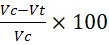
|
(Vc- absorbance of the positive control, Vt -absorbance of the test)
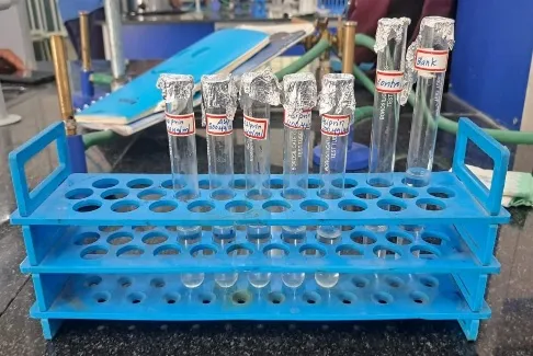
Fig.4: Reaction mixture of Standard
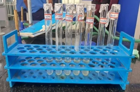
Fig.5: Reaction mixture of Ethanol
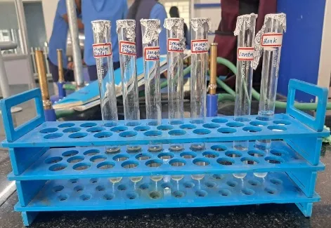
Fig.6: Reaction mixture of n-Hexane
Statistical Analysis
Results are expressed as Mean ± SEM (n = 3). Statistical analysis was performed using one-way analysis of variance (ANOVA) followed by Dunnett's multiple comparison test (Control vs test groups) using GraphPad Prism. Differences were considered statistically significant at P < 0.001
RESULTS AND DISCUSSION:
Table 1: Extractive Values Of Various Extracts.
|
S.NO |
Extracts |
Sample taken (g) |
Obtained yield (g) |
Percentage yield (%) |
|
1. |
Ethanol |
35 |
2.35 |
6.7 |
|
2.
|
n-hexane |
35 |
0.85 |
2.4 |
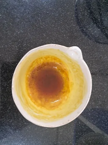
Fig.7: n-Hexane Residue
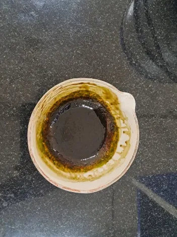
Fig.8: Ethanol Residue
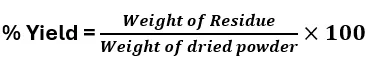
Table 2: Phytochemical Screening Of Various Extracts
|
S.N0 |
Phytoconstituents
|
Ethanol |
N-Hexane |
|
1. |
Alkaloids |
Present(+) |
Absent(-)
|
|
2. |
Carbohydrates |
Present(+) |
Absent(-)
|
|
3. |
Flavonoids |
Present(+) |
Absent(-)
|
|
4.
|
Tannins |
Present(+) |
Absent(-)
|
|
5. |
Saponins |
Absent (-) |
Absent(-)
|
|
6. |
Glycosides |
Present(+) |
Absent(-)
|
|
7. |
Proteins and amino acids |
Absent(-) |
Absent(-)
|
|
8. |
Phenolic compounds
|
Present(+) |
Present(+) |
|
9. |
Terpenoids
|
Present(+) |
Present(+) |
|
10. |
Steroids
|
Present(+) |
Present(+) |
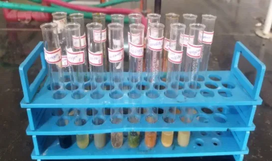
Fig.9:Test for Ethanolic extract
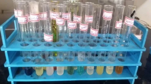
Fig.10: Test for n-Hexane extract
Table 3: In Vitro Anti-Inflammatory Activity Of Mangifera Indica L. Extracts By Protein Denaturation Assay.
|
S.NO |
TREATMENT
|
CONC.(µg\mL) |
MEAN ±SEM |
PERCENTAGE INHIBITION(%) |
|
1. |
Control
|
_ |
0.300±0.000 |
_ |
|
2. |
Ethanol
|
100 |
0.084±0.000 |
71.87
|
|
3. |
Ethanol
|
200 |
0.065±0.001 |
78.31 |
|
4. |
Ethanol
|
300 |
0.054±0.000 |
81.87 |
|
5. |
Ethanol
|
400 |
0.036±0.001 |
87.99 |
|
6. |
Ethanol
|
500 |
0.026±0.000 |
91.21 |
|
7. |
n-Hexane
|
100 |
0.473±0.001 |
-57.82 |
|
8. |
n-Hexane
|
200 |
0.396±0.000
|
-32.24 |
|
9. |
n-Hexane
|
300 |
0.359±0.000 |
-19.92 |
|
10. |
n-Hexane
|
400 |
0.290±0.001 |
3.22 |
|
11. |
n-Hexane
|
500 |
0.193±0.001 |
35.60 |
|
12. |
Aspirin
|
100 |
0.001±0.000 |
99.56 |
Each value represents Mean ± SEM (n = 3). Experimental groups were compared with the control by one-way Variance ( ANOVA ) followed by Dunnett's multiple comparison test. P < 0.001 was considered statistically significant.
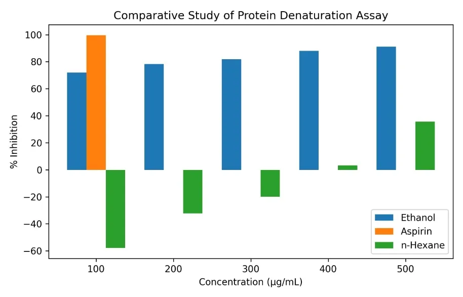
Fig.11: Percentage inhibition of protein denaturation by ethanol extract, n-hexane extract, and aspirin.
CONCLUSION :
The present study demonstrated that the ethanol extract of Mangifera indica L. flowers possesses significant in vitro anti-inflammatory activity as evaluated by the heat-induced protein denaturation assay. The ethanol extract exhibited a dose-dependent inhibitory effect, with the percentage inhibition increasing from 71.86% at 100 µg/mL to 91.21% at 500 µg/mL, indicating strong anti-inflammatory potential. In comparison, the n-hexane extract exhibited negative inhibition at lower concentrations and only mild activity at higher concentrations, showing 3.23% inhibition at 400 µg/mL and 35.60% inhibition at 500 µg/mL. The standard drug aspirin (100 µg/mL) produced 99.56% inhibition, confirming the validity of the assay. The superior activity of the ethanol extract suggests that polar phytoconstituents, extracted by ethanol may contribute to the observed anti-inflammatory effect. These findings indicate that the ethanol extract of Mangifera indica L. flowers is a promising natural source of anti-inflammatory compounds.
REFERENCES


A.Sarala*, Dr.S.K.Senthil Kumar, M.Kaviya, A.S.Kaviya Sri, S.Kiruthiya, A Comprehensive Review Of Water Quality Analysis: Parameters, Analytical Methods, And Emerging Perspectives, Int. J. of Pharm. Sci., 2026, Vol 4, Issue 7, 5854-5865. https://doi.org/ 10.5281/zenodo.21705133
 10.5281/zenodo.21705133