

We use cookies to ensure our website works properly and to personalise your experience. Cookies policy
1-Karmayogi Tatyasaheb Bondre Institute of Pharmacy, Chikhli
The ocular drug delivery system is crucial due to the isolation of the human eye and its challenges in administering drugs. Traditional opthalmic formulations have a short pre-corneal residence time and poor bioavailability, as medications are eliminated from the pre-corneal lachrymal fluid quickly and extensive. Newer research aims to develop stable sustained release in-situ gels to overcome these limitations. In situ gel systems are formulated as liquid preparations suitable for instillation into the eyes, which transform into gels when exposed to the physiologic environment. This increases the pre-corneal residence time of the delivery system, increasing the ocular bioavailability of the drug. The production of gels depends on changes in physicochemical parameters such as pH, temperature, or ion-sensitivity, allowing for controlled and sustained delivery of medication. Evaluations of these systems include drug content, clarity, pH, gelling capacity, viscosity, in vitro drug release tests, texture analysis, sterility testing, isotonicity assessment, accelerated studies, and irritancy tests. FT-IR spectroscopy was used to determine incompatibilities between drugs and polymers. Various innovative dosage forms are available, such as insitu gel, collagen shield, minidisc, ocular film, ocusert, nanosuspension, nanoparticulate system, liposomes, niosomes, dendrimers, ocular iontophoresis, and more. However, the development of ophthalmic drug delivery systems has been challenging due to difficulties associated with the ocular route, such as non-productive absorption, drainage, induced lacrimation, tear turn over, and impermeability of medications to the cornea. In situ gel formation can be triggered by various physical and chemical stimuli, including temperature, pH, electric field, magnetic field, and light. Stimuli-responsive polymers, both naturally occurring and manufactured, are viable options for developing in situ gels. Oral, ophthalmic, rectal, vaginal, injectable, and intra-peritoneal administration methods can be used to give in situ gels. In situ gel systems address limitations related to conventional methods of both solutions and gels.In situ gel systems address limitations related to conventional methods of both solutions and gels, such as the inability to accurately dose and the difficulty of administration. This review discusses the introduction, advantages, disadvantages, suitable polymer characteristics, approaches, applications, evaluation, and marketing products of in situ gels, as well as reported studies and recent advancements.
The ocular drug delivery system (Figure 1) is regarded to be both essential and problematic due to the fact that the human eye is an isolated organ that presents a number of challenges when it comes to the administration of drugs. Furthermore, the typical opthalmic formulations have a short precorneal residence time and poor bioavailability. This is because the medications are eliminated from the pre-corneal lachrymal fluid in a quick and extensive manner through the processes of solution drainage, lachrymation, and non-productive absorption by the conjunctiva. It is (1).
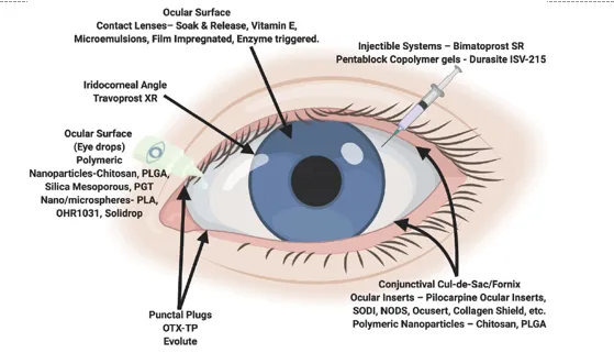
Figure 1: Ocular Drug Delivery System
There have been a number of different attempts made towards the creation of stable sustained release in-situ gels in order to overcome the limitations that are connected with the current formulations of ophthalmic medications. Newer research in ophthalmic drug delivery systems is aimed at incorporating multiple drug delivery technologies. This includes the development of systems that not only extend the contact time of the vehicle at the ocular surface, but also slow down the removal of the drug. This is an important factor in the development of these systems. Specifically, the in situ gel system is formulated as a liquid preparation that is suitable for instillation into the eyes. When the liquid is exposed to the physiologic environment, it transforms into gel, which results in the formation of in-situ gel. This results in an increase in the precorneal residence time of the delivery system, which in turn increases the ocular bioavailability of the drug (2). The production of gels is dependent on elements such as changes in a particular physicochemical parameter (such as pH, temperature, or ion-sensitivity), which allows for the medication to be delivered in a controlled and sustained way. The following characteristics of these systems were evaluated: drug content, clarity, pH, gelling capacity, viscosity, in vitro drug release tests, texture analysis, sterility testing, isotonicity assessment, accelerated studies, and irritancy test. In order to determine the incompatibilities between drugs and polymers, FT-IR spectroscopy was utilised (3). There are many different innovative dosage forms available, such as insitu gel, collagen shield, minidisc, ocular film, ocusert, nanosuspension, nanoparticulate system, liposomes, niosomes, dendrimers, ocular iontophoresis, and so on. Because of the difficulties associated with the ocular route, such as non-productive absorption, drainage, induced lacrimation, tear turn over, and impermeability of medications to the cornea, the development of ophthalmic drug delivery systems has always been a hard endeavour. When it comes to the treatment of a variety of eye disorders, such as dryness, conjunctivitis, keratitis, eye flu, and others, the highly established method of administration for administering pharmaceuticals to the eye is the topical application of the drug. The use of polymers, which play a significant role in the transport of medications to the pre and intra ocular tissues, has been the subject of research for the purpose of developing new methods for the delivery of pharmaceuticals to the eye (4). These consistent efforts have resulted in the achievement of the goal of increasing the bioavailability of the ocular medicine and extending the duration of its therapeutic activity. The use of intelligent polymeric systems has been demonstrated to be a potential method of administering the medications. Immediately upon administration, these polymers go through a sol-gel transition. Before they are administered, they are in the solution phase, but when they are under physiological conditions, they are gels. By raising the corneal permeability of the medications and extending the amount of time they spend in the cul-de-sac, it is possible to increase the ocular bioavailability of the pharmaceuticals (5). In situ gel formation can be triggered by a number of different physical and chemical stimuli, including temperature, pH, electric field, magnetic field, and light, among others. A stimuliresponsive polymer is a type of polymer that resembles biological systems in a primitive manner (6). This type of polymer is characterised by the fact that an external stimulus, such as temperature and pH, alters the characteristics of the formulation. Naturally occurring polymers as well as manufactured polymers are both viable options for the development of in situ gels. Oral, ophthalmic, rectal, vaginal, injectable, and intra-peritoneal administration are the many methods that may be utilised to give in situ gels (7). In situ gels and the numerous methods for in situ gelling systems are discussed in this review, which provides a concise synopsis of the subject matter. The evaluation of polymeric in situ gel, as well as the many types of smart polymers, their methods of gel production from sol forms, and the evaluation of the gels themselves. It is well acknowledged that the ocular medication delivery system is both essential and difficult to implement since the distribution of drugs is highly hard. Moreover, the traditional opthalmic formulations demonstrate a short pre-corneal residence period and limited absorption. The creation of stable sustained release in-situ gels has been the subject of a number of different attempts on several occasions (8). Newer research in ophthalmic drug delivery systems is aimed at incorporating multiple drug delivery technologies. This includes the development of systems that not only extend the contact time of the vehicle at the ocular surface, but also slow down the removal of the drug. This is an example of how the research is being directed. The evaluation that is now being conducted on in situ gelling systems develops become one of the most well- known and famous (9). It was a delivery method that had a significant potential advantage due to various benefits, such as being easy to use and simple to manufacture; improving both adherence and patient comfort by minimising the frequency of drug administration through its distinctive features feature of transitioning from sol to gel. In addition to that, it offers nanoemulsions that gel in situ, nanospheres, microspheres, and liposome technologies. The in situ gelling systems are able to address the limitations that are connected with conventional methods of both solutions and gels (10). These problems include the inability to accurately dose and the difficulty of administration. Within the scope of this review, the definitions, kinds, benefits, drawbacks, polymers employed, and appropriate features of polymers were the primary topics of discussion. In this review, they mainly focussed on introduction, advantages, disadvantages, suitable polymer characteristics, approaches, applications, evaluation, and marketing products of in situ gels. It also focused on some reported studies as well as recent advancements of in situ gels. The intention of writing this review article to describes every aspect of in situ gels, which near the readers a specific feature and might contribute to research and development.
2. In Situ Gels
A gel (Figure 2) is a substance that is soft, stable, or solid-like in nature, and it is composed of at least two components, one of which is a liquid, that are present in a significant number (11). Gels, which are a transitory state of matter, are composed of liquid as well as dependable components, which may be classified as semi-liquids or semi-liquids. Gels combine the cohesive qualities of solids with the diffusive transport characteristics of fluids (12). Gels are a special type of material. In addition to being stable and secure, it is composed of a component network that is threedimensional (13). Gels are characterised by the production of a polymer network by the process of cross-linking polymer chains. This can be accomplished through the formation of covalent bonds (chemical cross-linking) or non-covalent links (physical cross-linking). Gels may be divided into two categories, namely physical and chemical, according to their natural characteristics. Physical gels are characterised by the presence of weak links such as electrostatic, hydrogen, and Vander Waal linkages (14). There is a growing interest in physically crosslinked gels, which are chemical gels that arise when strong covalent connections are formed (15). This is because of the worry of toxicity. The chains of polymeric molecules that make up three-dimensional (3-D) formations are known as hydrogels. Therefore, they are straightforward to produce in a wide range of sizes and shapes (16). These hydrogels are a sort of hydrophilic preparations, and they have a high absorption capacity that allows them to shift between being liquid-gel and becoming themselves (17).
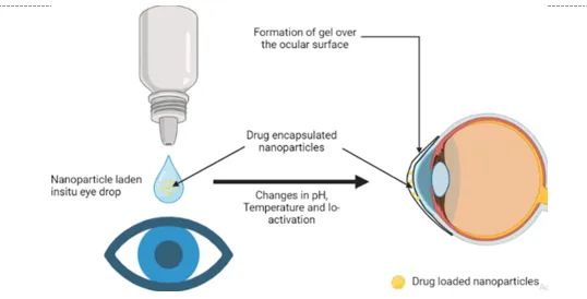
Figure 2: In situ gel for Ocular Drug Delivery System
For the purpose of accommodating a significant quantity of air, hydro-gels are made up of cross-links. Additionally, it is able to hold a substantial amount of water and biological fluids, which allows it to expand. There are also two forms of hydrogels, which are referred to as preformed hydrogels and in situ gels (18). In situ gels are the solutions or suspensions that undergo gelation after reaching the particular site due to contact with body fluids or physicochemical changes such as pH, temperature, ionic concentration, UV radiation, presence of specific molecules, or ions, external triggers, etc. (19). In situ gel produces a constant plasma drug profile in the body by extending the release of a drug, so it is attached and absorbed in gel form and is known to prolong the life of the drug in the mucosa (20). The drug delivery systems having the properties, as mentioned earlier, of sol to gel transition can be widely used for sustained delivery vehicle preparation of bioactive molecules (21). In situ gels, potentially used for oral, buccal, subcutaneous, transdermal, intraperitoneal, ocular, nasal, rectal, vaginal, and parenteral routes (22). From a manufacturing point of view, less complicated and thus lowers the investment and manufacturing cost (23). In the discovery phase, the gel formulations are used to enhance the local and systemic exposure of potential lead compounds, which is ideal for establishing animal models for various conditions quickly and cost-effective (24). Despite the massive diversity of gels, a particular class of gels, namely smart polymer gels, are in the focus of pharmaceutical research during the last decades (25). These intelligent polymers change their physicochemical properties in response to an altered environment. In recent advances, in situ gels have made it possible to exploit the changes in physiological uniqueness (26). Comprehensive research has been carried in designing of in situ gels, emerged as one of the best novel drug delivery systems (NDDS) (27).
3. Mechanism of Sol-Gel Formulation
In situ forming hydrogels are liquid preparations that, following instillation, undergo phase transition in the ocular cul-de-sac leading to the formation of viscoelastic hydrogels, gel, and with this, a reaction to changes in the surrounding environment is provided (28). In situ gel- forming ocular drug delivery systems are made from polymers that exhibit reversible phase transitions (sol–gel–sol) and pseudoplastic behavior (29). These methods are designed to minimise interference with blinking. The formulation of such a system can be in the form of a liquid dosage form that is suited for instillation into the eye (30). This liquid dosage form, when subjected to physiological circumstances, transforms into a gel phase, which results in an increase in the pre-corneal residence period of the delivery system (31). Polymeric polymers, which, when administered, create gel matrices, are the foundation of the vast majority of the in situ forming drug delivery systems that have been documented. Polysaccharides such as alginate, gellan, and xyloglucan; (32) polyesters such as PLA and PLGA; (33) polyethers such as PEGPPG-PEG (Poloxamers); and (34) mixed polyesters and polyethers such as PEG-PLGAPEG are examples of polymers that have been explored before (Figure 3).
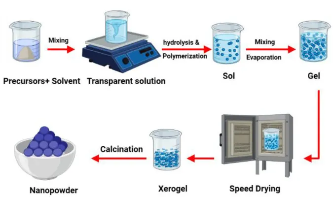
Figure 3: Mechanism of Sol-Gel Formulation
4. Advantages over Conventional Eye Preparations
Because of the quick turnover of tear fluid and the dynamics that induce rapid precorneal removal of the medication, eye drops that are known as traditional ophthalmic administration methods frequently result in low bioavailability and therapeutic response (35). There is a correlation between the patient's non-compliance and the high frequency of eye drop instillation performed (36). If the medication solution that is drained from the eye is systemically absorbed from the naso lacrimal duct, then the inclusion of extra drug in the formulation is an attempt to solve the bioavailability problem (37). This might possibly be harmful. There are many different types of ophthalmic vehicles that have been created in order to increase the ocular bioavailability and extend the residence duration of the dosage that has been injected (38). These vehicles include inserts, ointments, suspensions, and aqueous gels (39). Like ointments, gels are also difficult to administer for some patients. In this respect In situ gels are interesting since these are conveniently dropped as a solution into the conjunctival sac, where they undergo a transition into a gel with its favourable residence. The sol-gel-sol transition occurs as a result of chemical and physical change induced by the physiological environment. Liquid-gel phase transition dependent delivery system vary according to the particular polymer employed and their mechanism for triggering the transition to gel phase in the eye take advantage of change in temperature, pH, ion sensitivity or lysozymes upon contact with tear fluid. However, due to a number of limitations, such as hazy vision caused by ointments or limited patient compliance with inserts, these ocular drug delivery methods have not been utilised to a significant extent (40).
5. Classification of In Situ Gel forming Hydrogels
5.1 Temperature triggered:
As a result of an increase in temperature, formulations become gelled when they come into contact with the application site, which is between 35 and 37 degrees Celsius. Formulations are liquid at room temperature (20-25 degrees Celsius). Temperature-sensitive hydrogels go through a volume phase transition or a sol–gel phase transition when they reach a critical temperature, often known as the lower critical solution temperature (LCST) or the upper critical solution temperature (UCST). After an increase in temperature, there is a progressive desolvation of the polymer and an increase in micellar aggregation, which is entanglement of the polymeric network. This is the process that is involved in the solto-gel transition. Micelles are formed as a result of the dehydration of the polyoxy propylene block; at a certain point, micelles come into contact with one another and cease to move (41). Examples: Pluronic (Poloxamer), Cellulose derivatives, Polymethacrylates.
5.2 PH triggered:
After application at the target location, formulations are polymeric dispersions in an aqueous solution that spontaneously gelate in response to changes in pH. This occurs after the formulation has been applied. At a particular pH, electrostatic and hydrophobic interactions, as well as hydrogen bonding, take place, which ultimately results in interdiffusion. The ionisation of carbopol polymer is responsible for the phase transition that was seen for carbopol solution. This phase transition was mediated by the change in pH from 4.0 to 7.4 and may be attributed to the change (42). Example: Cellulose acetate phthalate, Polyacrylic acid (Carbopol), Polycarbophils
5.3 Ion activated:
In this particular category of in situ hydrogels, the sol-to-gel transition is brought about by the presence of monovalent or divalent cations, such as sodium ions, potassium ions, calcium ions, and magnesium ions. After being injected into the cul-de-sac in the form of a liquid solution, the electrolytes that are present in the tear fluid, and in particular the cations of sodium, calcium, and magnesium, are particularly well-suited to commence the gelation of the polymer. The negatively charged polysaccharide and the cations are linked together in a cross-linking process (43). Examples: Gellan gum (Gelrite R), Sodium Alginate.
6. Evaluations
If Insitu Gel System Clearness, pH measurement, gelling capability, drug content, rheological research, in vitro diffusion study, isotonicity, antibacterial activity, in vivo visual testing in rabbits, and accelerated stability tests are some of the evaluation factors that are utilised for insitu gel formulations (44, 45). It is essential that the formulation possess an optimal viscosity that would facilitate the instillation of the liquid into the eye in the form of drops (46). This liquid would then undergo a quick transition from a sol-to-gel state, which might be triggered by pH, temperature, or ion exchange (47-53).
6.1 Physical parameters
Physical parameters to be tested for insitu gel solution are clarity, pH, gelling capacity, and drug content estimation.
6.2 Gelling capacity
The gelling capacity of the prepared formulation is determined by placing a drop of the formulation in a vial containing 2.0 ml of freshly prepared simulated tear fluid and visually observe.
6.3 Rheological studies
The viscosity measurements can be calculated using Brookfield viscometer, Cone and Plate viscometer. In-situ gel formulation is placed in sample tube. Formulation should have viscosity of 5-1000 m Pas, before gelling & after formation of gel should have viscosity from about 50- 50,000 m Pas.
6.4 In vitro drug release studies
In vitro release study of insitu gel solution is carried out by using Franz diffusion cell. The best fit model is check for Krosmeyers Peppas and Fickinian diffusion mechanism for their kinetics.
6.5 Texture analysis
The consistency, firmness and cohesiveness of insitu gel are assessed by using texture profile analyzer which mainly indicated gel strength and easiness in administration in vivo. Higher values of adhesiveness of gels are needed to maintain an intimate contact with mucus surface.
6.6 Isotonicity evaluation
Isotonicity is important characteristic of the ophthalmic preparations. Isotonicity has to be maintained to prevent tissue damage or irritation of eye. All ophthalmic preparations are subjected to isotonicity testing, since they exhibited good release characteristics and gelling capacity and the requisite viscosity.
6.7 Drug-polymer interaction study and thermal analysis
Interaction study should be performed with Fourier Transform Infra Red (FTIR). 6.8 Antibacterial activity The microbiological growth of bacteria is measured by concentration of antibiotics and this has to be compared with that produced by known concentration of standard preparation of antibiotic (56).
6.9 Ocular irritancy test
The Draize irritancy test should designed for the ocular irritation potential of the ophthalmic product prior to marketing. According to the Draize test, the amount of substance applied to the eye is normally 100µl placed into the lower culdesac with observation of the various criteria made at a designed required time interval of 1hr, 24hrs, 48 hrs, 72hrs, and 1week after administration. Three rabbits (male) weighing 1.5 to 2kg are used for the study (57). The sterile formulation is instilled twice a day for a period of 7 days, and a cross‐over study is carried out (a 3 day washing period with saline was carried out before the cross‐over study). Rabbits are observed periodically for redness, swelling, watering of the eye (58).
7. Applications
7.1 Oral drug delivery systems
Gellan gum, pectin, xyloglucan, etc., are used for oral in situ gels. The pH-sensitive gels have potential use in site-specific delivery of drugs to specific regions of the gastrointestinal tract (GIT), and some of the reported polymers and drugs, including the route of administration of in situ gelling systems (59). Gellan gum has the tendency of gelation, which is temperaturedependent or cations induced. The in situ gelling systems consisted of a gellan solution with calcium chloride and sodium citrate complexes. When it’s administered orally, the calcium ions were released in the acidic environment of the stomach leading to the gelation of gellan, thus form an in situ gel (60). Gelation of pectin will occur in the presence of H+ions, a source of divalent cations; generally, calcium ions are required to produce the gels that are suitable as vehicles for drug delivery (61). The main advantage is that it is water-soluble, so there is no need for organic solvents in the formulation. Divalent cations present in the stomach carry us the transition of pectin to gel state when it is administered orally (62). Xyloglucan is partially degraded by β-galactosidase, resultant product exhibits thermally reversible gelation by the lateral stacking of the rod-like chains or on warming to body temperature (63). Depends on the degree of galactose elimination, sol-gel transition temperature also varies. Its potential application in oral drug delivery exploits the proposed slow gelation time that would allow in situ gelations in the stomach following the oral administration of chilled xyloglucan solution (64). The gelation behavior of xyloglucan is similar to Pluronic F127, but it forms a ‘gel’ at a much lower concentration (65).
7.2 Ocular Drug Delivery Systems
Because of the high tear fluids that are produced by conventional administration methods, the bioavailability and therapeutic impact of the medication are frequently diminished. On the other hand, dynamics lead to the quick drug removal (66). When it comes to ocular medication administration, the most frequent ingredients employed are alginic acid, gellan gum, and xyloglucan. There are medications that have a local effect, including as antibacterial, anti- inflammatory, and autonomic medicines, that are used to alleviate intraocular tension in patients with glaucoma (67). There are several other water-soluble polymers and pH-induced in situ precipitating polymeric systems, such as carbopol, HPMC, and PMA-PEG (68).
7.3 Nasal drug delivery systems
Nasal drug administration has been considered as an alternative route for systems use of drugs restricted to intravenous administration. Nasal drug delivery can also provide a way of entry to the brain that circumvents the blood-brain barrier (BBB) because the olfactory receptor cells are in direct contact with the central nervous system (69). Because of the large absorptive surface and low proteolytic activity, the nasal mucosa is considered an attractive site for the delivery of vaccines. Nasal vaccines will improve patient compliance and reduce production costs compared with parental products. Mostly protein and peptides can deliver by this route (70).
7.4 Parenteral drug delivery systems
Chitosan is a pH-dependent cationic polymer that remains dissolved in an aqueous solution under conditions exceeding 6.2, resulting in a hydrated gel-like precipitate. However, its nonbiodegradability is a major issue. To address this, polyol salts with a single anionic head, such as glycerol, sorbitol, fructose, and glucose phosphate salts, are added to the chitosan solution to transform it into a thermally sensitive solution without chemical modification or cross-linking. This resolves the problem of chitosan's inability to biodegrade (71). Synthetic polymers, such as aliphatic polyesters like PLA, PGA, PLCG, PDL, and PCL, have been the focus of extensive research (72). Lactide or glycolide polymers have been shown to be viable options for controlled release of bioactive substances. Despite thorough testing on both animals and humans, these materials have no adverse consequences. Upon implantation, the polymers do not show any signs of inflammatory response or other detrimental consequences when produced under GMP conditions from purified monomers (73). The systems are positioned in complex-shaped volumes, leading to the development of implants. Photoreactions enable fast polymerization rates at physiological temperatures (74). When heated, thermosetting systems transform into their final form, a "gel." The curing process involves the development of covalent cross-links between polymer chains, producing a macromolecular network. However, further heating may result in degeneration (75). In situ precipitating polymeric systems can result in the formation of a gel due to temperature changes, solvent removal, or pH changes (76). NIPAAm is an example of a thermosensitive polymer with a phase separation at a lower critical solution temperature of 32 degrees Celsius (77). Poloxamers and pluronic are examples of triblock copolymers with POE and POP units, which are subject to variations in solubility due to temperature (78).
7.5 Dermal And Transdermal Drug Delivery Systems
Skin is considered an essential route of administration of drugs for both local and systemic effects. Common preparations for topical and dermatological administration of drugs have certain limitations like poor adherence, reduced permeability, and compromised patient compliance (79). In vivo studies suggest that 20 % w/w aqueous gel maybe it is used as a base for topical administration of the drug. The combination of iontophoresis and chemical enhancers results in a synergistic enhancement of insulin permeation (80).
7.6 Rectal Drug Delivery Systems
Although the oral route is the most convenient route for drug administration, this is not possible from either a clinical or pharmaceutical perspective. In these cases, the rectal way may represent a practical alternative and can be used to administer drugs for both local and systemic effects. The environment in the rectum is considered relatively constant and stable and has low enzyme activity in comparison to other sections of the GIT. However, the rectal cavity can be challenged by erratic drug absorption due to the low adhesion to the rectal membrane and the potential expulsion of the dosage form. These can prevent dosage form leakage, which is familiar with rectal suspensions and enemas (81-84).
8. CONCLUSIONS
The ocular drug delivery system is crucial due to the isolation of the human eye and its challenges in administering drugs. Traditional opthalmic formulations have a short pre-corneal residence time and poor bioavailability, as medications are eliminated from the pre-corneal lachrymal fluid quickly and extensive. Newer research aims to develop stable sustained release in-situ gels to overcome these limitations. In situ gel systems are liquid preparations suitable for instillation into the eyes, which transform into gels when exposed to the physiologic environment, increasing the pre-corneal residence time and ocular bioavailability of the drug. The production of gels depends on changes in physicochemical parameters such as pH, temperature, or ion-sensitivity, allowing for controlled and sustained delivery of medication. Evaluations of these systems include drug content, clarity, pH, gelling capacity, viscosity, in vitro drug release tests, texture analysis, sterility testing, isotonicity assessment, accelerated studies, and irritancy tests. FT-IR spectroscopy was used to determine incompatibilities between drugs and polymers. Various innovative dosage forms are available, such as insitu gel, collagen shield, minidisc, ocular film, ocusert, nanosuspension, nanoparticulate system, liposomes, niosomes, dendrimers, and ocular iontophoresis.
9. Conflict of Interest
None
REFERENCES

Gaurav Haware1*, A Review On Ocular Drug Delivery System, Int. J. of Pharm. Sci., 2026, Vol 4, Issue 7, 5803-5817. https://doi.org/10.5281/zenodo.21701863
 10.5281/zenodo.21701863