

We use cookies to ensure our website works properly and to personalise your experience. Cookies policy
Karnataka college of Pharmacy, Bengaluru 560064
Melasma is a common acquired hyperpigmentary disorder characterized by symmetrical brown-to-gray patches on sun-exposed areas of the skin, particularly the face. It is a multifactorial condition influenced by genetic predisposition, ultraviolet (UV) radiation, hormonal changes, inflammation, and oxidative stress. Although several conventional treatment options, including topical depigmenting agents, chemical peels, oral therapies, and laser-based procedures, are available, their long-term effectiveness is often limited by poor skin penetration, frequent recurrence, local irritation, systemic adverse effects, and reduced patient compliance. Consequently, there is a growing need for advanced topical drug delivery systems that improve therapeutic efficacy while minimizing unwanted effects. Nanotechnology-based drug delivery systems have emerged as promising alternatives for melasma management, among which niosomal gel has gained considerable attention. Niosomes are non-ionic surfactant-based vesicular carriers capable of encapsulating both hydrophilic and lipophilic drugs, thereby enhancing drug stability, bioavailability, skin permeation, and controlled drug release. Incorporation of niosomes into a topical gel further improves drug retention at the target site, prolongs therapeutic action, reduces systemic absorption, and enhances patient acceptability due to its non-greasy and convenient application. This review comprehensively discusses the epidemiology, risk factors, pathogenesis, diagnosis, and conventional treatment strategies for melasma, followed by an overview of recent nanotechnology-based approaches, including liposomes, solid lipid nanoparticles, nanostructured lipid carriers, transferosomes, Nano emulsions, metallic nanoparticles, nanocrystals, and niosomes. Special emphasis is placed on the formulation, mechanism of action, methods of preparation, characterization, evaluation parameters, therapeutic advantages, applications, challenges, recent patents, and future perspectives of niosomal gel systems. Future research should focus on the development of targeted, stimuli-responsive, and personalized niosomal formulations with improved stability and clinical efficacy. Overall, niosomal gel represents a promising and effective topical drug delivery platform for melasma, offering enhanced therapeutic outcomes, improved safety, sustained drug release, and better patient compliance compared with conventional treatment approaches.
Melasma is a common acquired hyperpigmentary disorder characterised by light to dark brown macules and patches occurring on sun-exposed areas of the face and neck. The condition is multifactorial in origin, with major etiological factors including genetic predisposition, ultraviolet (UV) radiation, and hormonal influences. It predominantly affects women of reproductive age, especially those with Fitzpatrick skin types IV–VI, although men can also be affected. In addition to its physical presentation, melasma has a significant psychological impact and is often associated with conditions such as depression, anxiety, and adjustment disorders1,2.
A recent global survey involving 324 women with melasma highlighted those multiple triggers—including pregnancy, hormonal contraceptive use, family history, and chronic sun exposure—play a crucial role in the onset and progression of the condition. Despite extensive research, the management of melasma remains challenging3Various therapeutic approaches, including topical agents, chemical peels, and laser-based treatments, have been explored. Among these, a triple fixed-combination cream containing hydroquinone, retinoic acid, and a corticosteroid is widely recommended and has shown better efficacy than hydroquinone monotherapy when used for a duration of up to 8 weeks4.
Several topical depigmenting agents such as hydroquinone, arbutin, kojic acid, vitamin C, and azelaic acid, along with advanced energy-based techniques like lasers, microneedling, and iontophoresis, have been introduced for enhanced drug delivery5 However, inconsistent therapeutic outcomes and adverse effects, including post-inflammatory hyperpigmentation, erythema, and skin barrier disruption, limit their long-term use.4.
In recent years, there has been an increasing interest in the creation of new drug delivery systems that can deliver targeted, sustained, and safe Melasma treatment. Liposomes were the initial vesicular drug delivery systems, but they possess numerous drawbacks like toxicity and pH-dependent stability; for this reason, the research interest moved towards Niosomes6. Niosomes are ionic surfactant vesicles created by the self-assembly of non-ionic amphiphiles in aqueous systems, leading to closed bilayered structures.
Niosomal gel, a topical drug delivery system, has shown great promise in the treatment for melasma. Niosomal gels can withstand the physiological stress developed due to skin flexion and thus extend controlled release of the drug to the site of action7.The gel drug delivery system offers an easy and patient-friendly delivery platform for transdermal administrations.
1.1 EPIDEMIOLOGY:
According to numerous epidemiologic studies, the prevalence of melasma is approximately 1% in the general population and ranges from 9% to 50% among individuals at higher risk 8,9These wide-ranging estimates are likely due to differences in study populations and methodologies.
There are significant differences in melasma prevalence by skin type, ethnic background, and UV exposure across regions. Consequently, the true global prevalence remains uncertain. The condition most commonly affects individuals aged 20 to 30, although the precise age of onset is unclear. Notably, one study found that individuals with mandibular melasma typically develop the condition in their 40s9.
Melasma is more common in women, according to research. Despite the widely recognised 9:1 female-to-male ratio, a 39:1 ratio was discovered in a more recent large, multicenter investigation of 953 melasma patients in Brazil. A 4:1 female-to-male ratio was discovered in an Indian study involving 312 melasma patients10 [21,22].
The incidence probably rises during pregnancy, as demonstrated by a cross-sectional study conducted in Tehran that found a 15.8% prevalence among pregnant women. Similarly, a 50.8% frequency was found in a randomly chosen sample of 2000 pregnant women in India 8,11 ,12
1.3 Classification of melasma:13, 14
1. Epidermal Melasma
Epidermal melasma is characterized by increased melanin deposition in the superficial layers of the skin. It shows enhancement under Wood’s lamp examination, indicating a more superficial pigment location. This is the most responsive type to topical therapies such as depigmenting agents and generally has a better prognosis.
2. Dermal Melasma
Dermal melasma involves melanin deposition in the deeper dermal layers. It does not show enhancement under Wood’s lamp and often appears bluish due to the Tyndall effect. This type is more resistant to treatment and typically shows a slower or minimal response to conventional therapies.
3. Mixed Melasma
Mixed melasma is the most common type and involves both epidermal and dermal pigmentation. It shows partial enhancement under Wood’s lamp examination. Due to the combined involvement of skin layers, treatment response is variable and may require combination therapies for effective management.
1.4 PATHOGENESIS OF MELASMA:
Melasma is now viewed as chronic photoaging-related, multifactorial hypermelanosis rather than a simple melanocyte overactivity. It arises from the interaction of environmental triggers (especially light), hormones, genetics, and complex changes across the epidermis, basement membrane, and dermis.
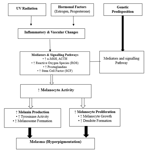
Fig. 5: Pathogenesis of Melasma
1.5 DIAGNOSIS OF MELASMA:
Morphologically, melasma appears as symmetric, reticulated patches of hyperpigmentation with irregular borders on the centrofacial region, malar cheeks, mandible, and, less commonly, the upper chest and extremities. Although melasma is more prevalent among individuals with darker skin types, it can occur in all skin types17,18
1.5.1 Wood’s lamp examination:
Wood’s lamp aids in assessing melasma depth, particularly in lighter skin. Epidermal pigmentation shows enhancement, while dermal pigmentation does not. Mixed patterns are common, and many cases show both components despite appearing superficia19.
1.5.2 Dermoscopy
Dermoscopy reveals pigment distribution and depth through colour and network patterns. Superficial melanin appears dark brown and well-defined, whereas deeper or dermal pigment shows lighter brown to bluish-grey hues2.
1.5.3 Histopathology
Histology shows increased epidermal melanin with normal but hyperactive melanocytes. Dermal features may include melanophages, mild inflammation, increased vascularity, and solar elastosis20.
1.5.4 Reflectance Confocal Microscopy
RCM is a non-invasive imaging tool providing near-histological resolution. It detects hyperpigmented keratinocytes in the epidermis and melanophages with vascular changes in the dermis.21.
1.6 Conventional Treatment Approaches for Melasma:
Conventional management of melasma primarily focuses on reducing melanogenesis, removing existing melanin, and preventing disease exacerbation. Treatment strategies mainly include photoprotection, topical depigmenting agents, chemical peels, and procedural therapies, either alone or in combination.
Table 3: Conventional Treatment Approaches for Melasma
|
Treatment Modality |
Drugs Used |
Examples / Methods |
Mechanism of Action |
Limitations |
Reference (Author et al.) |
|
Photoprotection |
Zinc oxide, titanium dioxide, and iron oxides |
Broad-spectrum sunscreens, protective clothing |
Blocks UV and visible light; reduces ROS-mediated melanocyte activation |
Inadequate protection against visible light alone, poor adherence in daily life, cannot reverse existing pigmentation |
21,22 |
|
Topical depigmenting agents |
Hydroquinone, azelaic acid, kojic acid, arbutin, liquorice extract |
Creams, gels, lotions |
Inhibition of tyrosinase and melanin synthesis |
Skin irritation, sensitization, post-inflammatory hyperpigmentation, and limited long-term efficacy |
23,24 |
|
Combination therapy |
Hydroquinone + tretinoin + corticosteroid (fluocinolone acetonide) |
Triple combination cream |
Decreases melanogenesis, increases epidermal turnover, and reduces inflammation |
Risk of skin atrophy, steroid-related side effects, rebound pigmentation, and poor patient compliance |
23,24 |
|
Topical retinoids |
Tretinoin, adapalene, tazarotene |
Creams and gels |
Enhances keratinocyte turnover and drug penetration |
Irritation, erythema, peeling; slow onset of effect; not sufficient as monotherapy |
25, |
|
Chemical peels |
Glycolic acid, salicylic acid, lactic acid, low-strength TCA |
Superficial chemical peeling |
Exfoliation of pigmented epidermis |
Risk of post-inflammatory hyperpigmentation, recurrence is common; not suitable for all skin types |
23,24 |
|
Oral therapy |
Tranexamic acid |
Oral tablets |
Inhibits plasmin-mediated melanocyte stimulation |
Systemic side effects, contraindicated in some patients; limited long-term data; variable efficacy |
25 |
|
Laser and light therapies |
— |
Q-switched Nd: YAG laser, fractional laser, IPL |
Selective photothermolysis of melanin |
High cost, risk of rebound hyperpigmentation, and limited efficacy in darker skin phototypes |
23,24 |
1.7 Novel Approaches for Melasma Treatment:
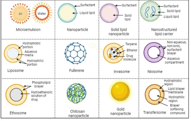
Fig no 6: Novel approaches for treatment of Melasma
To address these challenges, recent years have witnessed growing interest in advanced drug-delivery strategies, especially the application of nanotechnology for targeted topical delivery. Nanocarrier-based systems offer improved drug stability, enhanced skin permeation, controlled release, and reduced systemic exposure, thereby leading to better therapeutic outcomes 26.
Consequently, a wide range of nanoparticulate systems—such as lipid nanoparticles, nano emulsions and microemulsions, vesicular nanocarriers, polymeric nanoparticles, nanocrystals, and metal nanoparticles—have been extensively explored for topical delivery in melasma management.
1.7.1 SLNs
Solid lipid nanoparticles (SLNs) are lipid-based carriers with particle sizes typically ranging from 50 to 1000 nm, composed of lipids that remain solid at body temperature and stabilised by emulsifiers. Due to their solid core, SLNs help improve drug stability and promote better retention of drugs within the skin27.
In melasma management, SLNs have shown significant potential for delivering depigmenting agents such as hydroquinone and kojic acid. Encapsulation of hydroquinone in SLNs improves its stability by preventing oxidative degradation, enhances epidermal deposition, and reduces systemic absorption. Studies have reported significantly higher skin deposition (~46%) compared to conventional gels (~15%), along with increased drug retention and reduced transdermal flux, indicating lower systemic exposure28.
Similarly, kojic acid-loaded SLNs exhibit enhanced dermal retention, controlled drug release, and improved tyrosinase inhibition activity. Overall, SLNs offer an effective and safer approach for topical delivery of anti-melanogenic agents in the management of melasma5.
1.7.2 Nanostructured Lipid Carriers (NLCs)
Nanostructured lipid carriers (NLCs), the second generation of lipid nanoparticles, have also shown considerable advantages in topical depigmenting therapy. Studies have reported that encapsulation of hydroquinone within NLCs leads to improved drug stability, targeted delivery, and reduced skin irritation (52). Hydroquinone-loaded NLCs have shown better skin retention and improved protection against UVA and UVB radiation compared to conventional formulations.
Azelaic acid-loaded NLCs have also been shown to possess enhanced occlusive properties, improved skin permeation, and targeted delivery to melanocytes, resulting in improved clinical efficacy29. Moreover, the sustained drug-release behaviour of azelaic acid-loaded NLCs offers an additional advantage by prolonging localised drug deposition within the skin layers, thereby enhancing therapeutic outcomes while minimising systemic exposure.
1.7.3 Liposomes
Liposomes are vesicular drug-delivery systems composed of phospholipid bilayers, often stabilised with cholesterol, capable of encapsulating both hydrophilic and lipophilic drugs. Their structural similarity to biological membranes enables them to interact with skin cells, enhancing drug penetration and promoting uniform distribution within the skin layers30.
The application of liposomes in topical drug delivery is particularly attractive due to several advantages, including improved penetration through the stratum corneum, skin-moisturizing and barrier-restoring effects, controlled and sustained drug release, and excellent biocompatibility and biodegradability 31.
1.7.4 Niosomes:
Niosomes are vesicular nanocarriers composed of nonionic surfactants that possess inherent skin–permeation–enhancing properties. These vesicles are typically prepared through the self-assembly of nonionic surfactants in an aqueous medium, most commonly using techniques such as thin-film hydration or solvent injection methods 33.
Niosomes have been widely investigated for the topical delivery of depigmenting agents. In particular, the simultaneous encapsulation of kojic acid and hydroquinone within niosomal systems resulted in a topical formulation with sustained and prolonged drug-release behaviors, which is desirable for maintaining therapeutic drug levels in the skin while minimizing systemic exposure34.
1.7.5 Transferosomes:
Transferosomes are highly flexible vesicular carriers composed of a phospholipid bilayer and an edge activator, which enhances their deformability. This unique structure allows them to squeeze through the narrow pores of the stratum corneum, resulting in improved skin penetration and prolonged drug retention.35.
Loading niacinamide into transfersomal carriers significantly improved its skin permeation and depigmenting activity when compared with conventional liposomal formulations. Similarly, the encapsulation of arbutin within transferosomes led to increased transdermal penetration and superior depigmenting efficacy of the drug36.
1.7.6 Metal nanoparticles:
Gold nanoparticles, owing to their excellent aqueous stability, biocompatibility, and chemical inertness, have been widely recognised as suitable nanocarriers for topical drug delivery applications37
Arbutin incorporated into gold nanoparticle-based nanocomplexes demonstrated improved depigmenting efficacy by suppressing melanin synthesis, reducing inflammatory responses, and minimising toxicity, thereby offering advantages over the free drug for melasma management38.
1.7.7 Nanocrystals:
Nanocrystals have gained attention in topical drug delivery due to their ability to enhance drug solubility, accelerate dissolution, and improve adhesion to the skin surface39.
Although azelaic acid is water-soluble, the presence of two carboxylic acid groups limits its penetration through the skin. To overcome this drawback, azelaic acid nanocrystals formulated with Pluronic F127 and hyaluronic acid demonstrated improved solubility, faster dissolution, enhanced drug stability, and increased skin permeation40.
1.7.8 Nano emulsions/microemulsions:
Nano emulsions are effective carriers for dermal drug delivery due to their ability to penetrate the skin's lipophilic barrier. Like microemulsions, they consist of oil and water phases stabilized by surfactants. They enhance drug solubility, improve skin penetration, and increase bioavailability, making them valuable in topical and cosmeceutical formulations41.
In addition, incorporating hydroquinone into microemulsion systems significantly improved its permeability across the stratum corneum and enhanced its photostability42.
1.7.9 Fullerenes
Fullerene and its analogues, particularly water-soluble fullerene analogues, have been dubbed cutting-edge, potent antioxidants that are supposed to minimize intracellular ROS and avoid oxidative cell injury while exhibiting reduced cytotoxicity 43.
Types of Nanocarrier Systems Used in Nanotechnology-Based Therapy for Melasma
Table 5: Novel treatment approaches for melasma
|
Nanocarrier Type |
Advantages |
Disadvantages |
Reference(s) |
|
Liposomes |
Biocompatible, improves skin penetration, reduces drug irritation |
Physical instability, possible drug leakage |
44 |
|
Niosomes |
High chemical stability, cost-effective, non-ionic surfactants |
Lower penetration compared to ethosomes |
45 |
|
Solid Lipid Nanoparticles (SLNs) |
Controlled drug release, good physical stability, occlusive effect |
Low drug loading, lipid crystallization |
27 |
|
Nanostructured Lipid Carriers (NLCs) |
Higher drug loading, improved stability, prolonged release |
Complex formulation process |
46,47 |
|
Transferosomes |
Highly deformable vesicles, superior skin penetration |
High production cost, stability concerns |
48 |
|
Nano emulsions / Microemulsions |
High drug solubilization, uniform skin distribution |
Surfactant-induced irritation |
49 |
|
Metallic Nanoparticles |
Intrinsic antioxidant and anti-melanogenic activity |
Risk of cytotoxicity and accumulation |
50 |
2. Recent Patents on nanocarriers for Melasma Treatments:
Table 6: Recent Patents on nanocarriers for Melasma Treatments
|
Publication No. |
Year |
Title of Invention |
Summary of the Invention |
Reference |
|
WO2024237777A1 |
2024 |
Compositions and Methods for Preventing and/or Treating Skin Pigmentation |
Compositions to prevent/treat pigmentation disorders, including melasma, by modulating melanin synthesis or degradation pathways. |
51 |
|
US20250090443A1 |
2025 |
Skin Care Compositions and Their Uses |
Topical formulations for depigmentation and treatment of melasma/dark spots; include combinations of antioxidants and tyrosinase inhibitors. |
52 |
|
US20250127700A1 |
2025 |
Methods of Treating Hyperpigmentation Disorders |
Methods and compositions targeting tyrosinase or melanogenesis pathways to reduce hyperpigmentation like melasma. |
53 |
3. Niosomes:
Niosomes are bilayered structures formed by non-ionic surface-active molecules. These thermodynamically stable, two-layered vesicles form only under specific conditions: the temperature exceeds the gel-liquid transition point, and surfactants and cholesterol are combined in the proper ratio54. The bilayered structure encloses an internal aqueous compartment, allowing niosomes to encapsulate both hydrophilic and hydrophobic drugs.
While hydrophobic medications partition into the bilayer matrix to enter it, hydrophilic drugs can be adsorbed onto the bilayer surface or trapped in the central watery core of niosomes 55.
3.1 Structure and Composition of Niosomes:
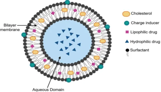
Fig No 7: Structure of Niosomes
The basic components of Niosomes include non-ionic surfactant, cholesterol hydration medium and charge inducer molecules.
3.1.1 Non-Ionic Surfactant:
Non-ionic surfactants are uncharged amphiphilic molecules. These are the most important parts of niosomes, which organise themselves into bilayer structures in a liquid medium. Typical non-ionic surfactants used in niosome formulation include Tween (polysorbates) and Span (sorbitan esters). Both hydrophilic and lipophilic medications can be trapped by the surfactants, which also stabilise the vesicles. By reducing the surface tension between the lipid phase and the aqueous phase, they facilitate the production of vesicles and enable the niosomal structure to form stable structures56,57,58.
3.1.2 Cholesterol
The Niosomal bilayer frequently contains cholesterol to improve the stability of the membrane. It is regarded as a "mortar" that strengthens the bilayer by sealing gaps between surfactant molecules. The inclusion of cholesterol can improve stability, decrease membrane fluidity, and change membrane permeability, all of which can improve the effectiveness of drug trapping in Niosomes.
The structural integrity of Niosomes depends on cholesterol, especially when the environment is changing58,59,60.
3.1.3 Charged Molecules:
Charged molecules are additives that impart either a positive or negative charge to the surface of Niosomes. This surface charge not only helps stabilise the vesicles but also influences their interactions with biological membranes. Among the most used charge-inducing agents are stearyl amine, which provides a positive charge, and diacetyl phosphate, which imparts a negative charge. Incorporating these molecules can prevent noisome aggregation, extend their circulation time in the bloodstream, and improve the cellular delivery of negatively charged drugs, such as polynucleotides 61,62,63.
3.2 Mechanism of action of Niosomes on melasma
Niosomes are vesicles made of non-ionic surfactants and cholesterol that self-assemble into bilayers, encapsulating both hydrophilic (e.g., glutathione, magnesium ascorbyl phosphate, hydroquinone) and lipophilic agents64,65 . They disrupt and “loosen” the stratum corneum lipids and can fuse/adhere to the skin surface, increasing permeability and drug flux into the viable epidermis where melanocytes reside 43,66
When dispersed in a Carbopol or similar gel, the gel acts as a reservoir, providing prolonged release and higher skin retention than the solution or cream form. This controlled, localised delivery raises drug concentration in epidermis while limiting deeper/systemic exposure and irritation 67.
3.3 Advantages of Niosomal gel over other formulations:
Niosomal gel merges the advantages of niosomes with the convenience of gel formulations, providing superior benefits compared to oral medications, conventional topical creams, and other vesicular systems like liposomes and ethosomes
3.4 Comparison table: Niosomal Gel vs Other Delivery Systems
Table 7: Comparison table: Niosomal Gel vs Other Delivery Systems
|
Feature |
Niosomal Gel |
Oral Formulations |
Topical Non-Vesicular Systems |
Vesicular Systems (Liposomes, Ethosomes) |
|
Stability |
High |
High |
Moderate |
Low to Moderate |
|
Skin Penetration |
High (enhanced epidermal retention) |
None |
Limited to superficial layers |
Moderate (Liposomes), High (Ethosomes) |
|
Avoids First-Pass Metabolism |
Yes |
No |
Yes |
Yes |
|
Controlled Release |
Yes |
No |
No |
Yes (limited by vesicle stability) |
|
Cost |
Moderate |
Low |
Low |
High |
|
Patient Compliance |
High |
Moderate |
High |
Moderate |
|
Irritation Potential |
Low |
Moderate (systemic side effects) |
Low to Moderate |
Moderate to High (Ethosomes) |
1. Thin Film Hydration Technique:
Thin-film hydration, also known as the lipid thin-film hydration or hand-shaking method, is a commonly employed conventional technique for the preparation of Niosomes. In this method, surfactants and lipids are initially dissolved in various ratios in a suitable organic solvent. The solvent is then evaporated under reduced pressure using a rotary evaporator, leading to the formation of a thin lipid film on the inner surface of a round-bottom flask73.
The resulting dry film is subsequently hydrated with an aqueous phase at a temperature slightly above the surfactants' phase-transition temperature. During the hydration period, gentle and occasional shaking is applied, allowing the lipid layers to swell and peel off from the flask wall, resulting in vesicle formation 74.
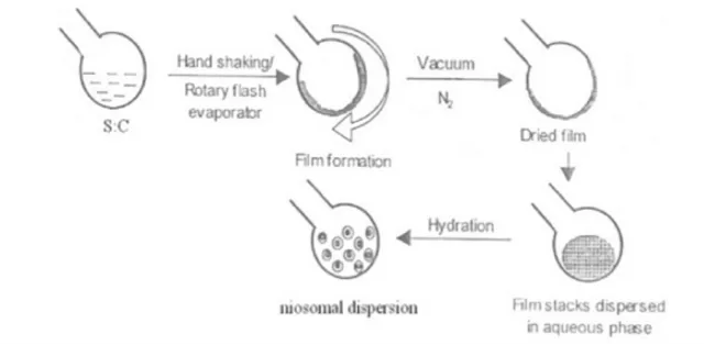
Fig. 7: Thin Film Hydration Technique
2. Ether Injection Method:
The ether injection method, first described by Deamer and Bangham in 1976 , involves dissolving non-ionic surfactants and other components in diethyl ether and then injecting this mixture into a preheated aqueous solution maintained at 60–65 °C. The temperature difference between the organic and aqueous phases facilitates the gradual evaporation of ether, leading to the formation of Niosome vesicles. This technique is particularly effective for economically producing large unilamellar Niosomes. One advantage of this method is its suitability for lipophilic drugs, which dissolve readily in ether and incorporate efficiently into the vesicle bilayer. However, a limitation is the potential presence of trace amounts of residual ether in the final suspension, which can be challenging to remove. This issue can be addressed by applying evaporation under reduced pressure, improving the safety profile for pharmaceutical use75.
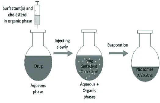
Fig. No 8: Ether Injection Method
3. Ethanol Injection Method:
The ethanol injection method is a quick and straightforward approach for preparing small unilamellar Niosomes. In this technique, an ethanolic solution of lipids is injected directly into an aqueous phase, where vesicles form spontaneously. To improve lipid solubility and enhance drug encapsulation, co-solvents such as isopropanol can be added to the ethanol. Additionally, parameters such as the injection rate and temperature can be adjusted to control vesicle size and reduce aggregation, making this method highly versatile76.
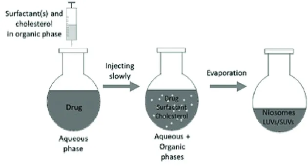
Fig. No 9: Ethanol Injection Method
4. Reverse Phase Evaporation Technique (REV)
The process of manufacturing Niosomes using the Reverse-Phase Evaporation Technique begins with dissolving the cholesterol and surfactant in a chloroform-ether mixture. A mixture. Next, a medication is introduced from the aqueous phase into the organic phase. After that, there are two stages, and to make it gel-like in phase, it is sonicated at low temperatures between 4 and 5°C.
Lastly, sonication is carried out once more after adding a tiny amount of phosphate-buffered saline. At 40°C, the organic solvent is removed under low pressure, creating a viscous suspension of Niosomes. After diluting the solution in PBS, it was incubated at 60°C to finish the production of niosomes77
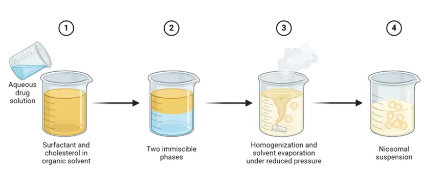
Fig no 10: Reverse Phase Evaporation Technique (REV)
5. Sonication Method:
Sonication is a simple and efficient method for the preparation of niosomes. In this technique, cholesterol and a non-ionic surfactant are first dissolved together, and the drug intended for encapsulation may be incorporated into the same solution. The resulting mixture is transferred into a glass vial, followed by the addition of a suitable buffer to obtain the desired concentration. The formulation is then subjected to probe sonication using a titanium sonicator, usually maintained at a controlled temperature of approximately 60 °C. The application of high-frequency ultrasonic waves generates intense shear forces, which break down surfactant aggregates and promote the formation of uniform niosomal vesicles78
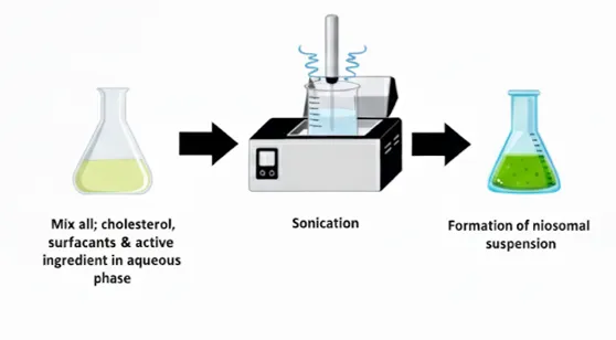
Fig no 11: Sonication Method
6. Micro fluidization method:
Niosomes can also be prepared using microfluidics-based techniques, which employ devices containing central channels flanked by side channels. In this approach, the aqueous and organic phases are separately pumped through different microchannels at controlled flow rates and pressures, allowing them to mix within a cooled interaction chamber, often maintained using ice. The formed niosomes are then collected from the mixing channels into a glass vial79. This method is known to produce niosomes with uniform morphology, small particle size, and well-defined cholesterol incorporation. niosomes prepared using microfluidics exhibit significantly smaller diameters compared to those obtained by the thin-film hydration technique80 .
Additional advantages of the microfluidics method include precise control over formulation and process variables, reduced consumption of chemicals, and the potential for scalable niosome production60.
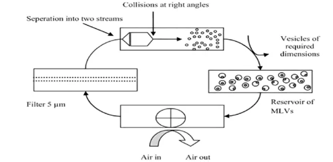
Fig no 12: Micro fluidization method
7. Formation of Niosomes from Proniosomes:
Preparation of niosomes from proniosomes involves a straightforward process of hydration. Proniosomes are powdered free-flowing systems which are a blend of surfactants, cholesterol, and a drug, often coated on a carrier, for example, maltodextrin or other hydrophilic substances. A suitable aqueous phase, typically phosphate buffer or distilled water, must be mixed with the proniosomes along with mild agitation or heat treatment for proniosome-to-niosome conversion. This process also hydrates the surfactant layer, resulting in the self-assembly of niosomes, which are vesicular structures that encapsulate the drug81.
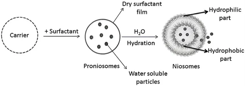
Fig no 13: niosomes from the proniosomes method
8. Bubble Method:
The Bubble Method for preparing niosomes involves introducing air bubbles into a mixture of surfactants and cholesterol in an aqueous solution. This technique takes advantage of the aeration provided by the bubbles to facilitate the formation of niosomes. The continuous agitation and collision of bubbles create shear forces that help in the dispersion of surfactants and promote the assembly of niosomal structures. The resulting niosomes can vary in size and encapsulation efficiency, depending on the parameters such as surfactant concentration and the size of the air bubbles introduced82.
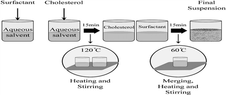
Fig no 14: Bubble Method
9. Freeze-Thawing Method:
The Freeze-Thawing Method for preparing niosomes involves alternating cycles of freezing and thawing the niosomal suspension. Initially, the niosomes are frozen at –20°C for a specified duration (typically 24 hours). Following this freezing period, the suspension can thaw back to ambient temperature. This process is usually repeated several times, which facilitates the fusion of niosomes and can increase vesicle diameter. The repeated freeze-thaw cycles help enhance the stability and uniformity of the niosomes. This method is appreciated for its simplicity and effectiveness in producing Niosomes with desired characteristics for drug delivery applications 83.
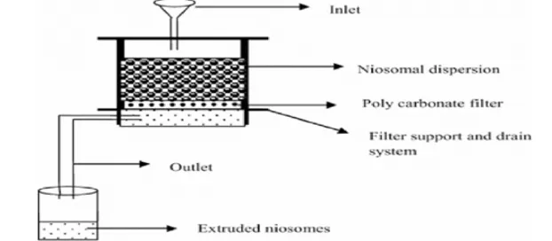
Fig. No 15: FREEZE THAWING METHOD
3.5 Evaluation of Niosomal Gel:
Table 8: Evaluation study for niosomal gel
|
Parameter |
Purpose / Significance |
Method / Technique |
References |
|
pH |
Ensures skin compatibility |
Digital pH meter |
84 |
|
Viscosity |
Determines consistency and ease of application |
Brookfield viscometer |
85 |
|
Spreadability |
Assesses ease of spreading on skin |
Glass plate/ slip & drag method |
86,87 |
|
Homogeneity |
Ensures uniform appearance and drug distribution |
Visual inspection |
88 |
|
Drug content uniformity |
Confirms consistent drug distribution |
UV–Vis spectrophotometry / HPLC |
86 |
|
In vitro drug release |
Evaluates release profile from gel |
Franz diffusion cell |
89 |
|
Ex vivo skin permeation |
Assesses penetration and skin retention |
Franz diffusion cell with excised skin |
65 |
|
Stability studies |
Assesses formulation stability over time |
ICH stability conditions |
90 |
3.6 Characterisation of Niosomal Gel:
Table 9: Characterisation studies of niosomal gel
|
Parameter |
Purpose / Significance |
Method / Instrument |
|
Vesicle size & PDI |
Determines uniformity, stability, and penetration ability |
Dynamic light scattering (DLS) |
|
Zeta potential |
Indicates surface charge and predicts vesicle stability |
Zeta potential analyser |
|
Morphology |
Confirms vesicular structure and shape |
TEM / SEM / Optical microscopy |
|
Entrapment efficiency |
Measures drug encapsulation capacity |
Centrifugation or dialysis + UV/HPLC |
|
Drug–excipient compatibility |
Detects interactions affecting stability |
FTIR, DSC |
|
Physical state of the drug |
Determines crystalline or amorphous nature |
XRD, DSC |
3.7 Application of Niosomes in Cosmetics91,75 :
Niosomes are widely used in topical and cosmeceutical formulations for the delivery of natural antioxidants such as resveratrol, curcumin, α-tocopherol, and mangostin. They enhance skin penetration and retention, improve the stability of sensitive compounds, and provide sustained drug release. These properties increase the effectiveness of antioxidants while reducing systemic exposure, making niosomes valuable in anti-ageing, photoprotective, and anti-inflammatory skincare applications
2. Niosomes as an Antioxidant and Whitening Delivery:
Niosomes enhance the topical delivery of antioxidants and whitening agents, such as ellagic acid, curcumin, resveratrol, and plant-derived oils, by improving skin penetration, retention, and stability. They offer controlled and biphasic release, prolonging therapeutic effects while protecting sensitive compounds from degradation. Niosomes also promote cellular uptake, increasing efficacy, making them highly suitable for cosmeceutical and dermatological applications.
3. Niosomes as an Anti-Scarring Delivery:
Elastic niosomes enhance the topical delivery of anti-scarring agents such as papain and gallic acid by improving dermal penetration, chemical stability, and therapeutic efficacy compared to conventional nanocarriers. Black tea extract, containing gallic acid and caffeine, was effectively delivered into the dermis via niosomes, demonstrating enhanced antioxidant and skin-protective effects. These findings highlight the potential of elastic niosomes as efficient carriers for anti-scarring and skin-repair formulations.
4. Niosomes as an Anti-Ageing Delivery:
Niosomes are effective carriers for anti-ageing compounds such as rice bran extract, morin, gallic acid, and purple glutinous rice extract, enhancing skin penetration, stability, and bioavailability. By encapsulating these antioxidants and flavonoids, niosomes improve skin hydration, elasticity, pigmentation, and protection against oxidative stress, while providing controlled and sustained release. The synergistic effects of entrapped and unentrapped bioactives further enhance anti-ageing benefits, making niosomal formulations ideal for cosmetic and dermatological applications.
3.8 Challenges and Limitations of Niosomal Formulations92,93 :
Physical Instability: Niosomes are prone to aggregation and fusion over time, which can alter their vesicle size and reduce drug entrapment efficiency. For instance, transferosomes loaded with papaverine hydrochloride were observed to increase in size and show decreased entrapment when stored at higher temperatures. Interestingly, incorporating them into a gel system improved their stability over 30 days at 25 °C
Chemical Degradation: The non-ionic surfactants used to form niosomes can undergo oxidation or hydrolysis, compromising vesicle integrity. Nevertheless, niosomes are generally more resistant to chemical degradation compared to liposomes, which allows for relatively longer storage periods.
Premature Release: Encapsulated drugs may slowly leak from niosomes into the surrounding gel matrix over time. For example, doxycycline-loaded niosomal gels were reported to show drug leakage, exposing the free drug to light and causing degradation. This highlights the importance of designing more stable niosomal formulations to prevent premature drug release.
Complex Preparation: Preparing niosomes involves multiple steps such as hydration, sonication, and size reduction. These procedures can be time-consuming and may introduce variability between batches.
Scalability Issues: Scaling up the production of niosomal gels remains difficult, which limits their commercial applicability.
Temperature Sensitivity: Niosomes are sensitive to temperature fluctuations, which can negatively affect their stability. Maintaining proper storage conditions is essential to preserving their integrity over time.
FUTURE DIRECTIONS94, 95, 96
1. Smart and Stimuli-Responsive Niosomes
Future melasma management is likely to benefit from stimuli-responsive niosomes capable of releasing drugs in response to pH changes, temperature, enzymatic activity, or UV exposure specific to diseased skin. These smart systems can enable controlled and on-demand drug release, reducing prolonged exposure to depigmenting agents and minimising adverse effects, thereby improving long-term treatment safety and patient compliance.
2. Targeted Niosomal Systems
Targeted niosomal systems are being explored to achieve epidermal-selective drug delivery by modifying vesicle surfaces with ligands such as hyaluronic acid or peptides. Such ligands interact with skin receptors like CD44, enhancing drug retention at melasma-affected sites while limiting deeper dermal penetration. This targeted approach may allow effective depigmentation at lower drug doses, reducing irritation and recurrence.
3.Combinatorial Nanocarrier Approaches
Combinatorial nanocarrier strategies, including niosomal gels, polymer-coated niosomes, and hybrid delivery systems, are emerging as advanced platforms for melasma therapy. Moreover, co-encapsulation of depigmenting agents with antioxidants or anti-inflammatory drugs within niosomes may address multiple pathogenic pathways simultaneously, potentially replacing aggressive multi-drug regimens.
4.Personalised Melasma Nanotherapy
Personalised nano therapy represents a future shift toward patient-specific melasma treatment, where niosomal formulations are tailored based on skin type, pigmentation depth, and disease severity. Optimization of vesicle size, surface charge, and drug loading may improve therapeutic outcomes and reduce relapse rates. Integration of dermal imaging and data-driven approaches could further support personalised niosomal treatment strategies.
CONCLUSION
Melasma is a chronic facial pigmentary disorder that carries a significant psychosocial burden, particularly due to its impact on appearance and quality of life. Although a variety of chemical and procedural treatment options are currently available, achieving consistent and long-lasting therapeutic outcomes remains challenging. In recent years, nanotechnology-based drug delivery systems have emerged as a promising strategy to enhance the physicochemical stability, skin penetration, and therapeutic efficacy of hypo pigmenting agents, enabling effective targeting of both epidermal and dermal melasma. While systemic therapies may occasionally serve as adjunctive treatment options due to their enhanced permeation, topical nanocarrier-based formulations are expected to remain the primary and most preferred approach for melasma management owing to their targeted action and improved safety pro
REFERENCES

Tejashree K C, Rashmi Mathews, Beny Baby, Pallavi A, Navya N, A Comprehensive Review: Recent Advances in Melasma Treatment Using Niosomes, Int. J. of Pharm. Sci., 2026, Vol 4, Issue 9, 2504-2529. https://doi.org/10.5281/zenodo.22877784
 10.5281/zenodo.22877784