

We use cookies to ensure our website works properly and to personalise your experience. Cookies policy
Dr. Gurachar Nargund College of pharmacy Muradi Koppal
The present study investigates the utilization of a provesicular system for enhancing skin permeation and retention of Lantana camara extract, while simultaneously improving its antimicrobial efficacy. Proniosomes were formulated using Soxhlet-extracted Lantana camara leaves combined with varying proportions of non-ionic surfactants specifically Span 60, Tween 80-and cholesterol as a membrane stabilizer. Four distinct formulations, designated LCI, LC2, LC3, and LC4, were developed, with formulation LCI demonstrating superior characteristics. A comprehensive evaluation of the proniosomes revealed notable properties, warranting further exploration of their antimicrobial potential. The vesicle size was observed from 2µm to 16µm, spreadability results were observed from 16 and 22gm.cm/sec, viscosity of prepared formulation was observed in the ranges from 2400 to 2712mPas, zeta potential values lied in between -5.4mV to -17.2mV suggest that proniosome particles have negative surface charge, as LC1 value closer to -17Mv, as particle would have less repulsive force keeping them apart and shows stable. Entrapment efficiency of proniosomes formulation ranges from 42.12% to56.21%. In vitro diffusion studies conducted over a 24-hour period indicated a linear release profile across all formulations, LC1 shows 61% to 94%, LC2 shows 79% to 99%, LC3 shows 74% to 98% and LC4 shows 81% to 98% and respectively with LC1 exhibiting a controlled release mechanism relative to the others. These findings underscore the promising application of proniosomes in dermatological formulations for enhanced delivery of bioactive compounds. Further research is warranted to fully elucidate the antimicrobial benefits and therapeutic applications of this innovative delivery system.
Pharmaceutical knowledge has grown exponentially over the last few years and great strides have been made in the management of diseases through the intervention of drugs and drug products. We now have a much clearer understanding of how drugs are absorbed into, distributed within and cleared from the body. Most conventional dosage forms release the drug initially at a faster rate, thus leading to a quick rise in the blood level of the drug and then falls exponentially until a further dose is administered. Moreover, when a drug is administered by conventional methods, it will be distributed throughout the body. Today, due to increased awareness in the safe, effective use of drugs, development of novel drug delivery systems are at forefront of research and development. New dosage forms and drug delivery.
Systems providing excellent improvement in drug therapy by controlled release or drug targeting are often referred to as “Novel Drug Delivery Systems”. The development of novel drug delivery systems is also the result of certain trends in thinking of the past decade or two: namely that the drug absorption and efficacy can be enhanced by redesigning the delivery processes rather than the drug molecules. Since there is increasing expense in bringing new drug entities to the market, greater emphasis is laid on improving existing dosage forms and developing drug delivery systems.
“Topical Drug Delivery Systems” have generated an interest in recent times as they provide continuous drug delivery through skin in a predetermined and controlled manner. However, the major obstacle in designing the formulation of drugs in topical drug delivery systems is their limited aqueous solubility. This problem can be overcome by entrapping the drug in a vesicular structure. Encapsulation of a drug in vesicular structure may enhance its permeation at target site and reduce toxicity, if selective uptake occurs. Novel vesicular systems that ensure topical sustained drug delivery include proniosomes, niosomes, liposomes, transferosomes, ethosomes etc. [1]
Though colloidal particulate carriers such as liposomes or niosomes have been widely employed in drug delivery systems, proniosomes offer a versatile vesicle drug delivery concept with potential for delivery of drugs via skin stratum corneum route. “Proniosomes are novel drug delivery systems that act as drug reservoirs, make use of surfactants, stabilizers and rate of drug release through them can be controlled by modification of their composition”.
These proniosomal vesicles can carry both hydrophilic drugs and hydrophobic drugs by encapsulation.
They are promising candidates for industrial applications as they can transport, distribute, store, and process easily. Therefore, proniosomes can be another alternative to liposomal and other vesicular drug delivery systems for the entrapment of both polar and non-polar medications. [2]
STRUCTURE OF PRONIOSOMES:-
Proniosomes are microscopic lamellar structures, hexangular structures, and blackish structures, where their location is clear, semi-transparent, and semi-solid gel-like structures (Figure 1). Consistent with their methodology of preparation, proniosomes are unilamellar or multi-lamellar. They even have bilayer in their structure having hydrophilic ends that are exposed on the surface and hydrophobic chains that face one another within the bilayer inside the vesicles. Bilayer consists of non-ionic surface-active agents. To create a bilayer surfactant molecule, offers direction in such a way that hydrophilic ends of the non-ionic surfactant are arranged toward the outside, whereas the hydrophobic ends exist in the opposite direction. Hydrophilic drugs are placed at intervals in the area encircled within the vesicle and the hydrophobic medication is implanted within the bilayer. For association, in liquid media, proniosomes attach to cholesterol with different categories of non-ionic surfactant like alkyl radical or dialkyl polyglycerol ether.
STRUCTURE OF A PRONIOSOME
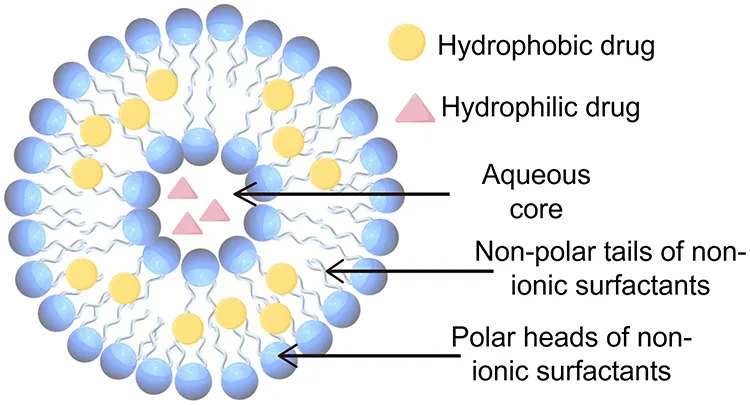
Figure 1: Structure of Proniosome
PREPARATION OF PRONIOSOMES
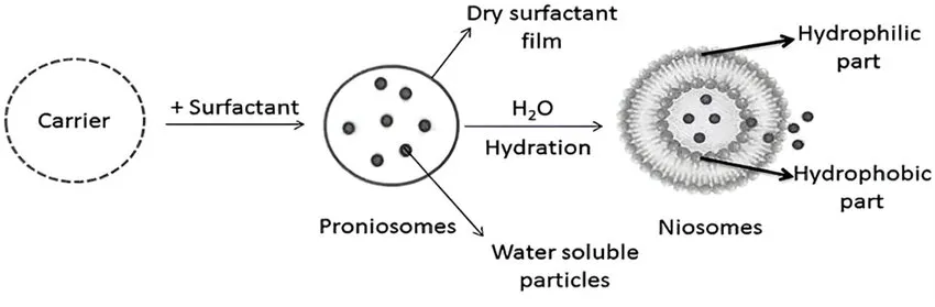
Figure 2: Preparation of Proniosomes
MATERIALS USED FOR THE PREPARATION OF PRONIOSOMES
SURFACTANT:
Surfactants, especially non-ionic surfactants are the key structural components in the preparation of proniosomes. These surfactants do not have any charge as they possess a polar head and non-polar tail. So, their stability, toxicity and compatibility is higher than other surfactants. The non-ionic surfactants have wet and emulsifying effects by which they improve the solubility and permeability of drugs. The hydrophilic-lipophilic balance (HLB) value is critical for selecting surfactants and HLB value between 4 and 8 is compatible with vesicle formation by proniosomes. It is difficult for hydrophilic surfactants to achieve a high concentration because of the high liquid solubility of hydrophilic surfactants.
CHOLESTEROL:
Cholesterol can interact with non-ionic surfactants and regulates the physical and structural properties of proniosomes. It improves the stability and rigidity of the proniosomal membrane and controls drug permeation through the membrane. Depending on the HLB value of the surfactants, the amount of cholesterol required for the preparation of proniosomes is determined. When the HLB value is above 10, the amount of cholesterol to be increased to cover the larger groups. But entrapment efficiency (EE) of the prepared formulation is decreased above a certain level of cholesterol, possibly due to a decrease in volume diameter.
HYDRATION MEDIUM:
Generally, the hydration medium used in proniosomes is phosphate buffer. Depending on the solubility of the encapsulated drug, the pH of the buffer is selected. Ascertained that drug leakage increased with the increase in the volume of hydration medium but simultaneously, EE increases, when the hydration time was increased from 20 to 45 min.
ORGANIC SOLVENT:
The solvent can act as a penetration enhancer. It also greatly affects the size of the vesicles formed. The size of the vesicle and permeation rate of the drug in a proniosomal formulation are influenced by the type of alcohol. Different sized vesicles are formed using different alcohols as they have the order: >> isopropanol < butanol < propanol < ethanol. [3]
METHOD OF PREPARATION OF PRONIOSOMES
COACERVATION PHASE SEPARATION METHOD:
In this method, accurately weighed amount of surfactant, carrier (lecithin), cholesterol and drug are taken in a clean and dry wide mouthed glass vial (5 ml) and solvent is be added to it followed by simple mixing. To prevent the loss of solvent, the open end of the glass vial can be covered with a lid and heated over water bath at 60-70ºC for 5 minutes until the surfactant dissolved completely. The mixture should be allowed to cool down at room temperature till the dispersion gets converted to a proniosomes. [4]
LANTANA CAMARA
Plants generally produce many secondary metabolites which are biosynthetically derived from primary metabolites and constitute an important source of chemicals which are used as pharmaceuticals, agrochemicals, flavours, fragrances, colours, biopesticides, and food additives. [5]
Lantana camara Linn, belongs to family Verbenaceae, commonly known as wild or red sage, is the most widespread species of this genus and regarded both as a notorious weed and a popular or ornamental garden plant. [6]
L. camara oil and extracts are used in herbal medicine for the treatment of various human diseases such as skin itches, leprosy, cancers, chicken pox, measles, asthma, ulcers, tumours, high blood pressure, tetanus, rheumatism etc.[7]
Lantana camara L. (Verbenaceae) is a woody straggling plant, commonly known as red or wild sage, with various flower colours, leaves with rounded tooth edges which comprises 650 species and indigenous to tropical and subtropical America.[8]
L. camara grows in tropical, subtropical, and temperate regions at a high altitude up to 2000 m. The plant has a woody stem with several different colours of flowers i.e., red, white, pink as well as the plant contain spines or prickles.
Lantana camara possesses therapeutic potential because of various bioactive components, including steroids, triterpenoids, oligosaccharides, iridoid glycosides, naphthoquinones, and phenylpropanoid glycosides. [9]
Lantana camara is an erect vigorous shrub that usually grows up to a height of 4 meters. The leaf is ovate or ovate-oblong shaped with a size 2-10 cm (length) and 2-6 cm (width). Leaves are green and tough with fine hairs and have a pungent odour, and it have the ability to climb up to 15 meters with the help of support. It can easily grow in favourable conditions, and flowers usually appear in the month of March and August. The colour of the fruit is green and drupaceous with two nutlets. Mature plants produce up to 2000 seeds annually. The roots of lantana camara are very strong, having a main taproot with many small side roots.
Parts Used: Apart from the whole plant, seeds, stem, root, leaves and flowers are also used.
LANTANA CAMARA LEAVES
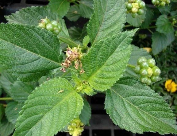
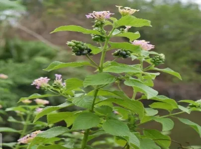
Figure 3: Lantana camara leaves
Although all the samples seemed to present homogeneous profile, a quantitative chemical variability was observed, with a significant variation of the contents of the major components. The chemical compositions of the samples were characterized by a high proportion of sesquiterpene hydrocarbons such as (E)-b- caryophyllene (40.8%) and a-humulene (21.2%), monoterpene hydrocarbons such as sabinene (up to 9.0%).It is noteworthy that the oil of Blokauss was poor in sabinene (0.4%) in opposition to others samples. Four other components were present at appreciable contents: bicyclogermacrene (7.9%), germacrene D (6.9%) and a- pinene (4.4%) as well as b-elemene (up to 3.5%). By contrast, oxygenated compounds are poorly represented: linalool (0.4–1.9%), sesquithuriferol (0.3–1.7%), panthenol (0.2–1.5%), (E)-nerolidol and t-cardanol (0.0–1.0%, respectively). The contents of caryophyllene oxide reached 4.9% in one sample.[10]
METHODOLOGY
COLLECTION OF PLANT MATERIAL
SOXHLET EXTRACTION PROCESS:
The leaves were dried under shade for 8 Days, done powder by using Mortar and Pestle and we passed leaves powder in Sieves (Mesh No 22).
The lantana camara leaves collected weighed (100gm), grinded leaves powder are added into the thimble apparatus and add (1000ml) methanol in a round bottom flask. Set up for Soxhlet apparatus for extraction and started to boil up to 40oc.
The set up Soxhlet apparatus for 6 to 7 cycles repeatedly until the complete extraction was done. Content was filtered out; filtrate was allowed to evaporate in evaporating pan until the desired concentration of extract was obtained.[11]
UV Spectroscopic Analysis: Selection of analytical wavelength of Lantana camara extract: With appropriate dilution of the standard stock solution with methanol, the solution was scanned using the double beam UV visible spectrophotometer (Labman LUV-2000T) in the spectrum mode between the wavelength ranges of 600 nm to 200 nm and the spectrum was recorded. From the spectrum, the wavelength of maximum absorbance (λmax) of the drug was identified against the blank. The λmax of pure drug was found to be 240 nm
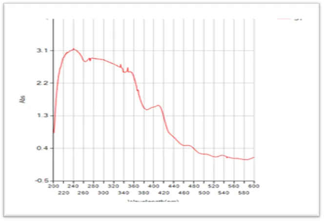
Figure 4: UV graph of lantana camera methanol extract
FORMULATION:
Table 1: Development of Formulation
|
Code |
Drug (mg) |
Ratio (C:S:T) |
Cholesterol (mg) |
Span (mg) |
Tween (mg) |
Ethanol (ml) |
Phosphate Ph 7.4 (ml) |
|
LC1 |
25 |
1:9:9 |
50 |
450 |
450 |
1.0 |
1.0 |
|
LC2 |
25 |
1:9:4:5 |
50 |
450 |
225 |
1.0 |
1.0 |
|
LC3 |
25 |
1:4:5:9 |
50 |
225 |
450 |
1.0 |
1.0 |
|
LC4 |
25 |
1:4.5:4.5 |
50 |
225 |
225 |
1.0 |
1.0 |
COACERVATION PHASE SEPARATION METHOD
EVALUATION OF PRONIOSOME
Organoleptic evaluation
Physical parameters such as colour, appearance and texture were carried out. Colour and texture were evaluated by vision and touch sensation respectively. For odour evaluation a team of five odour sensitive persons were selected. [13]
Physicochemical evaluation
Physicochemical parameters were determined, including the determination of pH, surface morphology and particle size by optical microscope, zeta potential, viscosity and spreadablity. [14]
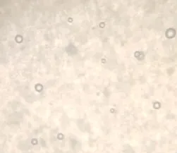
The proniosomes were mounted on glass slides and viewed under a microscope. The microscope has a magnification of 10x/40x used for morphological observation. The photomicrograph of the preparation was obtained from the microscope by using a digital Single lens reflex (SLR) camera. The particle size analysis of the formulations was performed using an optical microscope with eye micrometer. [15]
About 10 g of proniosomal was taken in a tube and 10 ml of distilled water was added on it at temperature 28.2℃ and spindle type 2 and 12 RPM. Proniosomal gel viscosity was determined by LMDV60 Viscometer. [16]
For the determination of spreadability, excess of sample was applied between the two glass slides and was compressed to uniform thickness by placing 500 gm weight for 5 min. Weight (50 gm) was added to the pan. The time required separating the two slides, i.e. the time in which the upper glass slide moves over the lower plate was taken as measure of spreadability (S).
Spreadability (g.cm/s) (S) = M×L/T
Where M = weight tied to upper slide, L = length moved on the glass slide, T= time taken.
Sample preparation: dilute approximately 3 ml of proniosome dispersion in 10 ml of distilled water. A zeta sizer to determine the surface charge [zeta potential] of the vesicles . Zeta potential value of + or – 30 mV or higher typically indicates good colloidal stability.
The formulation was taken in a test tube and reconstituted with 10ml of PBS [ph=7.4]. This aqueous solution was sonicated in a sonicator bath for 10 minutes; the drug containing proniosomes were separated from the unentrapped drug by centrifugation at 25,000 rpm for 30 min at 20 degree. The supernatant was removed and diluted with PBS. The drug concentration in the resulting solution was removed was assayed by the UV SPECTROSCOPY at 240 nm.
The percentage of drug encapsulation was calculated by using the following equation.
EP% = [Ct –Cr] \ [Ct] *100
Ct =is the concentration of total drug .
Cr = is the concentration of free drug.
Preparation of phosphate buffer of [ph 7.4]
Dissolve 6.8 g of potassium dihydrogen orthophosphate,1.56 g of sodium hydroxide was adjusted to 1000 ml with distilled water.
In Vitro Diffusion Study
The vitro release study using Franze diffusion cell assembly
It consists of two compartments , one of the receptor chamber contains a phosphate buffer of saline ph 7.4 and another donar compartment containing proniosomal gel of 25 mg of drug .
A dialysis membrane [molecular size 12000 -14000] was previously soaked for 24 hours.
The dialysis membrane was placed in contact with PBS filled in the receptor compartment to avoid disruption in the on going process. It was ensured that no air bubbles were seen between the dialysis membrane and the liquid surface of PBS.
The temperature was maintained at 37ºC + 0.5 at 50 rpm using a magnetic stirrer. 0.5 ml of sample was withdrawn from the receptor chamber slide tube at the time interval of 1 hr, 2 hr ,4 hr,6 hr ,8 hr, 12 hr and 24 hours and equilibrated with a new fresh dissolution medium to main to a sink state. Suitable dilution was carried out and spectroscopically analyzed at lamda max of 240 nm using UV visible spectroscopy.[16]
RESULT AND DISCUSSION
ORGANOLEPTIC EVALUATION RESULTS
Table 2: Organoleptic Evaluation Results
|
Sl. No |
Parameter |
L1 |
L2 |
L3 |
L4 |
|
1. |
Colour |
Light green colour |
Light green colour |
Light green colour |
Light green colour |
|
2. |
Odour |
Pleasant |
Pleasant |
Pleasant |
Pleasant |
|
3. |
Appearance |
Viscous & creamy |
Viscous & creamy |
Viscous & creamy |
Viscous & creamy |
|
4. |
Texture |
Fine |
Fine |
Fine |
Fine |
|
5. |
Smoothness |
Smooth |
Smooth |
Smooth |
Smooth |
|
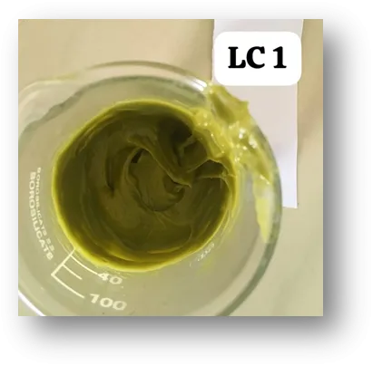
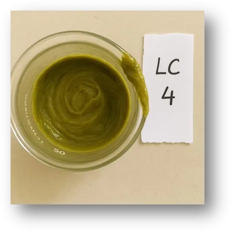
Figure 4: LC1 Proniosome Formulation |
|
|
|
|
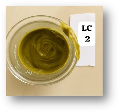
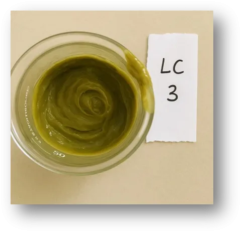
Proniosome was evaluated for organoleptic parameters showed in the Table 2. The colour of formulation was Light greenish. The odour of prepared formulations was pleasant. Appearance was smooth, Texture and smoothness was acceptable as per requirement of cosmetic formulations.
PHYSICOCHEMICAL EVALUATION RESULTS
Table 3: Physicochemical Evaluation Results
|
Sl. No |
Parameter |
L1 |
L2 |
L3 |
L4 |
|
1. |
pH |
5.61 |
5.34 |
5.65 |
5.56 |
|
2. |
Surface morphology By optical microscope |
Found vesicles entrapped of drug |
Found vesicles entrapped of drug |
Found vesicles entrapped of drug |
Found vesicles entrapped of drug |
|
3. |
Particle size (μm) |
2 - 12 |
3 – 15 |
7 – 11 |
4 -16 |
|
4. |
Spreadability (gm.cm/sec) |
22.0 |
26.2 |
18.0 |
16.70 |
|
5. |
Viscosity (mPa.s) |
2654 |
2712 |
2400 |
2466 |
|
6. |
Zeta potential (mV) |
-17.2 |
-13.0 |
-6.3 |
-5.4 |
Proniosome was evaluated for physicochemical parameters showed in the Table 3.
The pH of formulations was found close to neutral.
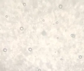
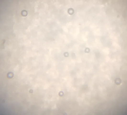
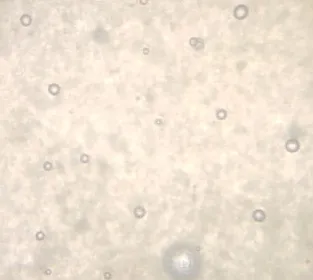
Surface morphology was observed through optical microscope, vesicles were observed in all formulations, understood that drug was entrapped clearly into vesicles. The particle size was measured by stage micrometer and size was found in the range of 2 - 16μm.
Spreadability is good in all the formulations.
Viscosity was found in the range of 2400 to 2712 mPas.
Zeta potential was found in the range of -5.4 mV to -17.2 mV suggests that the proniosome gel particles have a negative surface charge.
Surface morphology images:
|
LC1 |
LC2 |
LC3 |
|
Figure 8 : Proniosome vesicles under optical microscope
ZETA POTENTIAL GRAPHS:
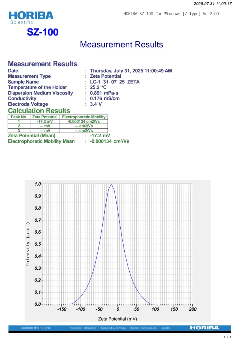
Figure 9: Zeta potential graph of LC1 Formulation
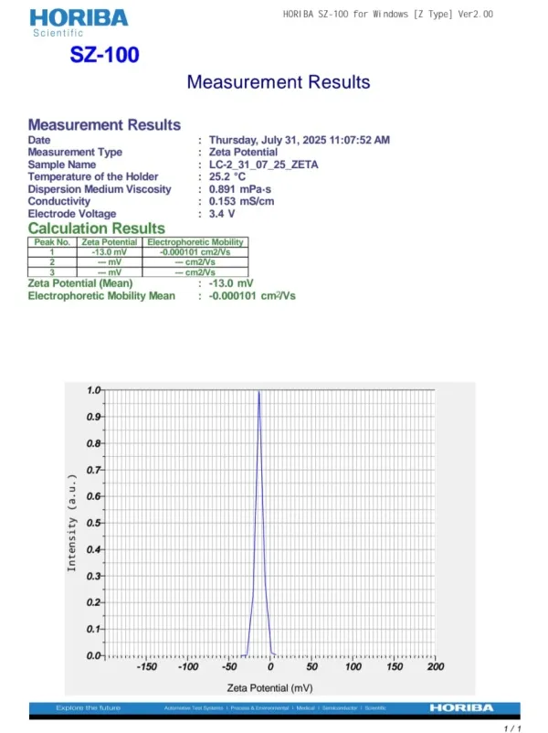
Figure 10: Zeta potential graph of LC2 Formulation
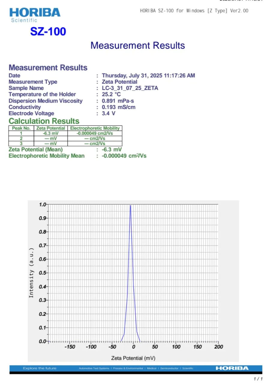
Figure 11: Zeta potential graph of LC3 Formulation
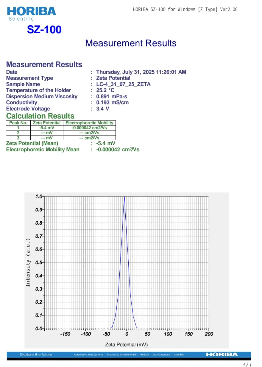
Figure 12: Zeta potential graph of LC4 Formulation
DRUG ENTRAPMENT
Table 4: Results of Drug entrapment efficiency Study
|
Formulations |
% ENTRAPMENT EFFICIENCY |
|
L1 |
56.21 + 1.71 |
|
L2 |
51.15 + 2.14 |
|
L3 |
42.12 + 1.62 |
|
L4 |
43.54 + 3.10 |
Entrapment efficiency of proniosomes formulations ranged from 42.12% to 56.21 %. Proniosomes formed from span 60 and tween 80 combination (1:1) proniosomal gel exhibits higher EE.
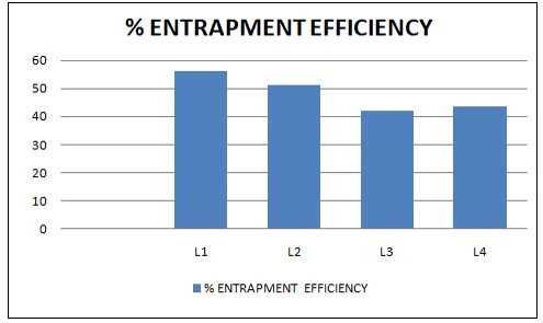
Figure 13: Drug entrapment efficiency graph
IN VITRO DIFFUSION STUDY
Table 5: Results of In-vitro Diffusion study
|
Time in hr |
L1 |
L2 |
L3 |
L4 |
|
0 |
0 |
0 |
0 |
0 |
|
1 |
61.024 |
79.714 |
74.632 |
81.112 |
|
2 |
64.456 |
80.436 |
78.145 |
84.214 |
|
3 |
70.356 |
81.399 |
82.133 |
84.689 |
|
4 |
74.964 |
82.671 |
84.256 |
86.214 |
|
6 |
75.454 |
84.742 |
84.645 |
88.415 |
|
8 |
79.621 |
91.414 |
93.294 |
90.284 |
|
12 |
88.787 |
96.456 |
94.165 |
96.451 |
|
24 |
94.771 |
99.649 |
98.127 |
98.465 |
The results of In Vitro diffusion were shown in Table 5. The percentage drug release of all prepared formulation is in the ranging from of 94.771 to 99.649% of 24 hr.
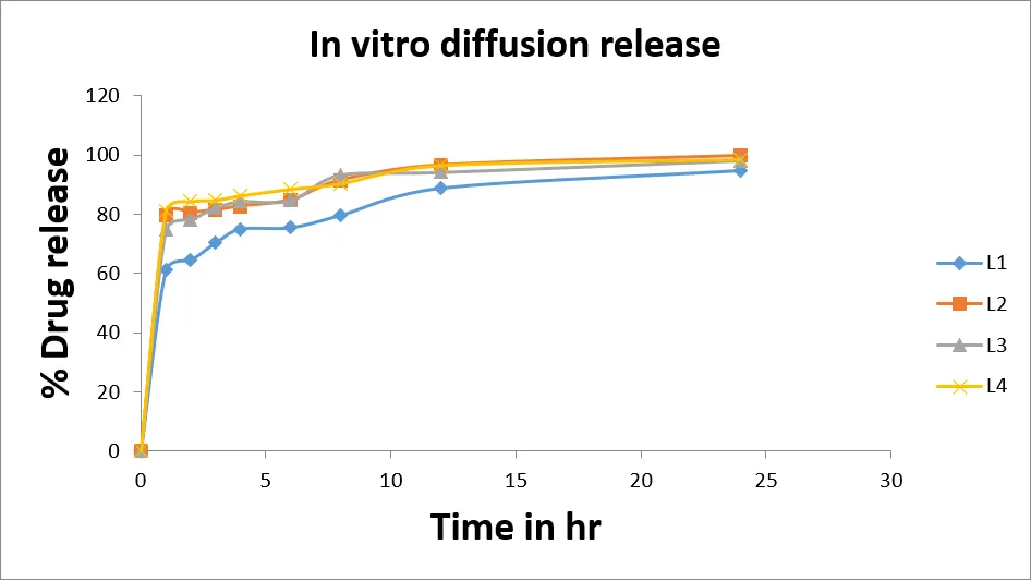
Figure 14: In vitro diffusion release graph
CONCLUSION
The present research investigation decidedly prove the huge potential of provesicular system in accomplishment of elevated skin permeation flux along with high skin retention of lantana camara and with improved pharmacodynamically antimicrobial activity.
The formulation and evaluation of proniosome were prepared successfully by using Lantana Camara leaves extract. Lantana camara leaves was extracted using the Soxhlet extraction method after that proniosome was prepared, which were prepared using different proportions
of non-ionic surfactant span 60, Tween 80 and cholesterol as membrane stabilizer, four formulations [LC1, LC2, LC3, LC4] were prepared in which the LC1 formulation has shown better results. We found excellent properties of the proniosome and further studies are needed to be performed to certain more useful benefits of antimicrobial property.
The physical appearance was observed Light greenish viscous creamy with pleasant Odor. Proniosomes were spherical and few being elongated shape while observed by optical microscope and given in figure 14. The smaller size may result from efficient hydration of a uniform and thin film of surfactant mixture at low surfactant loading.
Vesicle size and size distribution is an important parameter for the vesicular systems. The mean size of the vesicles was in the range of 2 - 12 μm in case of LC1 containing the ratio 1:9:9 (C:S:T) and 4-16 μm in case of LC4 containing the ratio 1:4.5:4.5 (C:S:T). Larger vesicle size was also a reason for higher entrapment of drug.
Surface morphology was observed through optical microscope, vesicles were observed in all formulations, understood that drug was entrapped clearly into vesicles.
A formulation with good spreadability ensures easy and uniform application over the target area, leading to consistent drug delivery. Spreadability results were observed from 16 to 22 gm.cm/sec, indicating that the pronisome gels are easily spreadable by small amount of shear. All formulations shows good spreadability.
Viscosity of all the prepared formulation in which vesicle formation was seen have shown optimum gel viscosity, ranges of 2400 to 2712 mPas.
Entrapment efficiency of proniosomes formulations ranged from 42.12% to 56.21 %. Proniosomes formed from LC1 formulations exhibits higher Entrapment efficiency.
The zeta potential analysis was performed to get information about the surface properties of the niosome derived from proniosomes. Zeta potential is an important parameter to maintain stability of niosomes. Zeta potential was determined using Horiba SZ-100 series and the values lied in between -5.4 mV to -17.2 mV suggests that the proniosome gel particles have a negative surface charge.
A zeta potential close to zero, such as -3 mV, might suggest a less stable system compared to one with a value closer to -17 mV, as the particles would have less repulsive force keeping them apart. So LC1 formulation considered as more stable than other formulations.
The in-vitro diffusion release study for proniosome was performed for 24 hrs, the release data of LC1 formula found 61% to 94%, LC2: 79 to 99, LC3: 74-98%, LC4: 81 to 98%.
Linear release was found in all formulations, LC1 shows control release compare to other formulations. LC4 shows more in-vitro drug release i.e. 81.11% in 1hr.
REFERENCES


Manjunath Halkeri, Dr. Shravan Nargund, Amitkumar, Veeresh k. A Novel Proniosomal Platform for Delivery of Lantana Camara Extract as A Natural Antimicrobial Agent, Int. J. of Pharm. Sci., 2026, Vol 4, Issue 8, 3274-3288, https://doi.org/10.5281/zenodo.22044619
 10.5281/zenodo.22044619