

We use cookies to ensure our website works properly and to personalise your experience. Cookies policy
Maharishi Markandeswar College of Pharmacy, Maharishi Markandeshwar (Deemed to be) University Mullana Ambala-133207 Haryana, India
Gastroesophageal reflux disease is a prevalent digestive disorder that occurs when stomach contents, including gastric acid, flow backwards into the esophagus, resulting in symptoms like nausea, vomiting, or chest pain. This review outlines current data on its rising global prevalence and highlights key pathophysiological mechanisms, including lower esophageal sphincter dysfunction and mucosal injury. We examine major contributing factors: dietary habits, lifestyle, medical conditions, physiological dysfunctions, and medications, and their roles in symptom development and disease progression. Histopathological changes and complications like Barrett’s esophagus and Hiatal hernia are also discussed. Diagnostic methods range from clinical assessment to advanced tools like pH monitoring and endoscopy. Treatment strategies are reviewed in detail, covering pharmacological options (e.g., proton pump inhibitors), surgical and endoscopic interventions, as well as complementary and alternative therapies. This review provides an integrated understanding of GERD’s causes, diagnosis, and treatment approaches, aiming to support more effective patient management.
Gastroesophageal reflux disease (GERD) is a persistent problem characterized by the retrograde movement of gastric contents into the esophagus, oral cavity, or pharynx, resulting in clinical manifestations such as heartburn and regurgitation (1). From a diagnostic perspective, GERD is categorized into non-erosive reflux disease (NERD) and erosive reflux disease (ERD) based on the presence or absence of mucosal damage observed via endoscopy. The pathophysiology of GERD primarily involves excessive esophageal acid exposure (EAE), which arises from a combination of physiological and anatomical abnormalities, including lower esophageal sphincter (LES) dysfunction, transient LES relaxations (TLESRs), the presence of a hiatal hernia (HH), impaired esophageal motility (IEM), duodenogastroesophageal reflux (DGER), delayed gastric emptying (DGE), and increased intra-abdominal pressure associated with obesity (2,3). GERD can lead to severe complications such as esophageal stricture (ES), Barrett’s esophagus (BE), and esophageal adenocarcinoma (EA) (4). Extra-esophageal symptoms like chronic cough, laryngitis, asthma, and dental erosion result from reflux-induced tissue injury (1). Persistent GERD impairs quality of life, disrupts sleep and diet, and increases anxiety, depression, and healthcare costs (5). Diagnosis is mainly based on symptoms such as heartburn, regurgitation, and nocturnal vomiting. Risk factors include age, sex, short dinner-to-sleep intervals, caffeine or carbonated drink intake, smoking, alcohol, NSAID use, poor sleep hygiene, and psychological stress (6–8). Dietary habits like fatty or spicy foods, carbonated drinks, and late meals further weaken LES function (9). Genetic, physiological, and behavioral factors also contribute, emphasizing the need for personalized prevention and treatment strategies (10).
Epidemiology and Prevalence
A increasing worldwide health concern, GERD is thought to impact approximately 1 billion people globally, or 14% of the population (11). Prevalence varies by region, with America reporting the highest rates (18%–30%), followed by the Middle East (9%–32%), Europe (98%–26%), and Australia (12%) (12,13). East Asia has lower rates (2.5%–7.8%) but shows an upward trend. Among university students, prevalence is higher (11.8%–52.6%) due to stress and lifestyle factors (5). Though women have slightly higher rates, GERD is not strongly associated with age, race, or ethnicity (14). Overall prevalence is 10–20%, with severe cases in about 6%, and around 5% in Asia.
Pathophysiology of GERD
GERD is majorly caused by malfunctioning of LES (15) shown in Figure 1. The LES is around 5 cm segment of smooth muscle located at the gastroesophageal junction (16), normally prevents reflux by maintaining tonic pressure. When compromised by hypotensive pressure, TLESRs, or anatomical issues like HH, reflux occurs (16,17). The refluxate may be acidic (hydrochloric acid) or non-acidic (bile, pepsin), both irritating the esophagus. Acid reflux often causes heartburn and esophagitis (18), while non-acid reflux can also cause damage, particularly in patients on acid-suppressive therapy (19). Conditions like gastroparesis, scleroderma (20), and neuromuscular diseases (21) further exacerbate these dysfunctions. The esophagus is less equipped than the stomach to handle reflux, relying on defenses like saliva, peristalsis, limited mucus, and epithelial integrity (22). GERD arises from anatomical, physiological, and molecular disruptions. Injury results from both direct chemical irritation and cytokine-mediated inflammation, involving breakdown of pre-epithelial, epithelial, and post-epithelial barriers. Inflammatory pathways like HIF and NF-κB are activated, exacerbating damage (23).
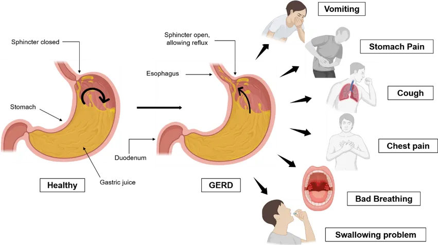
Figure 1: Representation of GERD pathophysiology and its related symptoms
Histopathological Alterations
GERD causes various histopathological changes in the distal esophageal squamous epithelium due to chronic reflux of gastric acid, pepsin, and bile salts (24). Even with normal endoscopic findings, early microscopic abnormalities like basal cell hyperplasia (15–25% thickness) (25) and elongation of lamina propria papillae (26) are common. Spongiosis, or dilated intercellular spaces, indicates increased permeability and is typical in non-erosive disease (25). Inflammatory infiltration by eosinophils, neutrophils, and lymphocytes near the basal zone, along with intercellular and subepithelial edema, is also seen (26) . Chronic injury may lead to squamous-to-columnar metaplasia, forming BE with goblet cells—a precancerous condition. BE, with positive goblet cells, carries a risk of dysplasia and adenocarcinoma, highlighting GERD’s potential progression to malignancy (27).
Etiology and Major Contributing Factors
GERD is influenced by multiple factors that impair LES function or increase stomach acid as shown in the Figure 2. Common dietary triggers include spicy foods, citrus, and carbonated drinks. Lifestyle habits like smoking, alcohol use further weaken the LES and delay gastric emptying. Obesity raises intra-abdominal pressure, worsening reflux. Medical conditions such as HH, asthma, and cancer can aggravate symptoms. Medications including NSAIDs, and antidepressants may relax the LES or irritate the esophagus. These factors collectively contribute to reflux by promoting LES dysfunction, acid overproduction, delayed gastric emptying, and heightened esophageal sensitivity Table 1.
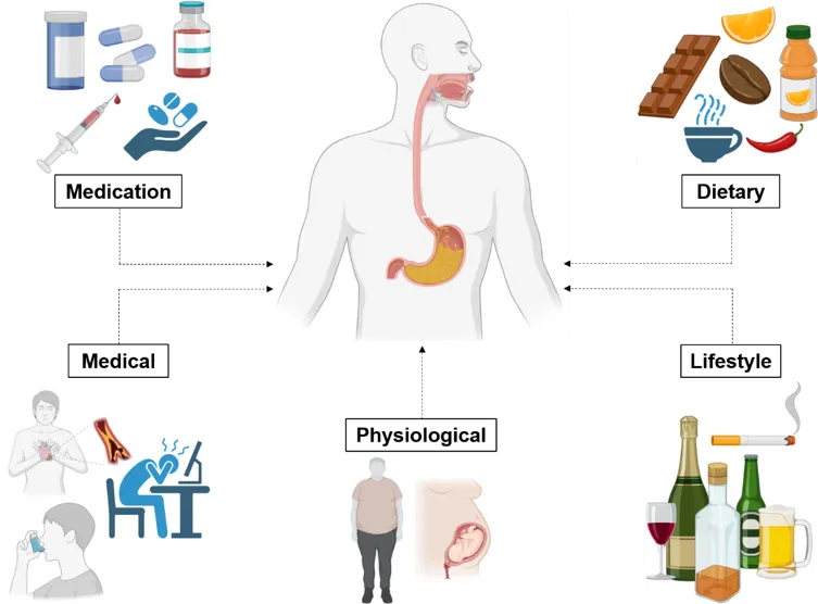
Figure 2: Primary factors behind the progression of GERD
Dietary factors Influences
Dietary components strongly influence GERD symptoms. Foods that delay gastric emptying, increase acid secretion, or irritate the esophagus worsen reflux. Sweet, fried, and spicy foods are common triggers, though evidence of increased acid exposure is limited (28). High-fat meals heighten symptom perception without raising acid exposure (29). Acidic foods and drinks—like citrus, tomatoes, and sodas—are often avoided for worsening symptoms. Most vitamins protect against GERD, except B12, which correlates with BE due to meat and dairy intake (30).
Lifestyle and Behavioral Habits
Recent studies highlight that lifestyle habits such as smoking, alcohol intake, overeating, late-night meals, and physical inactivity substantially contribute to the onset and severity of GERD. Smoking has been consistently associated with an elevated risk, with smokers and those exposed to secondhand smoke having up to a 1.26 times higher risk (31). Smoking reduces LES tone, increases transient relaxations, and impairs esophageal acid clearance, worsening GERD symptoms. Alcohol also contributes to GERD by relaxing the LES, increasing acid secretion, and delaying gastric emptying (32) with white wine particularly triggering reflux events. Although inconsistent findings exist, alcohol overall exacerbates GERD and should be minimized (33). Overeating and late-night meals increase intra-abdominal pressure and delay gastric emptying, promoting acid reflux. Studies show strong links between GERD and poor lifestyle habits, including midnight snacking (78.3%), eating close to bedtime (87.3%), and insufficient exercise (90.9%). These habits disturb digestion and LES function, increasing GERD incidence and symptom burden (34).
Physiological and Anatomical Factors
Obesity, especially central (visceral) fat, greatly increases GERD risk and severity. Abdominal fat raises intra-abdominal pressure, weakens the LES, and promotes hiatal hernia (35). Fat tissue also releases inflammatory cytokines and adipokines that damage esophageal mucosa (36), leading to complications such as erosive esophagitis, Barrett’s esophagus (BE), and esophageal adenocarcinoma (EA) (37). GERD is also common during pregnancy, affecting up to 90% of women by the third trimester (38). Slower gastric emptying and reduced esophageal clearance worsen reflux, peaking late in pregnancy but usually resolving after delivery (39,40). Though generally benign, pregnancy-related GERD can impair quality of life, especially in women with pre-existing GERD who may benefit from preconception counseling (40).
Medical Comorbidities
GERD has a multifactorial etiology, with psychological factors, especially anxiety and depression, contributing to its onset and progression (41). These mental health conditions not only increase GERD risk but also intensify symptom perception by lowering sensory thresholds and heightening esophageal sensitivity (42). GERD significantly impairs health-related quality of life, affecting physical comfort, emotional well-being, and social participation (43). Anxiety affects 34.4% and depression 24.2% of GERD patients, with anxiety increasing GERD risk nearly sevenfold and moderate to severe depression more than doubling the risk (44,45). Medical conditions like HH, diabetes, asthma, and systemic sclerosis also contribute to GERD. HH weakens the LES, allowing acid reflux (46). Asthma and systemic sclerosis further exacerbate GERD through mechanisms like increased abdominal pressure and LES dysfunction, with systemic sclerosis results in gastric reflux in approximately 90% of patients, frequently leading to complications such as ES and BE (47).
Medication-Induced GERD
Recent evidence shows that several medications—including NSAIDs, calcium channel blockers (CCBs), anticholinergics, sedatives, and antidepressants—significantly increase GERD risk and severity through multiple mechanisms. NSAIDs such as ibuprofen enhance gastric acid secretion, weaken the mucosal lining, and lower LES pressure, resulting in reflux and heartburn (48,49). CCBs like nifedipine, diltiazem, and verapamil relax smooth muscles, including the LES, impairing esophageal clearance; among them, diltiazem exerts a stronger effect, while cilnidipine presents lower risk (50). Anticholinergics, often used for respiratory or bladder conditions, reduce LES tone, slow gastric emptying, and decrease saliva production, collectively worsening reflux symptoms (51). Sedatives such as benzodiazepines, propofol, and midazolam decrease CNS activity and further reduce LES pressure, especially during sleep, increasing nocturnal reflux (52). Antidepressants including TCAs, SSRIs, and SNRIs alter esophageal motility and delay gastric emptying, with long-term use raising GERD risk by nearly 50% and linking to complications like erosive esophagitis and Barrett’s esophagus. Overall, these medications should be prescribed cautiously in GERD-prone individuals.
Table 1: Factors Influencing GERD: Effects and Management Strategies
|
Category |
Factors |
Effect on GERD |
Preventive/ Management Strategy |
References |
|
Dietary |
Acidic foods/ drinks |
Increase stomach acidity & irritate the esophagus |
Limit or avoid these items, especially on an empty stomach |
(53)
|
|
Fatty foods |
Delay gastric emptying & reduce LES pressure |
Reduce intake; choose low-fat alternatives |
(54) |
|
|
Lifestyle |
Smoking |
Reduces LES pressure & impairs esophageal clearance |
Quit smoking with cessation programs or support |
(55)
|
|
Alcohol consumption |
Relaxes LES |
Limit intake or abstain, especially during symptom flare-ups |
(32) |
|
|
Physiological |
Obesity |
Increases intra-abdominal pressure |
Weight loss through diet and exercise |
(35) |
|
Pregnancy |
Hormonal changes & uterine pressure reduce LES tone |
Eat smaller meals, avoid triggers, elevate head during sleep |
(56)
|
|
|
Medical |
Anxiety and depression |
Alter gastro-intestinal motility & increase sensitivity |
Manage with therapy, stress reduction, or medications under medical supervision |
(57) |
|
Asthma |
Coughing & meds can promote reflux |
Manage asthma effectively; review medications with a doctor |
(58) |
|
|
Medication |
NSAIDs |
Irritate esophageal lining |
Use alternatives (e.g., acetaminophen); take with food or protective meds |
(48) |
|
Calcium channel blockers |
Relax LES |
Discuss alternatives with healthcare provider |
(50) |
Symptoms and complications
GERD encompasses by common symptoms like heartburn, vomiting, or stomach pain, which typically worsen with improper diet or lack of physical activity (59). Other classic symptoms include chest pain, dysphagia, and excessive salivation. Typical symptoms include chronic cough, sore throat, hoarseness, asthma-like symptoms, and dental erosions, with children showing irritability and feeding issues. Improper management of GERD may progress to esophagitis, ES, BE, and EA. Extra-esophageal symptoms may result from reflux reaching the larynx, causing hoarseness and globus sensation (60), and acid reflux may exacerbate asthma, causing wheezing, coughing, and dyspnea. It is essential to evaluate GERD patients for alarm symptoms such as dysphagia, odynophagia, anemia, gastrointestinal bleeding, and unexplained weight loss, which warrant endoscopic investigation to screen for complications or malignancy. GERD-induced inflammation can lead to esophagitis, causing ulcers, bleeding, and scarring, potentially resulting in peptic strictures and swallowing difficulty (61).
Diagnostic Approaches
The diagnosis of GERD has shifted from a symptom-based to a more objective, personalized approach (62) as shown in Figure 3. Earlier, GERD was diagnosed clinically based on symptoms like chest pain or nausea, with improvement after acid-suppressive therapy (PPIs or H2RAs) considered diagnostic (63). Recent guidelines, such as the Lyon Consensus 2.0, now stress objective testing, especially in atypical, treatment-resistant, or complicated cases (64). Since normal EGD doesn’t exclude GERD, ambulatory pH or pH-impedance monitoring remains the gold standard, offering high sensitivity and specificity for both acid and non-acid reflux (65).
Beyond primary tools, advanced technologies improve diagnostic accuracy and outcomes. High-resolution manometry assesses motility and excludes disorders like achalasia (66). Salivary pepsin tests (e.g., Peptest) offer 73–85% sensitivity and 60–88.3% specificity but have variable reliability (67). Barium esophagram now mainly detects dysphagia or structural issues like strictures or hiatal hernia but has low sensitivity (26%) and specificity (50%) for mild esophagitis (68). AI now aids analysis of clinical and reflux data, enhancing accuracy and subtype classification (69). Overall, diagnosis is shifting toward a comprehensive, patient-specific approach.
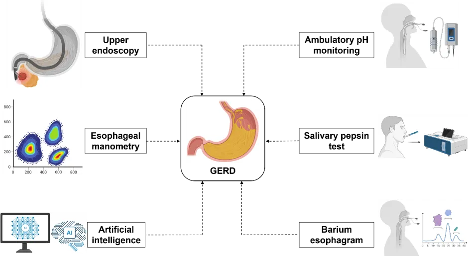
Figure 3: Various diagnostic methods for real time monitoring of GERD
Therapeutic Strategies for GERD
GERD is a chronic condition requiring structured management. Initial assessment should screen for alarm features (dysphagia, GI bleeding, unexplained weight loss) that necessitate urgent endoscopy (70). In the absence of these, first-line treatment focuses on lifestyle changes, which, though not always robustly supported by evidence, remain valuable due to safety and accessibility (71). Head-of-bed elevation markedly mitigates for night time reflux (72). Weight loss is advised for overweight/obese individuals, though its benefits in those with normal weight are unclear (73). Table 2 denomstrate that when lifestyle measures are insufficient, different therapies are often required for moderate to severe symptoms.
Pharmacological therapy is central to GERD management, with PPIs being the most effective for both erosive and non-erosive forms, providing symptom relief and esophagitis healing. They are usually prescribed for 8 weeks, with adjustments if symptoms persist (74). H2RAs are less potent and used mainly for mild or nocturnal symptoms (75). Antacids, alginates, and prokinetics offer short-term relief but limited healing. Long-term use may lead to bone fractures, kidney disease, and infections (76), with frequent relapse after discontinuation. Non-responders should be evaluated for alternative causes such as eosinophilic esophagitis (77).
When drug therapy fails or isn’t preferred, surgical and endoscopic options are used for GERD. Laparoscopic fundoplication, the standard surgery, wraps the stomach around the esophagus to strengthen the LES and prevent reflux (78). The LINX device, a magnetic ring, also reinforces LES closure (79). Endoscopic methods like Transoral Incisionless Fundoplication (TIF) tighten the LES without incisions (80), These alternatives carry risks such as dysphagia, complications, and reduced long-term durability (81). Endoscopic procedures also show limited long-term efficacy, especially in functional heartburn (82).
Complementary and alternative therapies, such as herbal remedies (e.g., ginger and deglycyrrhizinated licorice), acupuncture, probiotics (Lactobacillus and Bifidobacterium strains), aromatherapy (lavender), and cognitive behavioral therapy (CBT), are increasingly used alongside conventional treatments for GERD (83,84). These therapies aim to provide natural symptom relief, stress reduction, and improve gut health. Acupuncture and CBT may help reduce reflux symptoms and improve esophageal motility by addressing psychological stress, but large-scale, high-quality trials are lacking (85). Peppermint oil, while helpful for indigestion, may worsen GERD (86). Patients should seek professional medical consultation before using these therapies to ensure safety and compatibility with other treatments.
Table 2: Overview of GERD Treatments: Types, Mechanisms, and Limitations
|
Treatment |
Types |
Mode of Action/ Mechanism |
Limitations |
References |
|
Pharmacological |
PPIs |
Inhibit the proton pump in parietal cells, reducing gastric acid production. |
Prolonged use may result in nutrient deficiencies, renal complications, and heightened susceptibility to infections. |
(74) |
|
H2RAs |
Antagonize H? receptors on parietal cells, thereby suppressing gastric acid secretion. |
Less effective than PPIs, especially in severe GERD. May cause headaches, dizziness, and gastrointestinal discomfort. |
(75) |
|
|
Surgical and Endoscopic |
Laparoscopic Fundoplication |
A surgical procedure where the gastric fundus is wrapped around the LES to prevent reflux. |
Complications like dysphagia, bloating, and gas/burping are possible. Long recovery period. |
(80) |
|
LINX Device |
A magnetic annular implant encircles the distal esophagus to augment LES competency and suppress gastroesophageal reflux. |
Risks of dysphagia, device malfunction, and possible need for removal. |
(79) |
|
|
Complementary and alternative therapies |
Herbal Remedies |
Some herbs are thought to have anti-inflammatory or soothing properties that may help reduce reflux. |
Limited evidence for effectiveness; may interact with other medications. |
(87) |
|
Acupuncture |
Involves inserting needles into specific points on the body to improve digestive function and reduce GERD symptoms. |
Effectiveness varies; requires multiple sessions for potential benefit. |
(85) |
CONCLUSION
GERD represents a complex and growing global health concern significantly impacting quality of life and posing risks for severe complications. Its multifaceted etiology—spanning dietary patterns, lifestyle habits, physiological dysfunctions, comorbidities, and pharmacological influences—requires a broad and integrated clinical approach. Understanding how these risk factors contribute to disease initiation and persistence is vital for effective prevention and personalized management. Although significant advances have been made in diagnostic techniques and therapeutic modalities, challenges remain in treatment adherence, long-term efficacy, and recurrence prevention. Pharmacological interventions, while effective, often necessitate lifelong use, and surgical or endoscopic procedures may be limited by patient eligibility or procedural risks. Complementary and alternative therapies show promise but require further validation through evidence-based research. Comprehensive and individualized care, grounded in updated clinical evidence and patient-specific factors, is crucial for improving outcomes. This review underscores the need for continued research into the mechanisms, diagnostic tools, and novel treatment strategies for GERD to reduce its burden and enhance patient care.
ABBREVIATIONS
GERD: Gastroesophageal reflux disease
EAE: Esophageal acid exposure
LES: Lower esophageal sphincter
TLESR: Transient LES relaxations
HH: Hiatal Hernia
IEM: Impaired esophageal motility
DGER: Duodeno-gastroesophageal reflux
DGE: Delayed gastric emptying
NSAID: Nonsteroidal Anti-Inflammatory Drugs
BMI: Body mass index
CCB: Calcium channel blockers
TCA: Tricyclic antidepressants
SSRI: Selective serotonin reuptake inhibitors
SNRI: Serotonin-norepinephrine reuptake inhibitors
PPI: Proton pump inhibitors
EGD: Upper endoscopy
NERD: Non-erosive reflux disease
ERD: Erosive reflux disease
AI: Artificial intelligence
H2RA: Histamine-2 Receptor Antagonists
TIF: Transoral Incisionless Fundoplication
CBT: Cognitive behavioral therapy
BE: Barrett’s esophagus
ES: Esophageal stricture
EA: Esophageal adenocarcinoma
REFERENCES

Dr. Nitish Gautam, Dr. Inderjeet Verma, Dr. Rahul, A Systematic Review of GERD: From Risk Determinants to Therapeutic Limitations, Int. J. of Pharm. Sci., 2026, Vol 4, Issue 9, 4221-4234. https://doi.org/10.5281/zenodo.23061064
 10.5281/zenodo.23061064