

We use cookies to ensure our website works properly and to personalise your experience. Cookies policy
Department of Pharmacy, Lokmanya Tilak Institute of Pharmacy, Kharghar, Navi Mumbai, Maharashtra, India.
Wound healing can be compromised in a diabetic environment due to chronic hypoxia, lack of neo angiogenesis, inflammation and oxidative stress. Oxygen is a key component in many biological processes, such as collagen synthesis, anti-infective and neo angiogenesis. Oxygen releasing hydrogels have shown great potential at increasing the local oxygen levels. This review discusses oxygen releasing mechanisms, hydrogel design, materials and potential. This review focuses on the oxygen releasing mechanism by using peroxide based materials such as calcium peroxide. There are many other materials that can be used such as microneedles, nanofibers, catalytic biomaterials and photosynthesis. Many multifunctional platforms have shown great potential to deliver oxygen to the site, provide anti-infective properties, prevent biofilms and control reactive oxygen species. From pre-clinical studies it has been found that these multifunctional platforms can stimulate cellular activity, neo angiogenesis and extracellular matrix. Challenges were highlighted throughout this review such as controlling the oxygen release, cytotoxicity and stability. Research should focus on optimizing the oxygen release, dosing and biocompatibility. Overall oxygen releasing hydrogels have great potential to improve wound healing in a diabetic environment but further validation would be required to ensure they are safe and effective.
The skin consists of three layers; the epidermis, dermis and subcutaneous. Immediately after trauma there are several phases of healing to occur. Firstly, haemostasis and inflammation. During proliferation the wound will be closed by migration of keratinocytes. Fibroblasts produce extracellular matrix and new blood vessels are formed from endothelial cells. Finally, during remodelling, the provisional matrix is replaced with a structured collagen matrix. Tensile strength is improved. Communication between cells, adequate blood supply, extracellular matrix and control of microbes will help the healing process [1]. Chronic wounds do not progress through the stages of healing. These are often seen in patients that suffer from diabetes, vascular, pressure, neuropathy, infection or other co-morbidity. This can cause numerous issues such as pain, restricted movement, frequent visits to the clinic and a large impact on the patient and healthcare system [2].
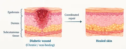
Figure 1: Diagram Showing the Anatomy of the Skin and Progression of a Diabetic Wound to Healed Skin
Modern wound dressings have evolved from passive sterile gauze dressings to active biomaterials with specific functionalities such as exudate control, moist wound healing, barrier properties and delivery of anti-infectives [3]. Hydrogels are particularly attractive as they consist of a hydrated polymeric matrix that can conform to the shape of the wound area. However, simply increasing the water content may not be enough to overcome the problems associated with wounds in diabetes such as hypoxia, chronic inflammation, poor neo angiogenesis, oxidative stress, and infection. Current reviews suggest that hydrogels should be designed with consideration to the pathophysiology of diabetic wounds and also the desired outcome [4].
Oxygen releasing or generating materials are a targeted approach to overcome hypoxia in a particular area. There are several approaches available including chemical oxygen generating systems, oxygen delivery from oxygen loaded materials or using biological photosynthesis. The benefit of using an oxygen source depends on how much oxygen is delivered, where it is delivered and how long it is released for. It is also important to consider safety of the material and any by-products [5-8]. This review will discuss the rationale behind oxygen generating materials, the various types of hydrogels and their design. Pre-clinical evidence will be discussed and Oxygenating patches compared in terms of their use in the management of chronic wounds.
WOUND HEALING, HYPOXIA AND DIABETIC WOUNDS
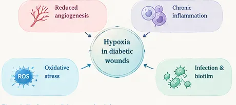
Figure 2: Main Phases of Wound Healing
Wound healing phases
Following injury there is a haemostatic phase. Vasoconstriction, platelet activation and coagulation occur. During the inflammatory stage neutrophils along with monocytes/macrophages will migrate to the wound site to destroy microbes and necrotic tissue. During the proliferative phase keratinocytes will migrate to cover the wound. Fibroblasts produce extracellular matrix and endothelial cells form new vessels. Tensile strength will increase as collagen is laid down in place of the provisional matrix. If any of these stages fail healing can be delayed causing abnormal scarring. Oxygen plays a role in many stages of wound healing including mitochondrial energy production, destruction of microbes using oxidative mechanisms, collagen hydroxylase and crosslinking and neo angiogenesis [6-8].
Hypoxia
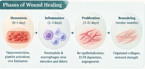
Figure 3: Pathological Factors That Contribute to Hypoxia in Diabetic Wounds
Oxygen delivery will depend on perfusion, diffusion distance, cellular consumption and physical properties of the wound. Diminished blood flow may be caused by injury to vasculature, oedema, infection and chronic inflammation. If there are cells that have a high metabolic activity, more oxygen will be required. A localised hypoxia can be beneficial as there will be a gradient in the oxygen concentration for a limited duration. The effectiveness of adaptation and survival and neo angiogenesis will be controlled via HIF pathways. However if hypoxia is prolonged then this can affect cell function. In diabetic wounds HIF pathways can become dysfunctional and vascular complications can occur meaning there is less of a response to hypoxia [9]. Oxygen should be viewed as an essential component of the wound healing process rather than a therapy in isolation to treat wounds caused by poor circulation, pressure loading, infection or metabolic disease.
Oxidative Stress, Inflammation and Infection
Reactive oxygen species (ROS) can have both positive as well as negative effect based on the situation. Moderate amounts of oxidation can be used to protect the host. However excessive amounts can cause lipid, protein and DNA damage. This will have a detrimental effect on fibroblasts and endothelial cells. Diabetic patients can cause increased oxidative stress via hyperglycaemic pathways causing long-term inflammation. Antioxidants may be used to reduce the harmful effects of ROS but if ROS is inhibited without consideration, it can cause disruption to the body’s natural anti-infective and signalling properties. Oxygen should therefore be regulated [10]. Angiogenesis is driven by VEGF and other neoangiogenic signals, however the timing and organisation of vascularisation will have an impact on functional tissue healing [11].
Chronic wounds often show bacterial colonisation and biofilms which can protect microbes, cause chronic inflammation and cause degradation. Some oxygen releasing strategies can be used alongside antibacterial, photothermal and biofilm disruption strategies. The anti-infective properties and safety of the host cell should be considered. If not carefully managed these strategies may cause damage to healing tissue due to their damaging effects on bacteria [10][12–14].
Clinical Considerations
Various factors are present that can affect chronic wound healing. Peripheral neuropathy may cause a lack of awareness of injuries to diabetic foot ulcers. Poor peripheral arterial disease may also cause poor circulation meaning less oxygen, nutrients and immune cells can reach the wound. Other damage can come from hyperglycaemia, infection, oedema and pressure. Increasing the levels of oxygen in the wound bed using an oxygen releasing dressing may not be enough if there is significant vascular compromise. The wound should not be left too long to assess the vascular system, offload, debride and treat infection. Localised oxygen would be best used if hypoxia was a significant and controllable factor in the wound along with appropriate standard care [2][4][9][10][15].
OXYGENATING PATCHES AND WOUND THERAPIES
Standard And Advanced Dressings
Gauze and standard contact dressings provide a protective barrier and absorb exudate. They are easily available and can be very useful in a wide variety of patient pathways. Gauze will not provide any oxygen. Foam dressings are soft and can absorb moderate to high amounts of exudate. Hydrocolloid dressings can hold onto moisture. Alginates absorb exudate and become a gel-like substance. Hydrogels can provide and/or absorb moisture and allow for delivery of agents. There is no one dressing type that can be used on all wounds. There is little evidence to suggest that any dressing is consistently better for the management of diabetic foot ulcers. Systematic reviews indicate that there is still some uncertainty about the effect of hydrocolloids, foams and alginates on wound healing. The dressing for wound is done on the basis of wound type and standard practice [16–19].
Definition and Types
Oxygenation patches are dressings or biomaterials designed to increase the amount of oxygen in a wound area. This can be achieved using different methods such as an oxygen releasing system, oxygen carrying system or a biological system. Oxygen releasing systems generally involve chemical reactions to give oxygen e.g. hydrolysis of peroxide and decomposition of hydrogen peroxide. Oxygen releasing systems can also have delivery systems and/or reservoirs. Oxygen carrying systems involve haemoglobin-based systems and perfluorocarbons. These will carry and dissolve oxygen. Dissolved oxygen systems provide oxygen that is already in a liquid form. Biological systems contain living microalgae or photosynthetic microbes which will create oxygen when given sufficient light [12][13][20–23].
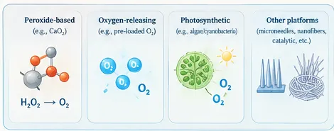
Figure 4: Examples of Oxygen Generation and Delivery
It can be seen these categories can be combined within one patch design. For example, a hydrogel can be used with calcium peroxide to generate oxygen via chemical reaction, catalase to react with hydrogen peroxide, an antioxidant or nanozyme to control ROS and an anti-infective. Microneedle arrays can be used to improve contact with the wound. Film, foam, cryogel or fibrous mats can be used to improve conformability, exudate control or mechanical support. Types should be specified based on what is providing the oxygen source, type of reaction, delivery method, intended duration and whether it has been tested as a patch, scaffold, injectable gel or other [12][21–23].
Oxygen Therapies and Oxygen Generating Biomaterials
Hyperbaric oxygen therapy uses high pressure oxygen. Topical oxygen therapies can provide targeted application. Oxygen releasing hydrogel patches contain components to help produce and/or release oxygen at the wound site. Equipment, method of application, control of area, dosing and evidence will all differ. Patches may be easier to incorporate into standard dressing change routines but may be limited by the reaction, availability of water and diffusion. Reviews of oxygenating dressings and clinical evidence from biomarker studies suggest oxygen-based therapies are an area of ongoing research. Patient needs to be selected with care and objectively assessed [14][15][21].
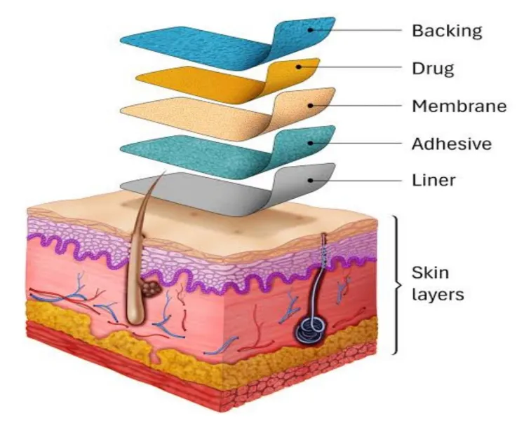
Figure 5: General Design of a Transdermal Patch
Patch Design and Structural Architecture
Different designs of patches exist. Film based patches can form a thin barrier to control transport of gas and liquid. Foam based patches can provide oxygen, absorb exudates and cushion. Both Fibrous mats and nanofiber patches provide a high surface area for delivering active agents. Consideration should be given to the wet structure and leaching of precursors. Hydrogel patches can provide a hydrated interface with the wound bed. Particles, enzymes and drugs can be encapsulated within the crosslinked hydrogel. Cryogels can provide macroporous structures and control of liquids. Microneedle arrays have ability to deliver the active formulation closer to the target tissue. Consideration should be given to oxygen delivery, exudate management, conformity, adhesion, ease of removal and compatibility with wound assessment [23–27].
A good specification for the product will include details about the wound type, duration of wear, source of oxygen, loading of active species, predicted oxygen delivery, moisture management and appropriate safety features. Once the dressing has been hydrated and activated it should remain stable. The attachment/fixation method should not cause any damage to the fragile skin around the wound. If multiple active functions are delivered by a patch, then all components should have an active role to play and have a measurable contribution, thus reducing the risk of creating a complex material that does not possess a clear clinical benefit over a simpler dressing [12][23][23][28][29].
MATERIALS AND MECHANISMS BEHIND OXYGEN GENERATION
Peroxides
Calcium peroxide (CaO2) has been widely studied due to its reaction with water to produce hydrogen peroxide and calcium hydroxide which can undergo decomposition to produce water and oxygen. This reaction can speed up in the presence of a catalase or any other catalysts. Other examples are magnesium peroxide (MgO2) and sodium percarbonate. The rate and amount of oxygen released depend upon the type of source, particle size, formulation, water availability, catalytic activity, pH and diffusion. As hydrogen peroxide is a reactive oxygen species too much can cause cytotoxicity. A formulation must therefore consider release of oxygen and production of peroxide by-products [24][30–33].
Peroxides can be incorporated into various materials such as polymer matrices, particles, fibres and microneedles. The peroxide may be encapsulated within a polymer matrix to help control the release however the oxygen may not penetrate through the polymer to reach the wound bed in a sufficient amount. This needs to be established experimentally. Research has been carried out to establish if sodium percarbonate and calcium peroxide have an effect on bio printed tissue models. Research has also been done into topical peroxide dressings to prove concept[24][31]. Calcium peroxide-based hydrogel patches show how formulations can be tailored to improve handling and oxygen availability [32][33].
Hydrogel Composition
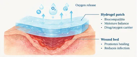
Figure 6: Conceptual Schematic Showing an Oxygen Generating Hydrogel Patch
The properties of the hydrogels depend on the type of polymer, crosslinking, porosity, swelling, water content, mechanical strength and degradation. Different hydrophilic polymers including chitosan, polyvinyl alcohol (PVA) and poloxamer based networks have been investigated for wound contacting applications. A highly swollen network provides a moist environment to allow diffusion to occur but even too much swelling or low mechanical strength can affect the suitability. Highly crosslinked networks may reduce particle release and increase structural integrity but can reduce water penetration and therefore oxygen delivery. The choice of polymer and crosslinking method should consider flexibility, moisture control, permeability, prevention of early release of the oxygen source and degradation [23][27][28][34].
Oxygen production will have no direct correlation with the dosage received by the wound cells. Oxygen must be produced, diffused through the hydrogel, cross the dressing-wound interface and remain available. There may be an initial rapid release of oxygen gas which levels off and then reduces due to the oxygen source running out or change in reaction conditions. The overall amount of oxygen produced, the rate of oxygen release, length of time oxygen is released for, the concentration of oxygen at the wound interface, and ability of the system to function in aqueous conditions should be considered. If only tested in a static buffer system it may not reflect the conditions seen in exudate rich or infected wounds. [27][31][32][35].
Catalysis And Regulation of Reactive Oxygen Species
Catalase can increase the rate of decomposition of hydrogen peroxide to make water and oxygen. If the enzyme remains active and is evenly dispersed there will be less excess hydrogen peroxide. Catalytic metal containing nanozymes and networks have been developed to provide oxygen production and regulation of reactive oxygen species. Hydrogels containing cross-linked iron porphyrin and hybrid hydrogels containing catalase mimetic nanozymes have been developed to overcome hypoxia and reactive oxygen species [36–38]. Quantification of ROS scavenging, oxygen production and biocompatibility is crucial. A positive outcome from any of these assays may not lead to an overall positive outcome.
Radical scavengers such as lignin-based antioxidants may be added to the hydrogel to control the level of oxygen produced in wounds with high levels of oxidative stress. Antimicrobials and photothermal agents have been included within hydrogels to control infection and biofilm. However, multifunctional systems may be used to produce O2 and NO. These must be considered as there may be additional biological effects. When using multifunctional systems, the system must perform at the correct levels without causing additional toxicities, increasing complexity during manufacture and unexpected release. [25][36][37][39–42].
Limitations Of Different Oxygen Sources
Chemical sources are the simplest option however they will rely upon hydration and chemical interactions. If the oxygen releasing compound is activated too soon too much oxygen may be released. By products may affect the pH and have an impact on cell viability. Enzymatic systems allow an efficient breakdown of Hydrogen Peroxide however the enzyme needs to remain active during production, storage and application. Many of the issues caused by chemical sources can be overcome by using delivery systems. The amount of oxygen being delivered and released will depend on this. Oxygen can be produced using photosynthetic systems however this will depend on the availability of light and biological stability. There will be issues with all systems and claims for long lasting oxygenation should be backed up with real data at point of wear [12][13][20–23][27][32][35].
Functionality And Quality Attributes
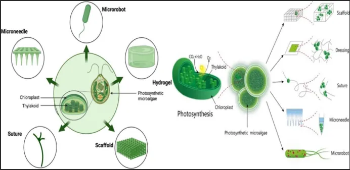
Figure 7: Conceptual Schematic Showing the Pathway of Photosynthetic Oxygen Generating Microalgae and How It Could Be Incorporated into Wound Care Biomaterials
Photosynthetic microalgae offer a sustainable source of producing oxygen. Microalgae hydrogel patches and photosynthetic gels have been investigated for the treatment of chronic and diabetes wounds. Bilayer systems have been developed to provide oxygen producing microalgae with antimicrobial and ROS modulating compounds [43–45]. The efficiency will depend upon the light intensity, viability of the organisms, gas transfer, stable formulation and biological containment. Oxygen delivery systems can be used to increase oxygen levels however oxygen loading, release, carrier stability and tissue compatibility will be important factors to consider [21][27][29][35].
Testing And Quality Attributes
Information should be provided about the oxygen source, polymer, oxygen production, delivery, swellability, biodegradability, mechanical properties, shelf life and by products. Biological data should show cell viability and functionality in normoxia and hypoxia using appropriate cells (fibroblasts, endothelial cells). If antimicrobial claims are being made then planktonic and biofilm testing should also be carried out. Host cell compatibility should be considered. In vivo testing should provide information about the wound model, control dressing, wound size, randomization, monitoring, wound healing and histology. There should be correlation between the concentration and duration of oxygen supply and biological outcomes [27][31][32][35][46].
Oxygen Delivery Systems
The design of the delivery system will determine how the oxygen generating composition interacts at the wound site. Thin films are highly flexible and could conform to a fairly flat wound. They can be adapted to allow controlled diffusion; however, they may not be ideal for wounds with a high level of exudate due to low fluid transport capabilities. Foam systems can aid cushioning and exudate control however may increase diffusion distances. Fiber and nanofiber mats can have a large surface area with tailored porosity but fiber packing, collapse in the presence of moisture, and oxygen source retention needs to be understood. Hydrogels and cryogels can act as a hydrated interface and conform to complex geometries. Microneedles provides a controlled release delivery system. The delivery systems cannot be interchangeable. Advantages of each system will depend on the type of wound, the oxygen profiles required, the amount of exudate and how easy they can be applied [23][24][26][27].
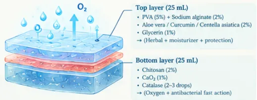
Figure 8: Conceptual Schematic Showing a Bilayer Hydrogel Patch for Generating Oxygen
Multi-layered and hybrid systems can be used to separate different functions within the product. This could include an oxygen delivery layer, a control layer for water/gas delivery, plus an interaction with the wound and management of moisture and antibacterial delivery. This will reduce the amount of direct contact tissue has with high concentrations of oxygen however it will increase the number of processing steps required. Adhesive properties, flexibility, ease of removal without causing pain and compatibility to sterilization should be considered [12][23][27][35].
Fundamental Formulation
Chitosan/PVA hydrogel patch: Chitosan and Poly Vinyl Alcohol (PVA) were combined with calcium peroxide (CaO2) nanoparticles to form an oxygen-releasing patch for healing diabetic wound. Glutaraldehyde and Freeze-thaw was used to crosslink the chitosan-PVA hydrogel [30].
PEG modified calcium peroxide hydrogel: Calcium peroxide microparticles were modified with PEG and incorporated into a hydrogel system to provide sustained oxygen delivery for diabetic wound healing. Oxygen could be released for up to 7 days [33].
PVA/Chitosan/Collagen hydrogel: Polycaprolactone-coated calcium peroxide particles were combined with PVA, chitosan and hydrolysed collagen peptides. Borax and freeze thaw provided physical crosslinking and self-healing properties [47].
Meth acryloyl Silk Fibroin (SilMA) microneedle patch: Calcium peroxide and catalase were incorporated into microneedle tips. Silver nanoparticles were used to supplement the base layer providing oxygen generation and antimicrobial properties [48].
Thermoresponsive hydrogel: Gelatin microspheres containing calcium peroxide and chitosan-catalase complexes were included within a thermoresponsive hydrogel composed of poly (organophosphazene). This provided sustained oxygenation and modulation of ROS [49].
Biofunctional Calcium peroxide: Calcium peroxide was functionalised with ε-Poly-L-Lysine and borosilicate glass in order to investigate an oxygen releasing bio-functional form of calcium peroxide for application to diabetic wounds [50].
Oxygen Dosing and Controlled Release
The oxygen delivery from the material depends upon the concentration of the oxygen source, particle distribution, water ingress, catalysis, temperature, pH, porosity and diffusion barriers. Increasing the concentration of the oxygen source may increase the amount of oxygen delivered; however, this may cause faster reactions, more peroxide by-products and altered pH. Encapsulation and crosslinking can reduce water ingress and loss of the oxygen source. The proportion of hydrogen peroxide to oxygen vary by their enzymatic reactions. Smart materials can be used to alter the delivery profile based on the local conditions for example glucose/pH or externally using near infrared. Control systems would be required to ensure the system only reacts to a real stimulus and is not triggered by hydration or temperature [31][32][35][37][40][41].
In order to optimise the oxygen delivery profile, a desired profile should be identified. A short burst of high release may be suitable for a targeted use however this may not provide continuous delivery. A longer duration of low release may be more appropriate. In studies, details of the volume should be provided, type of medium, temperature, agitation, oxygen sensor location, mass of oxygen source and normalisation. If only peak concentrations are given it will be hard to compare different studies. A clinically relevant profile should be considered, taking into account oxygen uptake by cells, microbes, loss of oxygen to the environment and changing fluid conditions [27][31][32][35].
Terminology
It is important to define whether the material provides new oxygen, delivers oxygen loaded into the carrier or just increases oxygen transport. Oxygen generating would be most appropriate if oxygen can be generated from a chemical or biochemical reaction within the formulation. Oxygen-releasing is a general term which may include both generation and releasing depending on your definition of oxygen source. Oxygen carrying generally refers to oxygen being bound and released. Oxygen delivery refers to the function of the system and may have a wide range of mechanisms. Oxygen dissolving or dissolved oxygen systems generally refer to the delivery of oxygen into a liquid medium. Photosynthetic systems will involve biological production. As terminology is not consistent, each study should state the source of the oxygen, what reaction/loading mechanism was used, how the oxygen was delivered and what was used to confirm delivery. This can help to avoid confusion between different systems and not consider a general oxygen delivery system to be the same as a chemically activated hydrogel patch [12][13][20–23][27][29].
PRECLINICAL EVIDENCE FOR OXYGEN-GIVING HYDROGEL PATCHES
Calcium Peroxide-Based Hydrogel Patches and Sustained Oxygen Delivery
Hydrogels with chitosan-PVA and calcium peroxide were studied as a treatment to heal diabetic wounds. The hydrogel used in this case was used as a scaffold and provided a controlled release of oxygen [30]. This study developed modified calcium peroxide microparticles which were incorporated into a hydrogel patch to sustain oxygen release for 7 days. Testing was carried out in diabetic mice and looked at cell migration, angiogenesis, extracellular matrix formation and wound healing [33]. This provides a good rationale for using a controlled release system. However, as this is in animals there is no guarantee it will show the same benefits in humans. It would be important to have independent validation of results and ensure this is better than the current standard. It would also be useful to look at the dose response.
Microneedle, Fiber and Dressing Based Oxygen Generation
Oxygen generating microneedles will provide the oxygen closer to the tissue. Some studies have looked at biodegradable gelatin meth acryloyl microneedles. Oxygen production in a solution similar to wound exudate was shown. Cell viability in hypoxia and biocompatibility in an acute mouse model was tested [46]. Other studies have looked at the antibacterial properties of microneedle patches which provide oxygen, calcium peroxide loaded microneedles and flexible microneedle arrays to improve oxygenation and reduce biofilm [48][51–53]. Due to the variety of microneedle designs, polymers, oxygen concentrations and catalase additions present this cannot be assumed to have a similar oxygen release and safety.
Peroxide dressings and oxygen generating polymeric nanofibers are good options when applied to the skin [24][26]. Cryogel dressings have been developed that provide oxygen generation alongside antioxidant, antibacterial and exosome properties[25]. However, it is very important to know which patch will provide the most oxygen but also that it will provide a useful amount. It is also important to know if the patch will fit the wound, manage any exudate and be easy to apply.
Multifunctional Hydrogels and Nanoenzyme Based Patches
Multifunctional hydrogels allow generation of oxygen but also have additional functionality. This can include biofilm reduction, photothermal antibacterial activity, reactive oxygen species scavenging and stimulus responsive oxygen generation. A multifunctional hydrogel patch was studied providing biofilm scavenging and oxygen generation in diabetic chronic wound models [39]. Crosslinked iron porphyrin hydrogels and non-enzymatic catalytic systems can provide oxygen generation alongside ROS regulation [36][37]. Bifunctional dressings have been developed to provide oxygen and nitric oxide release in response to glucose or near infrared light. Non-enzymatic pH responsive hydrogels have also been produced. These are quite complex so characterisation of each component should be done. It should be easy to replicate and the functions should be identifiable.
Photosynthetic Systems
A hydrogel patch containing microalgae has been reported to provide dissolved oxygen and improve chronic wound healing in a diabetic model[43]. More complex photosynthetic hydrogels have been studied. Live microalgae were used in combination with bilayer systems to provide oxygenation alongside antibacterial activity and ROS scavenging [44][45]. These systems are scientifically interesting as they are producing oxygen from live components. However, this will depend on the surroundings. It will be very important to know if the organisms are alive, what light they require, if they are stable during storage and use and how to prevent contamination. Is the oxygen being generated enough?
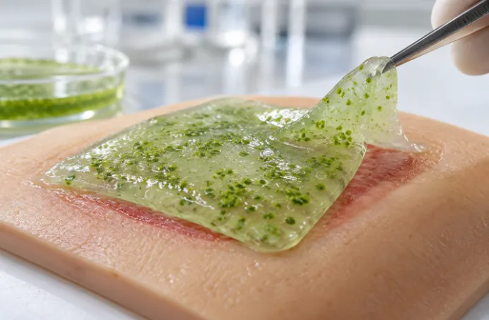
Figure 9: Conceptual Representation of a Microalgae-Containing Hydrogel Patch at the Wound Site
How strong is the evidence?
It seems there is some evidence to suggest developing oxygen generating/delivery biomaterials can improve oxygen availability or wound healing in vitro and in vivo. There seems to be many examples of oxygen delivery systems which have improved oxygen availability in either in vitro or in vivo. Different biomaterials have been used, different animal models and wound sizes, controls, oxygen and wound healing measured. Many of these systems are not patchy. They are hydrogels/scaffolds. Many have antibacterial, antioxidant and/or photothermal properties making it difficult to determine if it is due to the oxygen. Reviews have shown issues with design and sustained delivery of oxygen generating hydrogels [12][13][27][29][35]. Overall, there is promising evidence however most of this is pre-clinical.
Oxygen Sources and Trade-Offs
There seems to be many different examples of calcium peroxide-based patches. Chitosan-PVA and porous hydrogels have also been used. Calcium peroxide is a solid therefore can be incorporated into a polymer matrix and activated with water [30][33].Sodium percarbonate has been investigated as an alternative to calcium peroxide, for oxygenating bio-printed tissue models [31]. It is important to determine how much oxygen is being delivered and how this is interacting with the cells. Magnesium peroxide nanoparticles have been used with manganese oxide in animal models to create catalytic oxygenation for chronic diabetic wounds that is preclinical evidence[54].
Although biological and catalytic systems overcome some issues with the peroxide systems, they may create new issues. Photosynthetic gels require living organisms as well as the right lighting conditions. Nano enzymes will depend on their catalytic properties. Multifunctional hydrogels containing nitric oxide and oxygen may help overcome many wound healing factors. However, both gases will have their own safety and dosing parameters. Microneedles and antibacterial hydrogels (for targeting biofilms) could be useful in treating infected wounds but simply being antibacterial doesn’t mean they will help tissues heal. Decisions need to be made about if the system is the best choice. There will be interactions between oxygen generation, ROS, antibacterial and host cell interactions [25][26][36][37][39–41][43–45][48][51–53].
Therefore, it is vital to know how much evidence there is to support each system. The formulation should be consistent with oxygen being released. The cell studies should demonstrate oxygen helping with cell survival and function under hypoxia. The animal studies should show the effects of the system on wound healing, tissue organisation, vascularisation, inflammation and infection. Clinical trials should be undertaken to understand if there are any added benefits to the patient, who has a clear wound aetiology. If positive results are seen further testing can be done. However, the evidence cannot be replaced with evidence from the next level [13][15][27][29][31][46].
Interpretation Of Results
It is necessary to know how the evidence has been found. High oxygen levels show the system is working however does not show any improvement in healing. Cell migration, proliferation and viability can give some information but does not reflect the complex vascular, immune and microbial responses seen in chronic ulcers. Animal studies can provide evidence of wound healing and histology however many studies will investigate acute wounds or tightly controlled diabetic models meaning chronic wounds are not represented. Multifunctional dressing research should have controls to see if improvements are due to the oxygen source or other active ingredients. The strongest evidence from the literature would be formulations that are consistent with oxygen delivery, safe to the host cells, evidence of mechanisms, controlled animal studies and well-designed clinical trials [13][24–26][29–33][35–41][43–46][54][55].
COMPARISON TO STANDARD DRESSINGS AND OTHER ADVANCED WOUND DRESSINGS
Oxygen releasing hydrogel patches should be considered as ‘adjunct’ therapies to standard wound care. They cannot replace debridement, offloading, infection control, vasculature and glucose control and using the correct dressing. Standard dressings can provide protection and some absorption however they cannot provide oxygen. Hydrogels can provide long term moisture and drug delivery. The oxygen producing hydrogels can provide an oxygen source, however there may be issues with kinetics and by products. Foam, hydrocolloid and alginate dressings can provide many benefits depending on the exudate. There have been systematic reviews showing there is not one dressing that shows clear benefit for diabetic foot ulcer healing [3][16–19].
Topical oxygen and Hyperbaric oxygen cannot be directly compared to the chemically producing oxygen patches. There are different methods of delivering oxygen meaning different equipment, dosing and evidence. The patch has targeted and easily delivered oxygen however the dose will depend on the dressing formulation and wound status. Comparison should be based on how much oxygen is being delivered and what effect it is having on the patient [14][15][21][29].
Table 1: Comparison of Conventional Wound Dressing and Oxygen-Based Approaches
| Dressing/ Therapy |
Main Function and Oxygen Provision | Moisture Management and Applicability | Evidence, Limitations and Viability |
|---|---|---|---|
| Gauze/basic dressings | Protection and absorption of exudate | Simple to apply but can stick once dry. Often need changing frequently based on wound status | Easy to obtain but do not address local hypoxia[3] |
| Hydrocolloid dressings | Moisture and exudate management | Suitable for certain wounds and exudate levels | Inconclusive evidence for diabetic foot ulcers[17] |
| Foam/alginate dressings | Exudate management and protection | Often chosen for moderate to high exudate depending on product and wound status | No consistent evidence to show one type is better [16][18][19] |
| Hydrogel dressings | Hydration and contact with wound. Can act as a drug delivery system | These can conform to irregular surfaces and provide a moist environment. Strength and swelling will depend on formulation | This can provide a moist environment but an extra source of oxygen is required [15][28] |
| Topical oxygen delivery | Oxygen provided from external source | Will require application and following a protocol. | Different delivery and evidence from chemically generating patches [14][15] |
| Hyperbaric oxygen therapy | Oxygen provided systemically at high pressures | Requires specialist facility and careful patient selection | Cannot be compared to a patch. Oxygen is not being generated chemically at the site [5][21] |
| Oxygen generating hydrogel patch | Oxygen generated from incorporated sources or catalyzed systems | May be portable and conformable. Oxygen generation will depend on water, chemistry diffusion and dressing design | Promising pre-clinical evidence but concerns about burst release by-products standardization of dose and clinical evidence [12][24][30–33] |
| Photosynthetic/ hybrid patch | Photosynthetic components or oxygen-generating components with antibacterial and antioxidant properties | May require light, viable biological components or extra controls | This is innovative but may cause issues with stability, containment, identification of components and manufacture [20][43–45] |
Choosing The Right Dressing
Assess the wound. If it is dry or has low exudate a dressing to provide moisture would be needed. If there is a lot of exudates a dressing with high absorption and protection would be needed. If the surrounding tissue is fragile the dressing may need to be applied and removed carefully. If there is infection or suspected deep involvement clinical assessment and management would be needed. A dressing with in vitro antibacterial properties should not be used to diagnose or as a replacement for antimicrobials. If there is poor perfusion in the wound then assessment and correction of the vasculature would be needed. Oxygen producing patches maybe of use for a specific group of wounds in the future however research needs to be done to find what group and show there is an improvement over standard moisture and barrier properties [3]4][14][16–19].
A fair comparison of the oxygen producing patch to an advanced dressing should take into account ease of use, comfort, exudate management, duration of wear, assessment, frequency of changing, cost and any adverse effects. However, just because the device can produce oxygen it does not necessarily make it a better dressing. It needs to improve wound outcomes in a clinically significant way. This could be full healing, speed of healing, infection related complications, recurrence or patient burden. This is an important consideration as oxygen producing patches can have multiple active ingredients and be harder to produce [11][14][15][28][29].
Interpreting Claims
If the intention of the dressing was for exudate management, simple and cheap then a standard dressing would be the best option. An oxygen producing patch maybe another method but can have a reduced duration of wear, increased storage issues and complex formulations. Adding antimicrobials or bioactive to advanced dressings cannot claim to be improving due to the oxygen. If possible, use a factorial or matched control study to see if it is the base hydrogel, oxygen or additional active ingredient causing the improvement. This will help to see if the oxygen is providing an additional benefit over just moisture management or antimicrobials. This would apply if comparing to topical oxygen devices or hyperbaric oxygen. Consider the intensity of treatment, patient group and quality of care [3][11][14–19][28][29].
SAFETY, LIMITATIONS AND TRANSLATION
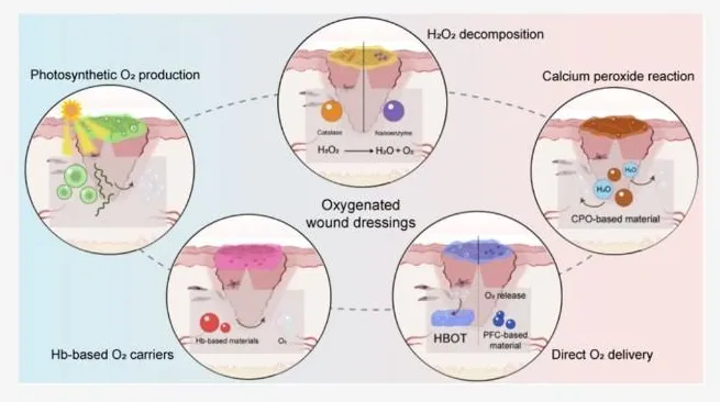
Figure 10: Conceptual Representation of Methods to Improve Oxygen Availability in wounds-Photosynthetic, Peroxide, Oxygen Carrying and Direct Delivery
Oxygen Dose, Kinetics, And By-Products
There will be limitations to oxygen delivery; however, this can be overcome by ensuring that oxygen is released slowly by the initial dose. If not, enough oxygen is released this may lead to issues with meeting requirements. Using calcium/Magnesium peroxides will affect the local environment. By looking at the oxygen, peroxide and pH levels, you will be able to determine what the optimal dose is. Oxygen can be controlled through encapsulation, catalase conversion and stimulus responsive systems. Advantages of each should be considered [24][30–33][36–38].
Biocompatibility And Wound Specificity
The concentration, degradation, by-products, contamination and exposure will depend on the materials used. Some may show good biocompatibility with a short-term cell study but if multiple dosing or long term, when in an inflamed wound, may not be good. The dressing must be considered to see if it is cytocompatabile, will cause irritation/inflammation (if required) and what the degradation profile is. Wounds can be very variable meaning it is unlikely that a dressing will work for all types of wounds. Oxygen should be used as an adjuvant therapy [4][15][28][46].
Manufacture And Translation
The precursors need to be evenly distributed during technology transfer. Oxygen delivery needs to be defined. To ensure good stability the dressing must be well packaged. A validated method of sterilisation needs to be used so that the enzyme and biological components are not affected. Characterisation needs to be done to determine how quickly it can be made, how long it will last, how much precursor will be left, what degradation products will be formed, physical properties and batch to batch variability. If any biological photochemical components are present considerations need to be made about viability and containment. Microneedle insertion and physical properties need to be considered. Clinical trials should be carried out to compare the device to standard treatment. Results need to be defined. Adverse events need to be noted. Patients should be divided into categories according to cause of wound and vascular. There is not enough pre-clinical data to determine what the optimal dose of oxygen is. Universal benefits have not been shown. [12][13][21][29][32][35][46][54][55].
Minimum Reporting Requirements
To ensure the results can be compared the following information needs to be known: source of oxygen generating particles, particle size, loading, polymer type, crosslinking, sterilisation and storage. The oxygen delivery method needs to be defined; medium, volume, temperature, calibration of device to measure oxygen and location. If biological studies were done the cell type, level of oxygenation, time period, number of replicates and suitable controls should be included. If animal studies were done the animal model, wound size, randomisation and blinding should be included. Calculations should be performed to determine wound closure, histology techniques and any adverse events. Include any adverse or ambiguous results. By including minimum information, the results will be more repeatable, showing whether differences are due to chemical, testing or biological factors [27][29][31][32][35][46].
Gaps In Evidence for Clinical Effectiveness
There is a large evidence gap as it would be hard to compare results between studies. This could be due to different animal models of diabetes, methods of wound induction, wound size, initial vascular, infection and follow up time. Wound healing can be calculated by determining the % reduction in wound area. Histology can be used to indicate re-epithelisation, collagen formation and angiogenesis. These are all linked but not the same. The wound may heal quickly but not necessarily have good healing or lower chance of recurrence. Any future clinical studies should have a clear primary outcome and suitable follow up times. Full healing should be recorded and any adverse events. Mechanistic outcomes have to be reported. Information on standard of care should be included to show any additional benefit [13][15][29][46][55].
RESEARCH GAPS AND FUTURE PERSPECTIVES
Further work should be completed to develop standardized protocols to measure oxygen generation in vitro. This should compare a variety of precursor materials, hydrogels, formulations and determine the level of oxygen generated at the tissue interface. This should also analyse the response of cells, angiogenic activity, collagen alignment, infection and wound healing. Factorial design can be used to understand the effect of oxygen in comparison to hydrogels, antimicrobials, antioxidants or photothermal agents. Long term tests should be performed to confirm the quality of the tissue, likelihood of disease reoccurrence and good local tolerance [27][29][31][32][46].
More work should be completed to determine the correct dose of oxygen to use. There are many types of wounds to consider such as ischemic wounds, neuropathic ulcers and highly infected wounds. Each will have different limitations meaning they may act differently. Biomarkers can be used to confirm that the patch has increased levels of oxygen in the tissue. Stimuli responsive systems have huge potential (pH, glucose, moisture, temperature) however, it is important to consider if they are specific, reliable and easy to manufacture [15][35][40][41][46].
Composite systems could be developed to increase oxygen generation, with control of ROS, biofilm, angiogenesis and accurate delivery of other therapeutics. The most effective systems will not necessarily be the most advanced, instead systems should provide a clear benefit without being too complex. Living photosynthetic patches and nano-enzyme hydrogels rely on biological interactions or catalytic activity so detailed quality control is needed. Careful clinical trials should be designed to ensure these systems are safe, feasible, well accepted by patients and cost-effective whilst providing an extra benefit [25][26][34][36][37][39–45].
Standardized methods should be developed to measure oxygen generation and biocompatibility. Different materials should be tested using the same testing methods to determine the best formulations. Sharing information with the field would be beneficial to know what formulations have not worked. If oxygen concentrations, by products, cytocompatibility and stability are standardized this can confirm that real improvements are being made rather than differences due to testing conditions. This can help find an effective and manufacturable patch [12][27][31][32][35][42].
CONCLUSION
Oxygen generating hydrogels have great potential in managing diabetic wounds and overcoming local hypoxia. Peroxides including calcium peroxide in a hydrogel carrier have had good results in pre-clinical models. Recent developments in hydrogel technology, micro-needles, polymer nanofibers and catalytic materials means there are many options for developing a controllable oxygen releasing system. Oxygen releasing materials can be combined with antimicrobial, antibiofilm and regulation of reactive oxygen species to combat the multifactorial issues of delayed wound healing.
There are still limitations with controlling oxygen release, cytotoxic effects and stability. There is limited data with most being gained from pre-clinical models. Future development should focus on standardized testing techniques, in depth safety information and thorough clinical trials to determine if these will improve wound healing. They should be viewed as an adjunctive treatment and cannot replace debridement, offloading, infection control, vascular assessment and glycemic control. Until they can show a safe and reliable benefit to patients with clinical significance, we will not know if they have any real value.
REFERENCES



Manasi Thakare, Natasha Crasto, Vinay Pal, Ajinkya Sonawane, Mohit Gautam, Shrutika Patil, A Review on Oxygen-Generating Hydrogel Patches and Other Related Oxygen-Based Technologies for Accelerated Diabetic Wound Closure, Int. J. of Pharm. Sci., 2026, Vol 4, Issue 11, 1168-1185. https://doi.org/10.5281/zenodo.23999438
 10.5281/zenodo.23999438