

We use cookies to ensure our website works properly and to personalise your experience. Cookies policy
Jagdamba Education Society’s SND College of Pharmacy (B.Pharm.), Babhulgaon, Tal. Yeola, Dist. Nashik, Maharashtra, India.
Fungal skin problems aren’t rare. Not even close. Most of the time they stay on the surface—think the epidermis, hard keratin areas like nails and hair, and those moist mucosal zones that fungi seem to love. You’ll keep running into the usual suspects: dermatophytosis, Candida infections on skin or mucosa, pityriasis-type eruptions, plus a grab bag of region-linked conditions that show up a lot in places like Malaysia. The share of cases they take up is… big.And yes, we’ve got plenty of antifungal drugs. That doesn’t mean treatment is smooth. People miss doses because dosing is too frequent. Medication doesn’t always “stick” where it’s needed. Absorption can be all over the map. Side effects turn some patients off fast. Resistance? It’s not theoretical anymore. So researchers keep circling back to one question: how do you get the drug where it matters, keep it there, and make the whole routine easier to live with?That’s where transdermal delivery starts looking tempting. A patch is non-invasive. It can drip-feed a medication at a controlled pace. Effects can last longer. You also dodge the stomach and the liver’s first-pass metabolism, which can chew up certain compounds before they do much good.But here’s the catch. Skin is stubborn. The stratum corneum is basically a bouncer at the door, and most drug molecules don’t get past it without a fight. So building an antifungal patch isn’t just “mix drug + stick-on backing.” You have to choose the right drug candidate, the right polymer base, the right permeation boosters, plasticizers, and whatever else the formulation needs so release stays predictable and penetration actually happens.Both classic synthetic antifungals and bioactive compounds pulled from natural sources have shown they can work in these patch-style systems. The newer designs get more creative too: polymer matrices tuned for steady release, chemical or plant-based permeation enhancers, nanocarrier-assisted setups, and other tweaks meant to push more drug into the right layer and keep it there longer.
Transdermal drug delivery systems—TDDS—keep showing up in pharma conversations for a reason. No needles. No swallowing pills. Just a patch that can sit on the skin and drip-feed a drug in a steady, predictable way. That matters. Oral meds get chewed up by stomach conditions or enzymes, then hammered again by first-pass metabolism in the liver. A transdermal route can dodge a lot of that mess, which often means better bioavailability, fewer stomach complaints, and less of that annoying “peak-and-crash” absorption swing.Now zoom out for a second. Humans don’t live alone. We’re basically walking ecosystems, carrying huge communities of microorganisms that usually mind their own business. Most of the time, nothing bad happens. But when immunity dips—or when microbial numbers spike—some of those organisms stop being polite. They cross barriers, settle into tissues, and infection starts. And it’s not just bacteria and viruses, either. Fungi, parasites, helminths, even prions can be in the mix.Fungal infections, in particular, are a public health headache. They can hit skin and nails, irritate mucosal surfaces, and in severe cases spread deeper into the body. The everyday kind—superficial and cutaneous infections—are everywhere, often tied to dermatophytes or opportunistic fungi that take advantage of a small opening. Treatments exist, sure, but the usual dosage forms come with strings attached: repeated dosing, inconsistent absorption, irritated skin, side effects that make people quit early, and sometimes not enough drug actually staying where it’s needed. And then there’s resistance. It keeps creeping upward, pushing researchers to rethink both strategy and delivery.Look at the antifungal toolbox and you’ll see a few big families: azoles, polyenes, allylamines, echinocandins, plus older or specialized agents like griseofulvin and flucytosine. They don’t all fight the same way. Some choke off ergosterol production. Some punch holes in membranes. Some mess with the fungal cell wall. Sounds straightforward, right? Except performance isn’t just about mechanism. The drug’s physicochemical profile, its ability to penetrate and linger in skin, and the way the formulation behaves all play huge roles. If the medicine can’t get in—and stay in—then the “right” drug can still deliver the wrong outcome.So yes, delivery systems matter. A lot. That’s why antifungal transdermal patches are getting real attention as a developing research area. They offer a tempting mix: more convenient use, longer therapeutic coverage, steadier exposure, and the possibility of better localized treatment. The big questions now revolve around what works best, what still limits the approach, and where these patch systems are headed next.
Advantages of Transdermal Drug Delivery System
Transdermal delivery lets a drug skip the whole GI gauntlet—no stomach acid drama, no enzymes chewing it up on the way in.And it can keep blood levels steady. Like an IV would. Just without the needle. Big difference.A patch can drip out an antifungal slowly, hour after hour, for days. That steady release matters. It helps keep the concentration more even instead of spiking and crashing.There’s another win people forget about: first-pass metabolism. Drugs that go through the gut often get “processed” by the liver right away, and you lose a chunk before it ever reaches circulation. Through the skin, you can often dodge that first pass and get better usable exposure—assuming the drug is a good fit for transdermal delivery.Now think about the patients who really need an easy route. Nauseated? Vomiting? Unconscious? Swallowing pills isn’t exactly an option. A patch doesn’t ask for much.Convenience is the quiet powerhouse here. Some patches are once-weekly. One application, then you stop thinking about it for days. That kind of simple schedule makes it easier to stick with treatment when real life gets messy.
Disadvantages of Transdermal Drug Delivery System
Sticking with it long-term is tough. Sometimes, after you apply it, the skin gets irritated right where it sits. It might just be mild. Still annoying. And here’s another snag: plenty of drugs—especially the more water-loving (hydrophilic) ones—crawl through skin at a painfully slow pace, so you may never reach a real therapeutic level. Ionic drugs? Forget it. These patches can’t deliver them. Then there’s the real-world mess. Sweat, movement, moisture, a rushed or sloppy application—any of that can make the patch lift or slip. When it doesn’t stick well, the dosing turns uneven. One day it’s fine, the next day it’s not.
Antifungal Agents
The agents used to treat fungal infections are called antifungals. Simple as that. They’re made to prevent fungal problems, treat them once they show up, and, when needed, wipe them out.So where do these drugs aim? Fungi have a few weak spots. The cell membrane. The cell wall. The internal machinery that builds the stuff a fungal cell can’t live without. One target gets a lot of attention: ergosterol. It’s a key sterol sitting in the fungal cell membrane, and many antifungal drugs either block how the fungus makes it or mess with the membrane directly. When that membrane can’t do its job, everything starts to fall apart—growth slows, stops, or the cell just dies.How you give an antifungal depends on what you’re dealing with. Mild, local, superficial infection? Topical treatment often makes the most sense because it puts the drug right where the fungus is. But topical products can be annoying. They wear off. You wash them away. Sweat happens. Clothes rub. Then you’re stuck reapplying again and again. That’s why people keep pushing newer delivery ideas—transdermal systems and patch-style formats that keep the medicine in contact longer and release it in a more controlled way.When you zoom out, antifungals fall into broad buckets based on what they target and what they’re made of: polyenes, azoles, echinocandins, allylamines, plus a grab-bag of other agents. Different chemistry, different targets, different trade-offs.
Classification of Antifungal Drugs
| Polyene Antibiotics | Amphotericin B, Nystatin,Natamycin | |
|---|---|---|
| Azole Antifungal | Imidazole Clotrimazole, Miconazole, Ketoconazole, Econazole, Luliconazole |
Triazoles Fluconazole, Itraconazole, Voricobazole Posaconazole |
| Allylamines | Terbinafine, Naftifine | |
| Antimetabolite | Flucytosine | |
| Echinocandins | Caspofungin, Micafungin, Anidulafungin | |
| Miscellaneous Antifungal | Tolnaftate, Ciclopirox, Amorolfine | |
Chemical properties of terbinafine hydrochloride
| Property | Description |
|---|---|
| Generic name | Terbinafine hydrochloride |
| Pharmacological class | Allylamine antifungal agent |
| Chemical form shown | Hydrochloride salt |
| IUPAC name | (E)-N,6,6-trimethyl-N-(naphthalen-1-ylmethyl)hept-2-en-4-yn-1-amine hydrochloride |
| Molecular formula | C₂₁H₂₆ClN |
| Molecular weight | Approximately 327.90 g/mol |
| Molecular weight of free base | Approximately 291.43 g/mol |
| Structural features | Naphthalene ring, tertiary amine, alkene, alkyne, and branched alkyl group |
| Nature of compound | Lipophilic, weakly basic organic compound |
| Salt formation | Protonation of the tertiary amine with hydrochloric acid |
| Mechanism of antifungal action | Inhibition of fungal squalene epoxidase, disrupting ergosterol biosynthesis |
| Therapeutic application | Treatment of susceptible fungal infections of the skin and nails |
Additional physicochemical properties
| Physicochemical property | Reported value or description |
|---|---|
| Appearance | White to off-white crystalline powder |
| Melting point | Approximately 195–208 °C, depending on the reported source and measurement |
| pKa | Approximately 7.1 |
| Aqueous solubility | Slightly soluble in water; one product monograph reports approximately 0.63% w/v |
| Solubility in organic solvents | Freely soluble in methanol and methylene chloride; soluble in ethanol |
| Ionization | Weakly basic tertiary amine; protonated in the hydrochloride salt |
| Lipophilicity | The free base is lipophilic; the hydrochloride salt has different ionization and solubility behavior |
| Solid-state characteristics | Crystalline; thermal behavior may vary with crystal form and experimental conditions |
Types of Fungal Infections
Fungal infections start with fungi. Simple as that. Dermatophytes, yeasts, moulds—different names, same annoying problem.They don’t pick just one spot, either. Skin gets hit a lot. So do hair and nails. Mucous membranes can be involved. And yes, sometimes the infection goes deeper and reaches internal organs, which is where things stop being “just a rash” and get serious fast.Why do they show up in the first place? A bunch of moving parts. The specific fungus matters. Your environment matters. Your skin barrier—how intact and resilient it is—matters. Your immune system matters most. Put all that together and you get the perfect setup, especially in warm, damp places where sweat hangs around.Think about it. Groin. Feet. Skin folds. Areas that stay moist, rub a lot, and don’t get much air. Fungi love that. They thrive there.The symptoms can be all over the map. Some cases are mild: a bit of itch, some redness, flaky scaling. Annoying, but manageable. Others keep smoldering, causing ongoing inflammation. Nails can get wrecked—thickened, brittle, discolored. And in the worst situations, certain fungi trigger severe illness throughout the body.Different fungi also have different “favorite” targets. Dermatophytes tend to go after keratin-rich tissue—skin, hair, nails. Candida, a common yeast, often shows up where it’s moist: skin creases and mucosal surfaces. Then there are fungi like Aspergillus. Those can invade and spread, especially when someone’s immune defenses are already down. That’s when a fungal infection stops being a nuisance and becomes a real threat.
| Type of Fungal Infection | Causitive Agent | Example | How it Affects the skin and body |
|---|---|---|---|
| Superficial mycosis | Malassezia spp., Hortaea werneckii | Pityriasis versicolor – skin | Causes small white, brown or pink patches on the skin. Usually affects the chest, back and shoulders. |
| Cutaneous | Trichophyton | Ringworm | Itching, redness, scaling |
| Tinea pedis | T. rubrum | Athlete’s foot | Itching, Cracked Skin |
| Tinea corporis | Dermatophytes | Body Ringworm | Red Circular lession |
| Tinea cruris | Epidermophyton | Jock Itch | Groin Itching and Redness |
| Tinea Capitis | Trichopyton | Scalp | Ringworm |
| Onychomycosis | Trichopyton | Nail infection | Thick Discoloration of Nail |
| Candidiasis Candida albicans Skin/ |
Candida albicans | Skin / Oral infection | Redness, itching |
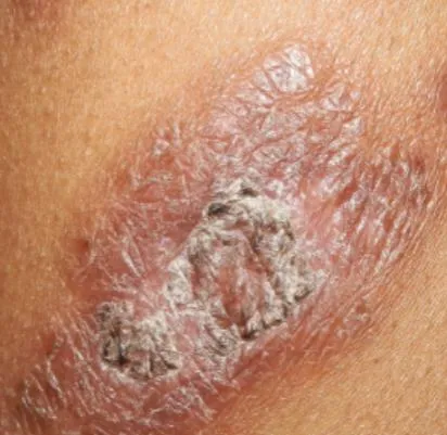
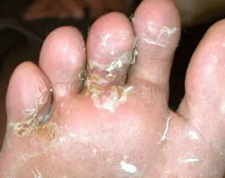
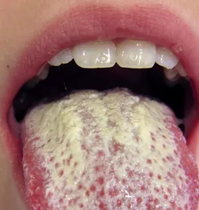
Transdermal Patch
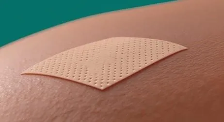
The medication sits inside a polymer-based setup and seeps out little by little once the patch hits your skin. Different builds handle this differently. Some stash the drug in a small reservoir, others embed it through a polymer matrix, and some mix it right into the adhesive layer itself. Either way, the patch keeps the drug in steady contact with the skin for a long stretch, which can mean you don’t have to reapply it all the time.
Skin
The skin is your body’s biggest organ. Full stop. It stands between you and everything outside—heat, cold, grime, microbes, friction, the whole mess. And it’s not just a shield, either. It helps you hold onto water, keeps your temperature from swinging all over the place, and feeds your brain a constant stream of sensations: touch, pain, pressure, itch.That “shield” part matters for medicine. People often want to deliver drugs through the skin because it can be convenient and steady. But the catch is obvious. The same barrier that keeps the outside world out also keeps a lot of drug molecules from getting in.Skin isn’t one uniform slab. It’s layered. Three main levels: the epidermis on top, the dermis underneath, and then the hypodermis (the deeper fatty layer). The real gatekeeper sits right at the surface of the epidermis: the stratum corneum.And this layer is stubborn.It’s mostly dead cells—corneocytes—packed with keratin, surrounded by a tidy lipid network that acts like mortar around bricks. Drugs trying to cross the skin have to negotiate this structure, either squeezing through the lipids or finding a route around and between cells. Many simply don’t make it far.Now bring fungal infections into the picture. Skin fungi are common. Annoyingly common. Creams and ointments can work, sure, but they come with practical headaches: you have to reapply, and the medication can be wiped off by sweat, washing, clothes, or just rubbing the area without thinking.That’s where a well-designed antifungal patch starts to sound appealing. Longer contact time. More consistent exposure at the exact spot that needs it. Less of the “did I put it on today?” routine.But here’s the part people sometimes miss: you can’t design a patch as if the only goal is “force the drug through at all costs.” Push penetration too hard and you may pay for it with irritation, a weakened barrier, or outright damage—especially when the skin is already inflamed.So you’re always juggling two needs:Get enough drug in.Don’t wreck the skin.Fungal infections themselves can change the skin’s structure and behavior. In superficial or cutaneous infections, fungi tend to live on or within keratinized layers. That can set off inflammation and a cascade of visible symptoms. Redness. Itching. Scaling. Dryness. Cracks. Sometimes soggy, softened skin (maceration), especially in folds. And pigmentation changes can linger during or after the infection.The “why” behind those signs isn’t mysterious, but it’s worth spelling out:- Redness (erythema): inflammation drives more blood flow to the area. - Itching (pruritus): inflammatory signals plus fungal activity irritate sensory nerves. - Scaling: the surface sheds faster, so flakes build up. - Cracks/fissures: dryness plus irritation can split the skin in tiny lines and breaks. - Maceration: too much moisture makes skin soft and fragile, often in warm, occluded places. - Lesions: some infections form recognizable rings, plaques, or patchy patterns. - Discoloration: the skin can look lighter or darker depending on inflammation and healing. - Barrier disturbance: once the outer layer is damaged, normal protection and water balance get shaky.And that last point loops right back to transdermal delivery. Healthy stratum corneum is a tight, reliable barrier. Infected skin isn’t always predictable. Inflammation, scaling, hydration shifts, and surface damage can all change how a drug penetrates—sometimes increasing entry, sometimes making distribution uneven, sometimes just making the skin more reactive.Think about the sequence:Inflammation, scaling, cracking → barrier gets altered → drug penetration may change.So when you build an antifungal patch, you don’t just chase maximum permeability. You aim for controlled delivery plus comfort. The drug’s physicochemical traits matter (size, solubility, affinity for lipids vs water). The condition of the skin matters (dry? macerated? broken? inflamed?). And the patch itself matters—polymers, adhesives, any permeation enhancers, all of it.Choose poorly and you can worsen irritation or strip the barrier further. Choose well and you can keep a steady drug level at the site while staying friendly to damaged, sensitive skin.Another Cpractical issue: untreated fungal infections can linger or spread. The longer they sit there, the more you see the same cycle—itching, scratching, more surface damage, more inflammation. Secondary problems can follow, simply because broken skin is easier to complicate.One more twist: fungal infections can look like other inflammatory conditions. People confuse them with eczema or psoriasis all the time. That’s why diagnosis matters before picking a treatment plan. Still, a fungal infection doesn’t “turn into” psoriasis or eczema in the usual sense; those conditions have different root causes, even if the redness and scaling can look similar from a distance.
Tinea Corporis
It Consists of Three Layer
1. Epidermis
2. Dermis
3. Hypodermis
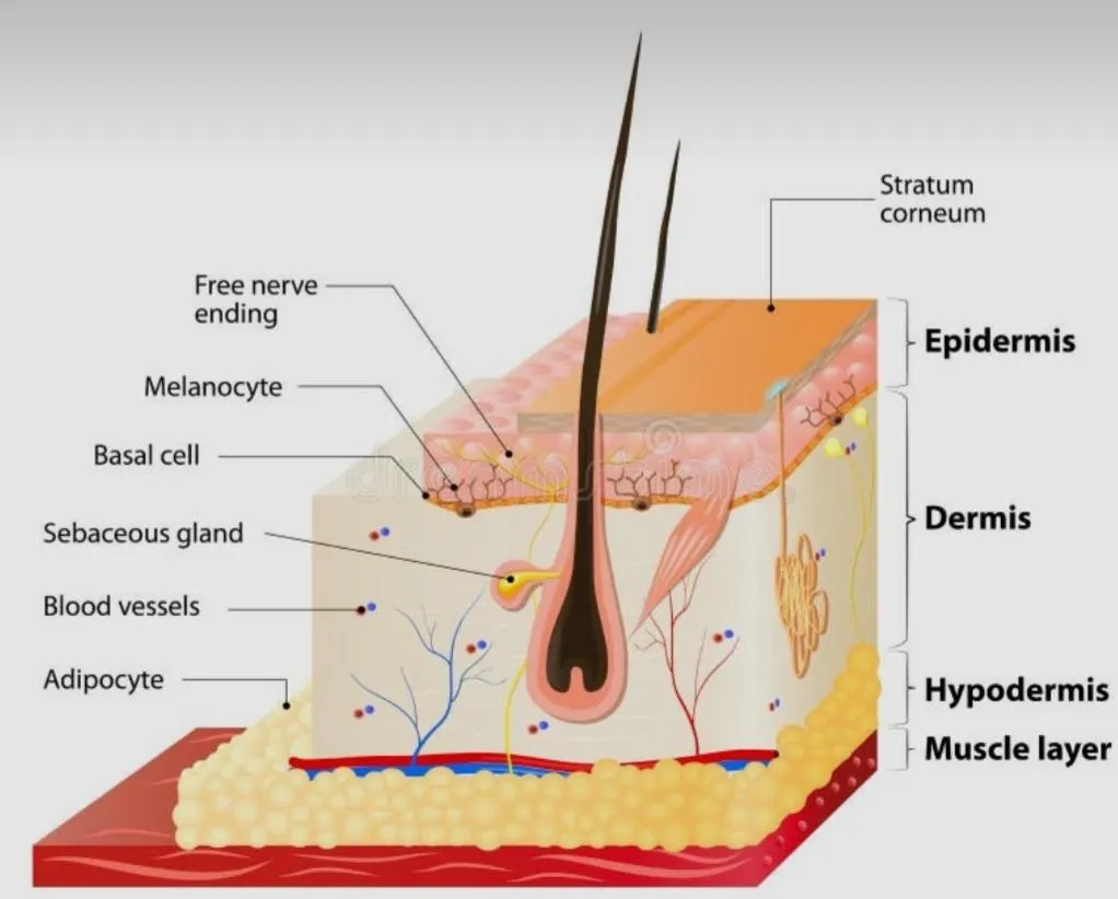
Figure of Skin
| Skin Layer | Description | Role in Drug Delivery | Diagram |
|---|---|---|---|
| 1.Epidermis | Outermost layer of Skin | Provides protection and acts as the main barrier to drug penetration. | |
| 2. Dermis | Middle Layer of Skin containing blood vessels, nerves and glands | Supports the skin and contains blood vessels that may carry absorbed drugs | |
| 3. Hypodermis |
Deep layer mainly composed of fat tissue | Provides insulation, cushioning and support to the skin. | |
| 4. Stratum corneum |
The stratum corneum is the outermost layer of the epidermis, | Acting as a Selective Permeability Barrier |
Mechanism of Transdermal Delivery system
The drug from a transdermal patch is released gradually and comes into contact with the skin surface. It then passes through the stratum corneum and deeper skin layers.
Flow Chart of Mechanism Transdermal Patches
Drug-loaded patch
↓
Drug release from the patch
↓
Drug reaches the skin surface
↓
Penetration through the stratum corneum
↓
Movement through the epidermis and dermis
↓
Drug reaches the target site or systemic circulation
↓
Therapeutic effect
A transdermal patch is a medicated adhesive preparation containing a drug that is designed to release the drug through the skin at a controlled rate.
Components of Transdermal Patches
1.Backing Membrane
2. Drug Reservoir or Matrix
3. Adhesive Layer
4. Polymer
5.Permeameation Enhancer
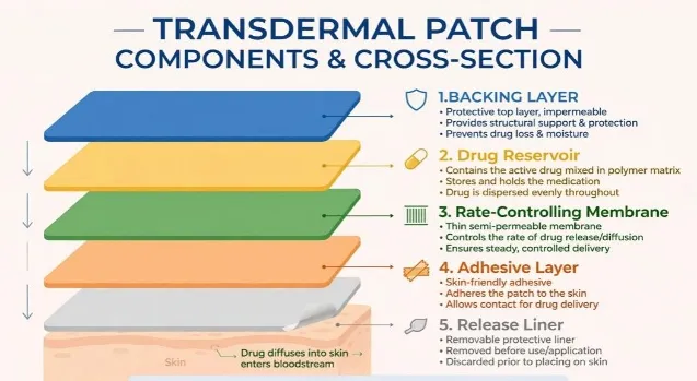
6. Release Liner
1. Backing Membrane
Protects the patch from the external environment.
Eg Polyethylene , Polyvinyl Chloride
2.Drug Reservoir Or Matrix
Contains the drug and controls its release pass through the skin more easily.
Example. Propylene glycol, oleic acid, ethanol.
3.Adhesive Layer
Keeps the patch attached to the skin during use.
Example . Polyacrylates, Siliconesi
4.Polymer
Forms the main Structure of the patch and controls the release of the drug.
The polymer should be stable..
The polymer should be easily of manufactured
The polymer should be inexpensive
The polymer and its deagration product must be non-toxic or non-antagonistic to the host.
There are Three Types of polymer
À) Natural polymers: Cellulose derivative, Gelatine, Waxes, Proteins, Gum.
B) Synthetic polymer:Hydrin rubber, silicone rubber, Nitrile, Acrylonitrile.
C) Semi Synthetic polymer: polyamide, polyurea, epoxy.
Example . HPMC, PVP, ethyl cellulose
5.Permeation Enhancer
Helps improve drug movement through the skin.
Example. Oleic acid, ethanol.
6.Release Liner
Covers the adhesive surface before use and is removed before applying the patch.
Example. Ethylene vinyl acetate (EVA, polypropylene
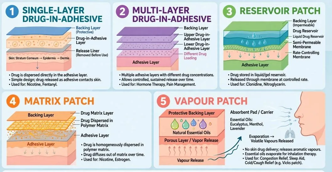
Types of Transdermal Patches
According to how the drug is stored and released from the patch.
Reservoir Type Patch
Multilayer Patch
Vapour Patch
Drug in adhesive Patch
Matrix Patch
| Type of Patch | Explanation | Main Feature |
|---|---|---|
| Reservoir-type patch | The drug is present in a separate liquid or gel reservoir. A membrane controls the rate of drug release | Controlled drug release |
| Matrix-type patch | The drug is mixed or dispersed uniformly in a polymeric matrix. The drug slowly diffuses out of the matrix. Simple design and easy preparation |
Simple design and easy preparation |
| Drug-in-adhesive patch | The drug is directly mixed with the adhesive layer, which both holds the patch to the skin and contains the drug. | No separate drug reservoir |
| Multilayer patch | Contains two or more drug-containing layers. Different layers can provide different drug-release rates. | Better control of drug release |
| Vapour Patch | Contains volatile substances that are released as vapour after application. | Mainly used for local vapour effects |
The Physiochemical Properties of Azoles, Polyenes, echinocandins allylamines and other Antifungal
| Class | Anti fungal agent | physical appearance |
|---|---|---|
| Azoles | Ketoconazole Miconazole Clotrimazole Econazole Itraconazole Posaconazole |
Colourless crystal or Powder White Powder White to Pale yellow Powder White Crystalline Powder White Powder White Powder |
| Polyenes | Nystatin Natamycin Amphotericin B |
Light Yellow Powder White Crystalline Powder Bright Yellow Powder |
| Echinocandins | Caspofungin Micafungin Anidulafungin |
Hygroscopic White to off White Solid Powder Solid Powder |
| Allylamines | Terbinafine Naftifine Butenafine |
Powder Powder White Crystalline Powder |
| Other antifungal agent | Flucytosine Griseofulvin Tolnaftate |
White Crystalline Powder White to Pale cream White Powder |
Factors Affecting Transdermal Patch
There are Several Factors That Affect Drug Delivery
Molecular weight
Smaller molecules generally cross the skin more easily.
Skin condition Damaged or diseased skin may show increased drug penetration.
Thickness of Patch Can affect the rate and duration of drug release.
Drug Concentration Higher concentration can increase the driving force for drug diffusion.
The barrier.
Partition Coefficient Good action requires the optimal partition coefficient High drugs are unable to access liquid layer of the skin additional medicines with low k would not penetrate
Evaluation of Transdermal Patches
Physical appearance
Checks colour, smoothness, flexibility and uniformity of the patchA good patch should have a uniform surface and should not show any visible defects.
Thickness
The thickness of the patch is measured at different points using a digital micrometer or vernier caliper. Measurements from different locations are compared to check whether the patch has a uniform thickness.Helps to obtain more consistent drug loading and drug release.
Weight Uniformity
Small pieces of the patch are cut from different areas and weighed individually. Their weights are compared with each other.It helps to determine whether the formulation has been distributed uniformly throughout the patch.
Drug Content
A known quantity of the patch is dissolved or extracted using a suitable solvent. The amount of antifungal drug present is then determined using an appropriate analytical method such as UV-visible spectrophotometry or HPLC.This test confirms that the patch contains the intended amount of drug and that the drug is distributed uniformly.
Folding Endurance
Folding endurance indicates the flexibility and mechanical strength of the patch. A small strip is repeatedly folded at the same place until it develops a crack or breaks.The number of folds the patch can tolerate before breaking is recorded.A patch with adequate folding endurance is less likely to break during handling or application.
Moisture Content
The patch is weighed before and after keeping it under suitable drying conditions. The change in weight is used to determine its moisture content.Moisture can affect the stability, flexibility and drug-release behaviour of the patch. Therefore, an appropriate moisture level is desirable
Moisture Uptake
The patch is exposed to an environment with controlled humidity and then weighed again. This test shows how much moisture the patch absorbs from the surroundings. Excessive moisture uptake may affect the physical properties and stability of the formulation.
Flatness
Flatness is checked to determine whether the patch remains flat or develops curling after preparation.A patch that remains flat can maintain better contact with the skin and provide more uniform drug delivery.
Tensile Strength
Tensile strength is the force required to break the patch when it is stretched. It is usually determined using a suitable tensile testing apparatus.It indicates the mechanical strength of the patch and its ability to withstand handling and application.
Percentage Elongation
Percentage elongation indicates how much the patch can stretch before breaking.Good elongation is useful because the patch needs to tolerate normal movement of the skin without tearing.
Adhesion Test
The adhesive property of the patch is evaluated to determine how effectively it remains attached to the skin.
Common parameters include:
Peel adhesion: force required to remove the patch from the surface.
Tack: ability of the adhesive to make immediate contact with the skin.
Shear adhesion: ability of the patch to resist sliding from the skin.Adequate adhesion is necessary to maintain drug delivery throughout the intended application period.
In-vitro Drug Release Study
In this test, the patch is placed in a suitable dissolution or diffusion medium, and samples are collected at predetermined time intervals. The amount of drug released is measured using an appropriate analytical method.It provides information about the rate and pattern of drug release from the patch.For an antifungal patch, the release profile is particularly important because the formulation should provide a suitable amount of drug over the intended period
Ex-vivo Skin Permeation Study
Ex-vivo studies are performed using suitable animal or human skin obtained according to ethical and regulatory requirements. The patch is placed on the skin, and the amount of drug passing through the skin is measured over time.A Franz diffusion cell is commonly used for this type of study.It helps determine how effectively the antifungal drug can cross the skin and provides information about parameters such as cumulative drug permeation and flux.
Skin Retention Study
In some antifungal formulations, it is important to know how much drug remains within the skin rather than only measuring the amount that passes through it.After the permeation experiment, the skin is analysed to determine the amount of drug retained in the skin layers.This is particularly relevant for cutaneous fungal infections, where maintaining the drug at the infected skin site may be desirable.
Skin Irritation Study
The formulation is checked for possible irritation such as redness, swelling, itching or other visible changes after application.Since the patch remains in contact with the skin for an extended period, it should have acceptable skin compatibility.
Stability Study
The prepared patches are stored under selected temperature and humidity conditions for a specified period. After storage, they are examined for changes in:
Appearance
Drug content
Thickness
Adhesion
Drug-release behaviour
Importance: The study helps determine whether the patch maintains its quality and performance during storage.
CONCLUSION
Transdermal drug delivery has become an interesting approach for improving the delivery of antifungal drugs, particularly when conventional topical preparations do not provide sufficient drug penetration or require repeated application. The main advantage of a transdermal patch is its ability to maintain prolonged contact between the formulation and the skin and to provide a more controlled release of the drug. For drugs intended for systemic delivery, the transdermal route can also avoid gastrointestinal degradation and hepatic first-pass metabolism. However, these advantages do not mean that every antifungal drug is suitable for transdermal delivery. Molecular size, lipophilicity, melting point, solubility and the ability of the drug to cross the stratum corneum have an important influence on its performance.
In antifungal therapy, transdermal systems may be useful for improving drug residence at the infected skin site and reducing some limitations associated with conventional formulations. Different approaches such as polymeric matrices, permeation enhancers, lipid-based carriers, nanoemulsions and microneedles have been investigated to improve movement of antifungal agents through the skin. Recent research has also shown interest in 3D-printed microneedle patches, which can create small pathways through the stratum corneum and allow better drug deposition within the skin.
Even with these developments, several challenges remain. The natural barrier function of the skin can restrict drug transport, while excessive use of penetration enhancers may produce irritation or affect skin integrity. Manufacturing complexity, drug loading, patch adhesion, stability and reproducibility also need to be considered before a formulation can be developed further. In addition, much of the recent work on advanced antifungal transdermal systems is still based on laboratory, ex-vivo or preclinical studies, so more clinical research is required to establish their practical value.
Overall, transdermal patches provide a useful research platform for exploring improved antifungal therapy. Further development of suitable polymers, safe permeation-enhancing methods and advanced technologies such as microneedles may help in designing antifungal patches with better skin penetration, controlled release and improved patient convenience
Future Prospects
Advanced microneedle patches: Microneedles may help antifungal drugs cross the stratum corneum and reach deeper skin layers more effectively.
3D-printed patches: 3D printing can be used to prepare microneedle patches with controlled shape, size and drug-loading characteristics.
Nanocarrier-based patches: Liposomes, Nano emulsions, nanogels and other carriers may improve drug penetration, skin retention and controlled release.
Targeted skin delivery: Future systems may be designed to keep more of the antifungal drug at the infected skin site and reduce unnecessary systemic exposure.
Patient-friendly systems: Thin, flexible and easy-to-apply patches may improve convenience and reduce the need for repeated application.
More clinical research: Most advanced antifungal transdermal systems are still supported mainly by laboratory, ex-vivo or preclinical studies. Human studies will be important before their wider clinical use.
REFERENCES


Purvesh Sharma, Maaz Aquil, A Review on Transdermal Drug Delivery System for Antifungal Agent Terbinafine, Int. J. of Pharm. Sci., 2026, Vol 4, Issue 10, 4738-4753. https://doi.org/10.5281/zenodo.23763129
 10.5281/zenodo.23763129