

We use cookies to ensure our website works properly and to personalise your experience. Cookies policy
1School of Pharmacy, Jagiellonian University.
2Faculty of Pharmaceutical Sciences, University of Port Harcourt.
3,4,5,6,7School of Pharmacy, RK University.
Sickle cell disease (SCD) remains a major global health burden and one of the most prevalent inherited blood disorders worldwide. For decades, hematopoietic stem cell transplantation (HSCT) has long been the only established curative therapy; however, donor scarcity, graft-versus-host disease, conditioning-related toxicity, and limited accessibility have restricted its use, prompting the search for donor-independent curative approaches. This review explores how advances in erythroid gene regulation and transcriptional reprogramming are reshaping curative therapy for SCD by tracing the evolution from HSCT to precision gene-based interventions. A deeper understanding of hemoglobin switching and the regulatory roles of erythroid transcription factors, including BCL11A, KLF1, ZBTB7A, and MYB, has enabled strategies that reactivate fetal hemoglobin or seek to directly correct the pathogenic HBB mutation. Gene addition, CRISPR-Cas9 gene editing, and emerging base- and prime-editing technologies offer autologous, donor-independent alternatives to transplantation, with early clinical studies demonstrating durable disease modification and encouraging therapeutic outcomes. Advances in genome editing, delivery systems, and long-term safety monitoring will determine the widespread clinical adoption of these therapies, particularly in low- and middle-income countries where the burden of SCD is greatest. Collectively, these advances demonstrate that while HSCT established the curability of SCD, erythroid gene regulation and transcriptional reprogramming are now redefining the concept of cure and expanding the potential for donor-independent curative therapy.
Sickle cell disease (SCD) remains one of the most prevalent monogenic disorders worldwide and continues to impose a substantial burden of morbidity and premature mortality, particularly in regions where access to disease modifying and curative therapies remain limited1,2. Global estimates indicate that annual births affected by SCD increased from approximately 305,800 in 2010 to over 515,000 in 2021, while the number of individuals living with the disease rose from 5.46 million to 7.74 million during the same period. Despite improvements in diagnosis and supportive care, the burden of SCD remains particularly high in low- and middle-income countries (LMICs), where delayed diagnosis, limited newborn screening, and restricted access to disease-modifying therapies remains major barriers to care3.
Historically, SCD management has focused on reducing disease complications rather than achieving cure. Hydroxyurea, chronic blood transfusion, iron chelation, and supportive measures have substantially improved survival and quality of life but do not correct the underlying genetic defect4. For decades, hematopoietic stem cell transplantation (HSCT) represented the only established curative therapy. However, its widespread application has been constrained by the availability of suitably matched donors, conditioning-related toxicities, graft-versus-host disease, and limited accessibility5.
The therapeutic landscape changed fundamentally in December 2023 with the approval of two autologous gene therapies for SCD: exagamglogene autotemcel (Casgevy), the first clinically approved CRISPR/Cas9-based gene-editing therapy, and lovotibeglogene autotemcel (Lyfgenia), a lentiviral gene-addition therapy. Rather than relying on donor-derived stem cells, these approaches genetically modify a patient's own hematopoietic stem cells to restore effective erythropoiesis through fetal hemoglobin reactivation or production of an anti-sickling β-globin variant6.
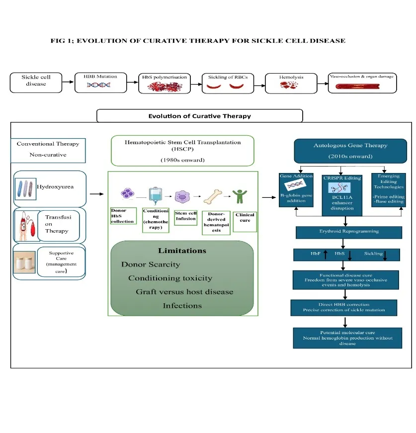
These advances represent more than technological innovation; they signal a conceptual shift in the definition of curative therapy. Instead of replacing the hematopoietic system, emerging strategies increasingly seek to modify erythroid gene regulation and reprogram gene expression, thereby suppressing the molecular and cellular processes responsible for sicklingFigure 1. Evolution of curative therapy for sickle cell disease from hematopoietic stem cell transplantation to autologous gene therapy and erythroid transcriptional reprogramming.
This review examines how advances in HSCT and erythroid gene regulation have transformed the therapeutic landscape of SCD, highlighting the transition from transplantation to transcriptional reprogramming and discussing how these innovations are redefining the concept of cure.
2. FROM SYMPTOM CONTROL TO CURATIVE THERAPY: THE HSCT ERA
2.1 Why conventional therapies were not enough
For decades, the management of sickle cell disease (SCD) focused primarily on alleviating symptoms and preventing complications rather than correcting the underlying genetic defect. Hydroxyurea, chronic red blood cell transfusion, iron chelation, and supportive care have substantially improved survival and quality of life. Hydroxyurea remains the cornerstone of disease-modifying therapy by inducing fetal hemoglobin (HbF), thereby reducing hemoglobin S polymerization, vaso-occlusive crises, and acute chest syndrome7,8. Likewise, chronic transfusion therapy lowers circulating HbS levels and effectively prevents primary and secondary stroke in high-risk patients9–11.
Despite these benefits, conventional therapies are not curative. Hydroxyurea requires lifelong adherence and exhibits variable clinical response, while chronic transfusions are associated with iron overload, alloimmunization, and transfusion-related complications. Importantly, neither strategy eliminates the causative β-globin mutation, and residual hemolysis, progressive vasculopathy, and cumulative organ damage may persist12. These limitations underscored the need for therapies capable of permanently correcting the disease rather than controlling its manifestations.
2.2 Hematopoietic Stem Cell Transplantation
A major turning point in curative therapy occurred in 1984, when Johnson and colleagues reported the successful bone marrow transplantation of an eight-year-old girl with both acute myeloid leukemia and SCD. Although the procedure was performed to treat leukemia, successful engraftment eliminated the patient's sickle phenotype, providing the first clinical evidence that replacement of the hematopoietic system could cure SCD13.
Subsequent clinical studies established hematopoietic stem cell transplantation (HSCT) as the first definitive curative therapy for SCD. In patients receiving grafts from HLA-matched sibling donors, overall and event-free survival rates are high, particularly in experienced transplant centers14. Importantly, complete donor hematopoiesis is not always required, as stable mixed donor chimerism is often sufficient to reverse the clinical manifestations of the disease15–17. To overcome donor limitations, alternative transplantation strategies—including matched unrelated donor, haploidentical donor, and umbilical cord blood transplantation—have been developed, expanding treatment opportunities while introducing additional challenges related to engraftment and immune complications18.
2.3 Remaining challenges
Despite its curative potential, HSCT remains inaccessible to the majority of individuals living with SCD. Fewer than 15% of patients possess an HLA-matched sibling donor, and outcomes with alternative donor transplantation are generally associated with higher risks of graft-versus-host disease, graft failure, delayed immune reconstitution, and treatment-related morbidity19. Myeloablative also remain an important source of treatment-related toxicity, although nonmyeloablative and reduced-intensity approaches have been developed to reduce these risks20.
Beyond biological limitations, HSCT requires highly specialized transplant centers, prolonged hospitalization, multidisciplinary expertise, and intensive long-term follow-up, making it difficult to implement in many low- and middle-income countries where the burden of SCD is greatest21. Consequently, although HSCT established that SCD could be cured through replacement of the hematopoietic system, its dependence on donors, conditioning, specialized infrastructure, and intensive follow-up highlighted the need for donor-independent approaches capable of achieving durable disease correction. This unmet need provided the foundation for the development of autologous gene therapies and, increasingly, strategies that manipulate erythroid gene regulation and transcriptional programs.
Table 1: Evolution of Disease-Modifying and Curative Therapeutic Strategies for Sickle Cell Disease.
| Therapeutic era/strategy | Principal intervention | Primary mechanism | Effect on disease | Curative potential | Major limitations |
|---|---|---|---|---|---|
| Conventional supportive care | Analgesia, hydration, infection prevention, supportive management | Management of acute and chronic complications | Reduces morbidity and improves quality of life | No | Does not modify the underlying genetic defect |
| Disease-modifying therapy | Hydroxyurea | Increases HbF and reduces HbS polymerization | Reduces vaso-occlusive crises and acute chest syndrome | No | Variable response; requires long-term treatment |
| Transfusion therapy | Chronic red blood cell transfusion | Reduces circulating HbS-containing erythrocytes | Particularly effective for stroke prevention | No | Iron overload, alloimmunization, transfusion dependence |
| Allogeneic HSCT | HLA-matched sibling HSCT | Replacement of recipient hematopoiesis with donor-derived erythropoiesis | Can eliminate sickle erythropoiesis and disease manifestations | Yes | Donor availability, GVHD, conditioning toxicity, infertility risk |
| Gene addition | Lovotibeglogene autotemcel (lovo-cel) | Lentiviral delivery of modified β-globin gene encoding HbAT87Q | Produces anti-sickling hemoglobin and reduces SCD manifestations | Potentially curative | Myeloablative conditioning, manufacturing complexity, cost, long-term monitoring |
| CRISPR-Cas9 gene editing | Exagamglogene autotemcel (exa-cel) | Editing of erythroid-specific BCL11A enhancer | Reactivates HbF and reduces HbS polymerization | Potentially curative | Conditioning toxicity, manufacturing complexity, cost, long-term safety monitoring |
| Base editing | BEAM-101 (risto-cel) | Precise editing of HBG1/HBG2 regulatory sequences to disrupt repression | Reactivates HbF without conventional double-strand DNA cleavage | Investigational | Clinical experience and long-term safety remain limited |
| Prime editing | HBB mutation correction | Precise correction of the pathogenic β-globin mutation | Aims to restore normal β-globin production | Potential molecular cure | Primarily preclinical/early development; editing efficiency, delivery and durability remain challenges |
3. ERYTHROID GENE REGULATION: THE FOUNDATION OF A NEW CURATIVE PARADIGM
3.1 Hemoglobin switching
Human hemoglobin production undergoes a tightly regulated developmental transition, progressing from embryonic to fetal and ultimately adult forms as erythropoiesis shifts from the yolk sac to the fetal liver and finally the bone marrow. Around birth, γ-globin-containing fetal hemoglobin (HbF) is progressively silenced and replaced by adult β-globin-containing hemoglobin (HbA), a process known as hemoglobin switching22.
In individuals with sickle cell disease (SCD), this developmental transition has profound clinical significance. Because HbF lacks the mutant β-globin chain responsible for hemoglobin S (HbS) polymerization, elevated HbF levels inhibit sickling by interfering with HbS polymer formation under deoxygenated conditions. Consequently, patients with persistently high HbF levels generally experience fewer vaso-occlusive crises, reduced hemolysis, slower progression of organ damage, and improved overall clinical outcomes.
The protective phenotype observed in individuals with hereditary persistence of fetal hemoglobin (HPFH) provided compelling natural evidence that sustained HbF expression can markedly ameliorate the clinical manifestations of SCD. This observation established HbF reactivation as a rational therapeutic strategy and laid the biological foundation for contemporary curative approaches. Today, reactivating the fetal hemoglobin program has become the cornerstone of erythroid-targeted therapies and underpins most modern gene-editing and transcriptional reprogramming strategies.
3.2 Key regulators of erythroid gene expression
Hemoglobin switching is orchestrated by a tightly coordinated network of transcription factors that regulate γ-globin repression during erythroid maturation. Among these, BCL11A functions as the principal transcriptional repressor of fetal hemoglobin by binding regulatory regions within the β-globin locus and recruiting chromatin-remodeling complexes, including the nucleosome remodeling and deacetylase (NuRD) complex23,24. The importance of BCL11A is highlighted by naturally occurring genetic variants associated with hereditary persistence of fetal hemoglobin, which preserve HbF expression into adulthood and provide compelling genetic validation for HbF-reactivation therapies25,26.
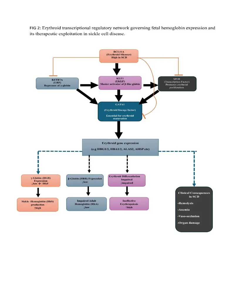
Figure 2: Erythroid Transcriptional Regulatory Network Governing Fetal Hemoglobin Expression and Its Therapeutic Exploitation in Sickle Cell Disease.
Acting through an independent regulatory pathway, ZBTB7A (also known as LRF) cooperates with chromatin-remodeling machinery to suppress γ-globin transcription independently of BCL11A. Experimental disruption of ZBTB7A has been shown to increase HbF expression to levels exceeding 60% in erythroid cell models, confirming that multiple complementary pathways contribute to fetal hemoglobin silencing27.
Upstream of these repressors, KLF1 serves as a central regulator of erythroid maturation by activating BCL11A expression while simultaneously promoting adult β-globin transcription, thereby coordinating the fetal-to-adult hemoglobin transition28,29. Additional regulators, including MYB and GATA1, further refine this transcriptional network. GATA1 cooperates with TAL1 at the erythroid enhancer of BCL11A, whereas MYB influences erythroid differentiation and indirectly modulates KLF1 activity30,31.
Table 2: Principal Erythroid Transcriptional Regulators Involved in Fetal Hemoglobin Regulation and Their Therapeutic Relevance in Sickle Cell Disease.
| Transcriptional regulator | Principal role | Effect on HbF | Relationship to erythroid regulation | Therapeutic relevance |
|---|---|---|---|---|
| BCL11A | Major developmental repressor of γ-globin expression | ↓ HbF | Recruits chromatin-remodeling machinery to repress fetal globin expression | Validated therapeutic target; erythroid-specific enhancer targeted by exa-cel |
| ZBTB7A (LRF) | Repressor of γ-globin transcription | ↓ HbF | Functions through a pathway partly independent of BCL11A | Potential target for HbF reactivation |
| KLF1 | Regulates erythroid differentiation and adult globin expression | Indirectly ↓ HbF | Activates BCL11A and promotes adult β-globin expression | Potential upstream regulatory target |
| MYB | Regulates erythroid proliferation and differentiation | Generally associated with ↓ HbF when highly expressed | Influences the erythroid transcriptional network and developmental globin expression | Potential target for therapeutic HbF induction |
| GATA1 | Master erythroid transcription factor | Indirect | Cooperates with other erythroid factors, including TAL1, at regulatory elements | Important component of the erythroid regulatory network |
| TAL1 | Regulates erythroid gene expression and enhancer activity | Indirect | Cooperates with GATA1 and other transcriptional complexes | Contributes to regulation of erythroid-specific enhancers |
| KLF1–BCL11A axis | Links erythroid maturation to developmental globin switching | ↓ HbF | KLF1 promotes BCL11A expression, reinforcing γ-globin repression | Provides an upstream regulatory framework for HbF reactivation |
Together, these factors form an integrated regulatory circuit that governs globin gene expression rather than functioning as isolated molecular switches.
3.3 Transcriptional reprogramming
Collectively, this regulatory network explains why erythroid cells have emerged as the principal therapeutic target in contemporary SCD research. Rather than replacing the entire hematopoietic system, as occurs with hematopoietic stem cell transplantation (HSCT), transcriptional reprogramming seeks to modify endogenous regulatory pathways within a patient's own hematopoietic stem cells. Current strategies predominantly target the erythroid-specific BCL11A enhancer, thereby reactivating the normally silenced fetal hemoglobin program while retaining erythroid differentiation and hematopoietic function32,33.
Clinical and experimental evidence indicates that increased HbF can substantially inhibit HbS polymerization and ameliorate the clinical manifestations of SCD. However, the protective effect of HbF depends not only on its overall level but also on its distribution among erythrocytes, as sufficient HbF within individual F-cells is required to effectively interfere with HbS polymerization34. Consequently, transcriptional reprogramming represents a mechanistically precise, potentially donor-independent therapeutic strategy that targets the erythroid processes underlying the sickling phenotype rather than simply treating its downstream clinical consequences.
This biological framework provides the rationale for the next generation of curative therapies discussed in the following section, where targeted gene addition, genome editing, and emerging nucleic acid engineering platforms exploit erythroid gene regulation to achieve durable disease modification.
4. ERYTHROID-TARGETED GENE THERAPIES
4.1 Gene addition
The first successful gene therapy platform for sickle cell disease (SCD) was based on lentiviral gene addition rather than direct genome editing. Lovotibeglogene autotemcel (lovo-cel), evaluated in the HGB-206 trial, involves the ex vivo transduction of autologous CD34⁺ hematopoietic stem cells using the BB305 lentiviral vector carrying a modified β-globin gene that encodes the anti-sickling hemoglobin variant HbAT87Q35. Following myeloablative conditioning, the genetically modified cells are reinfused to establish long-term production of anti-sickling erythrocytes.
In the pivotal HGB-206 cohort, treatment increased mean total hemoglobin concentrations from approximately 8.5 g/dL to 11 g/dL, with HbAT87Q constituting a substantial proportion of circulating hemoglobin. The principal advantage of this strategy is that it avoids nuclease-mediated genome editing and the generation of double-strand DNA breaks. However, because therapeutic efficacy depends on semi-random lentiviral integration into the host genome, long-term surveillance remains essential to monitor the rare but important risk of insertional mutagenesis,36 although advances in self-inactivating lentiviral vectors have substantially improved the safety profile of this approach.
4.2 Gene editing
Genome editing using the CRISPR-Cas9 system introduced a more precise therapeutic strategy by directly modifying endogenous regulatory elements controlling fetal hemoglobin expression. Exagamglogene autotemcel (exa-cel), the first CRISPR-Cas9-based therapy to receive regulatory approval for any disease,37 disrupts the erythroid-specific enhancer of BCL11A in autologous CD34⁺ hematopoietic stem cells, thereby relieving γ-globin repression and restoring sustained fetal hemoglobin production.
Clinical evaluation has demonstrated remarkable therapeutic efficacy. In the phase III trial supporting regulatory approval, the majority of treated patients remained free from severe vaso-occlusive crises for at least 12 consecutive months, extending the encouraging findings of earlier phase I/II studies that first established durable HbF induction following BCL11A enhancer editing38,39. By targeting an endogenous regulatory pathway rather than introducing an exogenous gene, exa-cel exemplifies the therapeutic potential of transcriptional reprogramming. Nevertheless, both exa-cel and lovo-cel continue to require myeloablative conditioning before cell infusion, meaning that although donor dependence has been eliminated, conditioning-related toxicity remains a significant limitation.
4.3 Emerging technologies
Base editing and prime editing represent the next generation of erythroid-targeted therapeutic platforms, designed to achieve precise genetic modification while minimizing or eliminating double-strand DNA breaks. BEAM-101 (risto-cel), an investigational adenine base-editing therapy, introduces naturally occurring A-to-G substitutions within the HBG1 and HBG2 promoters, disrupting BCL11A binding sites and reactivating fetal hemoglobin expression without inducing DNA cleavage. Interim findings from the ongoing BEACON trial have demonstrated HbF induction exceeding 60%, reduction of HbS to below 40%, and sustained resolution of anemia for up to 20 months of follow-up40.
Prime editing represents a further conceptual advance because it enables direct correction of the pathogenic HBB mutation without requiring double-strand DNA breaks or donor DNA templates. Ex vivo studies using patient-derived hematopoietic stem cells have achieved correction of the sickle mutation in 15–41% of edited cells while preserving normal engraftment and multilineage differentiation following transplantation into murine models41. Although these technologies remain in preclinical development or early-phase clinical evaluation, their ability to directly repair the causative mutation rather than compensating for its downstream effects through HbF induction, marks a fundamental shift toward precise molecular correction and further expands the curative landscape of SCD.
Table 3: Current and Emerging Gene-Based Therapeutic Strategies for Sickle Cell Disease: Mechanisms, Clinical Development, Therapeutic Outcomes, and Limitations.
| Therapy/platform | Genetic strategy | Primary target | Mechanism of therapeutic action | Development/clinical status | Major reported therapeutic outcome | Major limitations |
|---|---|---|---|---|---|---|
| Lovotibeglogene autotemcel (lovo-cel) | Lentiviral gene addition | β-globin pathway | Introduces a modified β-globin gene encoding anti-sickling HbAT87Q | Regulatory approved | Marked reduction in vaso-occlusive events in treated patients | Myeloablative conditioning, complex manufacturing, cost, long-term monitoring |
| Exagamglogene autotemcel (exa-cel) | CRISPR-Cas9 genome editing | Erythroid-specific BCL11A enhancer | Disrupts BCL11A enhancer activity, increasing γ-globin and HbF production | Regulatory approved | High proportion of treated patients achieving sustained freedom from severe vaso-occlusive crises | Conditioning toxicity, manufacturing complexity, cost, long-term safety surveillance |
| BEAM-101 (risto-cel) | Adenine base editing | HBG1/HBG2 promoter regulatory elements | Introduces precise nucleotide changes that reduce transcriptional repression and reactivate HbF | Investigational | Early clinical data indicate substantial HbF induction and reduction in HbS | Limited follow-up; long-term genomic safety and durability remain under evaluation |
| CRISPR-Cas12a approaches | Genome editing | HbF regulatory elements | Disrupts regulatory repression to induce HbF | Investigational/clinical development | Early studies demonstrate substantial HbF induction and reduction in disease manifestations | Conditioning, manufacturing, durability and long-term safety |
| Prime editing | Precision genome editing | Pathogenic HBB variant | Aims to directly correct the sickle mutation without conventional double-strand DNA breaks | Preclinical/early development | Demonstrated mutation correction in experimental hematopoietic stem/progenitor cells | Editing efficiency, delivery, engraftment, off-target effects and clinical translation |
| Future in vivo editing approaches | In vivo genome editing | HSCs and/or erythroid regulatory pathways | Direct delivery of editing machinery within the patient | Experimental | Potential to simplify manufacturing and reduce ex vivo processing | Efficient HSC targeting, delivery, safety and conditioning remain major challenges |
5. REDEFINING "CURE" IN SICKLE CELL DISEASE
5.1 The traditional definition of cure
For much of the therapeutic history of sickle cell disease (SCD), the concept of cure was synonymous with replacement of the defective hematopoietic system through allogeneic hematopoietic stem cell transplantation (HSCT). Under this paradigm, successful treatment was defined by durable donor engraftment and sustained elimination of sickle erythropoiesis. However, subsequent clinical experience demonstrated that complete donor hematopoiesis was not always necessary. Stable mixed chimerism, in which only a proportion of hematopoietic cells originate from the donor, has consistently been shown to reverse the clinical phenotype by substantially reducing the production of hemoglobin S (HbS)-containing erythrocytes. These observations established that complete biological replacement was not an absolute prerequisite for clinical cure; rather, sufficient correction of erythropoiesis was capable of fundamentally altering disease progression16,17.
5.2 The evolving concept of cure in the era of gene therapy
The emergence of gene-based therapies has broadened the definition of curative treatment beyond donor-derived cell replacement. Clinical trials evaluating exagamglogene autotemcel (exa-cel), lovotibeglogene autotemcel (lovo-cel), and emerging genome-editing platforms increasingly assess success using outcomes such as sustained freedom from vaso-occlusive crises, transfusion independence, durable fetal hemoglobin induction, normalization of hemolytic biomarkers, and improvements in health-related quality of life.
Results from the RUBY trial investigating an investigational CRISPR-Cas12a therapy further illustrate this conceptual transition42. Most treated participants achieved sustained elimination of painful vaso-occlusive crises, an outcome described as a functional cure because clinical manifestations were effectively abolished despite persistence of the underlying pathogenic mutation within portions of the hematopoietic compartment. In contrast, emerging gene-correction strategies such as prime editing seek to directly repair the causative HBB mutation, representing a potential molecular cure rather than functional disease compensation.
5.3 Comparing curative strategies
Although HSCT and gene-based therapies differ fundamentally in their mechanisms, both ultimately seek durable restoration of normal erythropoiesis43.
Table 4: Comparison of Hematopoietic Stem Cell Transplantation and Autologous Gene-Based Therapies for Sickle Cell Disease.
| Characteristic | Allogeneic HSCT | Autologous gene-based therapy |
|---|---|---|
| Cell source | Donor-derived hematopoietic stem cells | Patient-derived hematopoietic stem/progenitor cells |
| Donor required | Yes | No |
| HLA matching | Important for donor selection | Not required |
| Graft-versus-host disease | Possible | Not expected because cells are autologous |
| Genetic manipulation | No | Yes |
| Conditioning | Generally required | Currently required for approved ex vivo approaches |
| Principal mechanism | Replacement of sickle hematopoiesis with donor hematopoiesis | Genetic modification of autologous hematopoietic cells |
| HbF reactivation | Not the primary therapeutic mechanism | Major mechanism of exa-cel and several emerging approaches |
| Gene addition | No | Lovo-cel |
| Direct HBB correction | No | Emerging approaches such as prime editing |
| Major biological advantage | Established long-term curative potential | Eliminates donor dependence |
| Major biological limitation | GVHD, graft failure and donor-related complications | Conditioning toxicity and uncertainties regarding long-term genomic safety |
| Manufacturing requirement | Donor collection and transplant infrastructure | Personalized cell collection, genetic modification, quality control and reinfusion |
| Major access barrier | Suitable donor and transplant infrastructure | Cost, manufacturing infrastructure and specialized treatment centers |
| Long-term follow-up | Required | Required |
Despite these differences, important therapeutic challenges remain shared. Both HSCT and current ex vivo gene therapies require conditioning before stem-cell infusion and remain associated with procedure-related risks, prolonged hospitalization, and substantial healthcare costs. The major advance offered by gene therapy is therefore not the complete elimination of treatment-associated toxicity, but rather the removal of donor dependence, thereby expanding the potential pool of patients eligible for curative intervention.
5.4 Functional cure versus molecular cure
Current therapeutic platforms occupy different positions along the spectrum of curative intervention. HbF-reactivation strategies, including BCL11A enhancer editing and HBG1/HBG2 promoter base editing, achieve disease control by restoring fetal hemoglobin expression while leaving the pathogenic HBB mutation intact. These approaches are therefore best described as functional cures, as they eliminate or markedly reduce disease manifestations without correcting the underlying genetic defect37,40.
By contrast, prime-editing technologies aim to directly repair the sickle mutation within HBB, thereby restoring the normal β-globin sequence and offering the possibility of molecular correction. Although clinical evidence remains limited and long-term durability has yet to be established, these approaches represent the logical progression of precision genome engineering41.
Collectively, these advances suggest that cure in SCD should no longer be viewed as a single binary outcome. Instead, it represents a continuum defined by the mechanism of correction, durability of therapeutic benefit, and the extent to which the underlying genetic lesion has been functionally compensated for or permanently repaired.
6. CHALLENGES AND FUTURE PERSPECTIVES
6.1 Current barriers
Despite the remarkable progress achieved in curative therapy for sickle cell disease (SCD), several scientific, clinical, and socioeconomic barriers continue to limit widespread implementation. Among these, cost remains the most immediate obstacle. Exagamglogene autotemcel (Casgevy) and lovotibeglogene autotemcel (Lyfgenia) were introduced at list prices of approximately US$2.2 million and US$3.1 million per patient, respectively44. Although these costs have been justified relative to the estimated lifetime healthcare expenditure associated with SCD, they remain beyond the reach of most healthcare systems, particularly in low- and middle-income countries (LMICs).
The challenge is especially significant because the global burden of SCD is concentrated in regions with limited healthcare resources, including sub-Saharan Africa and India, where access to advanced cellular therapies remains extremely restricted45. In addition to financial barriers, current ex vivo gene therapies require individualized manufacturing processes involving stem cell mobilization, apheresis, genetic modification, quality-controlled cell processing, intensive conditioning, and specialized transplantation facilities. These complex manufacturing and clinical requirements substantially limit treatment availability outside highly specialized centers.
Long-term safety also remains an important consideration. Although clinical studies evaluating CRISPR-based therapies have thus far demonstrated encouraging safety profiles with no clinically significant off-target editing events detected,46 continued follow-up extending up to 15 years has been mandated to evaluate the long-term durability of gene editing and identify potential delayed adverse effects. Furthermore, regulatory agencies face the challenge of developing frameworks47 capable of evaluating rapidly evolving genome-editing technologies—including base editing and prime editing—which differ fundamentally from conventional gene-transfer approaches.
6.2 Future perspectives
Future progress in curative SCD therapy will depend not only on improving therapeutic efficacy but also on increasing accessibility, affordability, and global implementation. One promising direction is in vivo genome editing,48 which seeks to deliver editing machinery directly to hematopoietic stem cells within the patient, potentially eliminating the need for ex vivo cell manipulation, prolonged manufacturing, and, ultimately, less intensive conditioning regimens. Such advances could substantially simplify treatment delivery and expand access in resource-limited settings.
Continued innovation in genome engineering is also expected to improve editing precision, minimize off-target effects, and enable direct correction of pathogenic mutations. As multiple therapeutic platforms become clinically available, personalized approaches that tailor gene-editing strategies according to individual genetic profiles, disease severity, and treatment goals may further optimize long-term outcomes.
Equally important will be the development of sustainable manufacturing capacity within regions that bear the greatest burden of SCD. Establishing regional production facilities, strengthening clinical infrastructure, expanding newborn screening programs, and training specialized healthcare personnel will be essential for translating scientific advances into equitable clinical benefit45.
Ultimately, the long-term success of curative therapies will depend on robust post-marketing surveillance and extended clinical follow-up to confirm the durability, safety, and lifelong effectiveness of these interventions. Collectively, these efforts will determine whether the remarkable advances in erythroid gene regulation and genome engineering can evolve from highly specialized therapies into globally accessible standards of care.
CONCLUSION
The therapeutic landscape of sickle cell disease has undergone a fundamental transformation over the past two decades, evolving from a model of cure defined primarily by donor-derived hematopoietic stem cell transplantation to one encompassing an expanding array of gene-based therapies capable of modifying erythroid gene expression within the patient’s own hematopoietic system. Insights into the transcriptional regulation of the fetal-to-adult hemoglobin switch, particularly the central role of BCL11A and the interconnected contributions of KLF1, ZBTB7A, MYB, and GATA1 have provided the mechanistic foundation for gene addition, gene editing, and emerging base- and prime-editing strategies that reactivate fetal hemoglobin or seek to directly correct the pathogenic HBB mutation. These advances have not only removed the requirement for a matched donor in autologous approaches but have also reshaped how cure is conceptualized, highlighting a distinction between functional correction of disease manifestations and direct molecular correction of the underlying genetic lesion.
Despite this progress, the translation of these therapies into equitable and sustainable clinical practice remains incomplete. Substantial barriers persist, including prohibitive treatment costs, the complexity of individualized cell-manufacturing processes, the need for intensive conditioning, and the concentration of disease burden in regions with limited access to specialized transplantation and gene-therapy infrastructure. Continued innovation particularly toward in vivo genome editing, less toxic conditioning regimens, and regionally scalable manufacturing—together with rigorous long-term safety surveillance, will be essential to determine whether these transformative advances can evolve into durable and more accessible curative therapies for individuals affected by sickle cell disease, rather than remaining largely confined to patients able to reach highly specialized treatment centers.
ACKNOWLEDGEMENT
The authors sincerely acknowledge God Almighty for His grace, guidance, wisdom, and strength throughout the preparation of this manuscript.
Authors contribution
Ebimo-Moko Gloria Ebisintei: conceptualization, literature synthesis, manuscript drafting, critical revision, and overall supervision.
Mosopefoluwa Ayobami Adeuti: literature review and drafting of the hematopoietic stem cell transplantation section and preparation of Table 1.
Fathiah Oladele: literature review and drafting of the erythroid gene regulation section and preparation of Figure 2 and Table 2.
Jochebed Joel: literature review and drafting of the gene therapy and genome-editing section and preparation of Table 3.
Ebimo-Moko Victoria: literature review and drafting of the sections on the evolving concept of cure, challenges, and future perspectives and preparation of Table 4.
Jeffrey Emmanuel Sampson: design and preparation of the graphical abstract
Winner Madu : Literature review and critical supervision.
All authors critically reviewed the manuscript and approved the final version.
Funding
None to declare.
Conflict of interest
None to declare.
Ethics approval
None to declare.
REFERENCES

M. A. Adeuti, V. Ebimo-moko, J. D. Joel, F. O. Oladele, J. E. Sampson, G. E. Ebimo-moko, W. C. Madu, From Transplant to Transcriptional Reprogramming: How Erythroid Gene Regulation Is Transforming Curative Therapy in Sickle Cell Disease, Int. J. of Pharm. Sci., 2026, Vol 4, Issue 10, 4676-4691. https://doi.org/10.5281/zenodo.23762535
 10.5281/zenodo.23762535