

We use cookies to ensure our website works properly and to personalise your experience. Cookies policy
1,2Student, Ezhuthachan College Of Pharmaceutical Sciences, Marayamuttom, Thiruvananthapuram.
3Assistant Professor, Department Of Pharmacy Practice, Ezuthachan College Of Pharmaceutical Sciences Marayamuttom, Thiruvananthapuram.
4Consultant Gastroenterologist, NIMS Medicity, Thiruvananthapuram.
5Principal/ HOD, Department Of Pharmacy Practice, Ezuthachan College Of Pharmaceutical Principal/ HOD, Department Of Pharmacy Practice, Ezuthachan College Of Pharmaceutical.
Acute-on-chronic liver failure (ACLF) is a clinical syndrome characterized by an acute deterioration of liver function in patients with pre-existing chronic liver disease, leading to liver failure, organ dysfunction, and high short-term mortality. It is commonly associated with jaundice, coagulopathy, and the development of complications such as hepatic encephalopathy and ascites . Autoimmune hepatitis (AIH)–primary sclerosing cholangitis (PSC) overlap syndrome is a rare form of autoimmune liver disease in which features of both hepatocellular inflammation, typical of AIH, and cholestatic bile duct injury, characteristic of PSC, coexist. This overlap results in a complex clinical presentation and may predispose patients to rapid disease progression and liver failure . A 52 year old male was admitted to gastroenterology department with the complaints of fatigue, generalized pruritis . He has history of jaundice since last 3 months. The patient had past medical history of Type II Diabetes mellitus, CKD, DVT (around 8 months back). According to the physical examination, symptoms, diagnostic (Liver biopsy) and laboratory investigation (EBV PCR positive), the patient was diagnosed with Acute On Chronic Liver Failure – Overlap Syndrome (Autoimmune Hepatitis With Primary Sclerosing Cholangitis) and managed with IV antibiotic, oral steroid, and liver supportive medication.
DEFINITION
Acute-on-chronic liver failure (ACLF) is a distinct clinical syndrome characterized by acute deterioration of pre-existing chronic liver disease, resulting in hepatic and extrahepatic organ failure with a high risk of short-term mortality.[1] Unlike simple acute decompensation, ACLF is associated with an intense systemic inflammatory response that precipitates multiorgan dysfunction and rapidly worsens clinical outcomes. The syndrome is now recognized as a major cause of hospitalization and mortality among patients with advanced chronic liver disease, with reported 28-day mortality rates exceeding 30–40% depending on the number of organ failures.
Autoimmune hepatitis (AIH) is a chronic immune-mediated inflammatory liver disease characterized by elevated serum aminotransferases, hypergammaglobulinemia, increased serum immunoglobulin G (IgG), circulating autoantibodies, and interface hepatitis on liver histopathology.[3] The disease results from loss of immune tolerance against hepatocyte antigens, leading to persistent hepatic inflammation and progressive liver injury. Without appropriate treatment, AIH may progress to fibrosis, cirrhosis, portal hypertension, hepatic decompensation, and liver failure.
Primary sclerosing cholangitis (PSC) is a chronic cholestatic liver disease characterized by progressive inflammation and fibrosis involving both the intrahepatic and extrahepatic bile ducts, leading to multifocal biliary strictures, impaired bile flow, and chronic cholestasis.[7] PSC predominantly affects young and middle-aged adults and is strongly associated with inflammatory bowel disease, particularly ulcerative colitis. Progressive biliary fibrosis ultimately results in biliary cirrhosis, recurrent cholangitis, portal hypertension, and an increased lifetime risk of cholangiocarcinoma.
Autoimmune hepatitis–primary sclerosing cholangitis (AIH–PSC) overlap syndrome is an uncommon autoimmune liver disease in which patients simultaneously exhibit clinical, biochemical, serological, histopathological, and cholangiographic features of both AIH and PSC.[10] Although overlap syndrome is reported more frequently among children and young adults, it may occur in patients of all age groups. Owing to the absence of universally accepted diagnostic criteria, the condition remains a significant diagnostic challenge in clinical practice.
ETIOLOGY
The development of ACLF is commonly precipitated by acute insults superimposed on chronic liver disease.[1] Frequent precipitating factors include bacterial infections, gastrointestinal bleeding, active alcohol consumption, viral hepatitis, drug-induced liver injury, and major surgical procedures. In a considerable proportion of patients, however, no definite precipitating event can be identified, suggesting that dysregulated immune activation and systemic inflammation also contribute significantly to disease progression.
The exact etiology of AIH remains incompletely understood. Current evidence suggests that the disease develops in genetically susceptible individuals following exposure to environmental triggers such as viral infections, medications, toxins, or alterations in intestinal microbiota, which ultimately result in loss of immune tolerance toward hepatocyte-specific antigens.[3] Susceptibility is closely associated with specific human leukocyte antigen (HLA) class II alleles, particularly HLA-DR3 and HLA-DR4, which increase the risk of developing autoimmune liver disease.
Similarly, the etiology of PSC remains uncertain and is believed to involve a multifactorial interaction between genetic predisposition, immune dysregulation, environmental exposures, altered gut microbiota, and chronic intestinal inflammation.[7] The strong association between PSC and inflammatory bowel disease supports the concept of a gut–liver axis, whereby activated intestinal lymphocytes migrate to the liver and perpetuate chronic biliary inflammation.
The precise mechanism underlying AIH–PSC overlap syndrome remains unclear. Current evidence suggests that overlap syndrome represents a distinct clinical entity resulting from shared genetic susceptibility and immune-mediated injury affecting both hepatocytes and biliary epithelial cells.[10] Patients therefore develop simultaneous hepatocellular inflammation characteristic of AIH together with progressive cholangiopathy typical of PSC, producing a heterogeneous clinical presentation and variable disease progression.
PATHOPHYSIOLOGY
The hallmark of ACLF is an uncontrolled systemic inflammatory response that leads to widespread immune activation, oxidative stress, endothelial dysfunction, microcirculatory impairment, and progressive organ failure.[14] Release of pro-inflammatory cytokines, activation of innate immune cells, and mitochondrial dysfunction contribute to hepatocellular injury while simultaneously affecting extrahepatic organs including the kidneys, brain, lungs, and cardiovascular system. The resulting immune dysregulation predisposes patients to severe infections, circulatory dysfunction, hepatic encephalopathy, acute kidney injury, and multiple organ failure, explaining the high short-term mortality associated with ACLF.
In AIH, impaired immune tolerance results in activation of autoreactive CD4+ and CD8+ T lymphocytes directed against hepatocyte antigens.[3] This immune-mediated response promotes chronic hepatic inflammation through the production of pro-inflammatory cytokines and recruitment of inflammatory cells, leading to interface hepatitis, hepatocyte necrosis, progressive fibrosis, and eventual cirrhosis. Persistent inflammation further stimulates hepatic stellate cell activation and extracellular matrix deposition, ultimately resulting in irreversible architectural distortion of the liver.
In PSC, chronic inflammation of the biliary epithelium initiates repeated cycles of bile duct injury and repair, causing concentric periductal fibrosis, obliterative cholangitis, and progressive narrowing of the biliary tree.[7] Obstruction of bile flow results in chronic cholestasis, hepatocellular injury, biliary fibrosis, and secondary biliary cirrhosis. Recurrent bacterial cholangitis and prolonged inflammation further increase the risk of cholangiocarcinoma and end-stage liver disease.
In AIH–PSC overlap syndrome, immune-mediated hepatocellular injury coexists with chronic cholangiopathy, producing overlapping pathological mechanisms that accelerate hepatic fibrosis and increase the likelihood of hepatic decompensation.[10] When an acute precipitating event occurs, these patients may rapidly develop ACLF characterized by severe systemic inflammation, hepatic dysfunction, extrahepatic organ failure, and markedly increased short-term mortality.
CLINICAL MANIFESTATIONS
The clinical presentation of ACLF is typically abrupt and severe, occurring in a patient with known or previously unrecognized chronic liver disease. Common manifestations include jaundice, ascites, hepatic encephalopathy, gastrointestinal bleeding, coagulopathy, renal dysfunction, hypotension, and signs of systemic inflammation such as fever or leukocytosis.[1] Depending on the organs involved, patients may also present with respiratory distress, oliguria, altered mental status, or sepsis-related features. The severity of symptoms often reflects the number and extent of organ failures.
Patients with AIH may present insidiously with fatigue, malaise, anorexia, right upper quadrant discomfort, nausea, arthralgia, or jaundice.[3] Some individuals are asymptomatic and are diagnosed incidentally on the basis of abnormal liver biochemistry. In more advanced disease, clinical features of chronic liver disease such as spider angiomas, palmar erythema, hepatomegaly, splenomegaly, ascites, and encephalopathy may be present.
PSC commonly manifests with fatigue, pruritus, jaundice, and recurrent episodes of cholangitis characterized by fever, right upper quadrant pain, and worsening cholestasis.[7] Many patients are diagnosed during evaluation of persistently elevated cholestatic liver enzymes or after investigation of inflammatory bowel disease. As the disease progresses, portal hypertension, cirrhosis, fat-soluble vitamin deficiency, and complications of chronic liver failure may develop.
AIH–PSC overlap syndrome often presents with a mixed hepatocellular and cholestatic pattern of liver injury.[10] Clinical features may include fatigue, pruritus, jaundice, abdominal discomfort, hepatomegaly, and manifestations of portal hypertension. In patients with advanced disease, decompensation may occur with ascites, variceal bleeding, encephalopathy, or recurrent cholangitis. Because the presentation is variable, a high index of suspicion is required, particularly in younger patients or those with coexisting inflammatory bowel disease.
MANAGEMENT
Management of ACLF requires urgent hospitalization, identification and treatment of the precipitating event, and intensive supportive care.[1] Initial treatment includes hemodynamic stabilization, infection screening and empiric antibiotics when indicated, correction of electrolyte and metabolic abnormalities, management of encephalopathy, renal support, and treatment of gastrointestinal bleeding or respiratory failure as needed. Early involvement of intensive care and hepatology teams is essential. In selected patients, liver transplantation is the definitive treatment and should be considered early because prognosis is poor once multiple organ failures develop.
The mainstay of AIH treatment is immunosuppression.[3] Corticosteroids, usually prednisone or prednisolone, are used for induction of remission, often followed by azathioprine for maintenance therapy. Patients who are intolerant of or refractory to standard therapy may require alternative agents such as mycophenolate mofetil or calcineurin inhibitors. Regular monitoring of liver enzymes, IgG levels, and treatment-related adverse effects is necessary. In patients with cirrhosis or acute liver failure, prompt assessment for liver transplantation may be required.
Management of PSC is largely supportive because no medical therapy has consistently been shown to halt disease progression.[7] Treatment focuses on symptom control, including ursodeoxycholic acid in selected patients, pruritus management with agents such as cholestyramine or rifampicin, and endoscopic intervention for dominant strictures when indicated. Recurrent cholangitis requires antibiotics and biliary drainage when necessary. Surveillance for cholangiocarcinoma, gallbladder cancer, and colorectal cancer is important, especially in patients with concomitant inflammatory bowel disease. Liver transplantation is indicated for end-stage liver disease, recurrent cholangitis, intractable pruritus, or suspected malignancy.
Treatment of AIH–PSC overlap syndrome generally combines immunosuppressive therapy for the autoimmune hepatitis component with cholestatic and endoscopic management for the PSC component.[10] Corticosteroids with or without azathioprine are commonly used when hepatitic features predominate, while ursodeoxycholic acid and endoscopic therapy may be considered for cholangiopathic disease. Patients require close biochemical and radiologic follow-up because progression to cirrhosis, portal hypertension, and liver failure may occur despite therapy. Liver transplantation is the definitive option for advanced or refractory disease and should be considered when decompensation or ACLF develops.We present a case of acute-on-chronic liver failure secondary to autoimmune hepatitis–primary sclerosing cholangitis overlap syndrome, emphasizing the importance of early recognition, comprehensive diagnostic evaluation, and prompt multidisciplinary management in improving patient outcomes.
CASE PRESENTATION
A 52 year old male patient was admitted to gastroenterology department with complaints of fatigue, generalised pruritic days duration. patient had past medical history of jaundice since last 3 months, and is known case of type II diabetes mellitus, CKD, DVT around 8 months back .patient past medications include TAB GLYCIPHAGE SR, TAB JUSLINA, TAB GLIZID XR, TAB PANTOCID, TAB ASYNAC, TAB NYSOLONE, TAB URSETOR, TAB HEPTAGON, INJ HUMINSULIN.
On admission, the patient was conscious and oriented, moderately built, with pallor and icterus. The patient had no clubbing, cyanosis, pedal edema, or generalized lymphadenopathy. His vital signs were stable with a pulse rate of 80 beats/min, respiratory rate of 18 breaths/min, blood pressure of 120/80 mmHg, oxygen saturation of 99.4% on room air, and he was afebrile. Systemic examination revealed a distended abdomen with minimal ascites, which was soft and non-tender with audible bowel sounds. Respiratory, cardiovascular, and neurological examinations were unremarkable.
Laboratory investigations showed macrocytic anemia (Hemoglobin 8–9.4 g/dL), leukopenia (lowest total leukocyte count 1120 cells/cumm), mild thrombocytopenia (platelet count 1.4–1.8 lakh/cumm), elevated ESR (92–98 mm/hr), elevated C-reactive protein (28.6 mg/L), prolonged prothrombin time with INR ranging from 1.3–1.9, persistent predominantly direct hyperbilirubinemia (total bilirubin up to 10.51 mg/dL, direct bilirubin up to 9.19 mg/dL), elevated liver enzymes (AST up to 109 U/L and ALT up to 134 U/L), hypoalbuminemia (2.31–2.74 g/dL) with reversal of albumin-globulin ratio, and hypokalemia (lowest 2.94 mmol/L). Renal function remained within normal limits. Glycosylated hemoglobin (HbA1c) was elevated at 8.5%, indicating poorly controlled diabetes mellitus. Viral serology for hepatitis B, hepatitis C, HIV, and CMV were negative, while EBV PCR was positive (576 IU/mL).
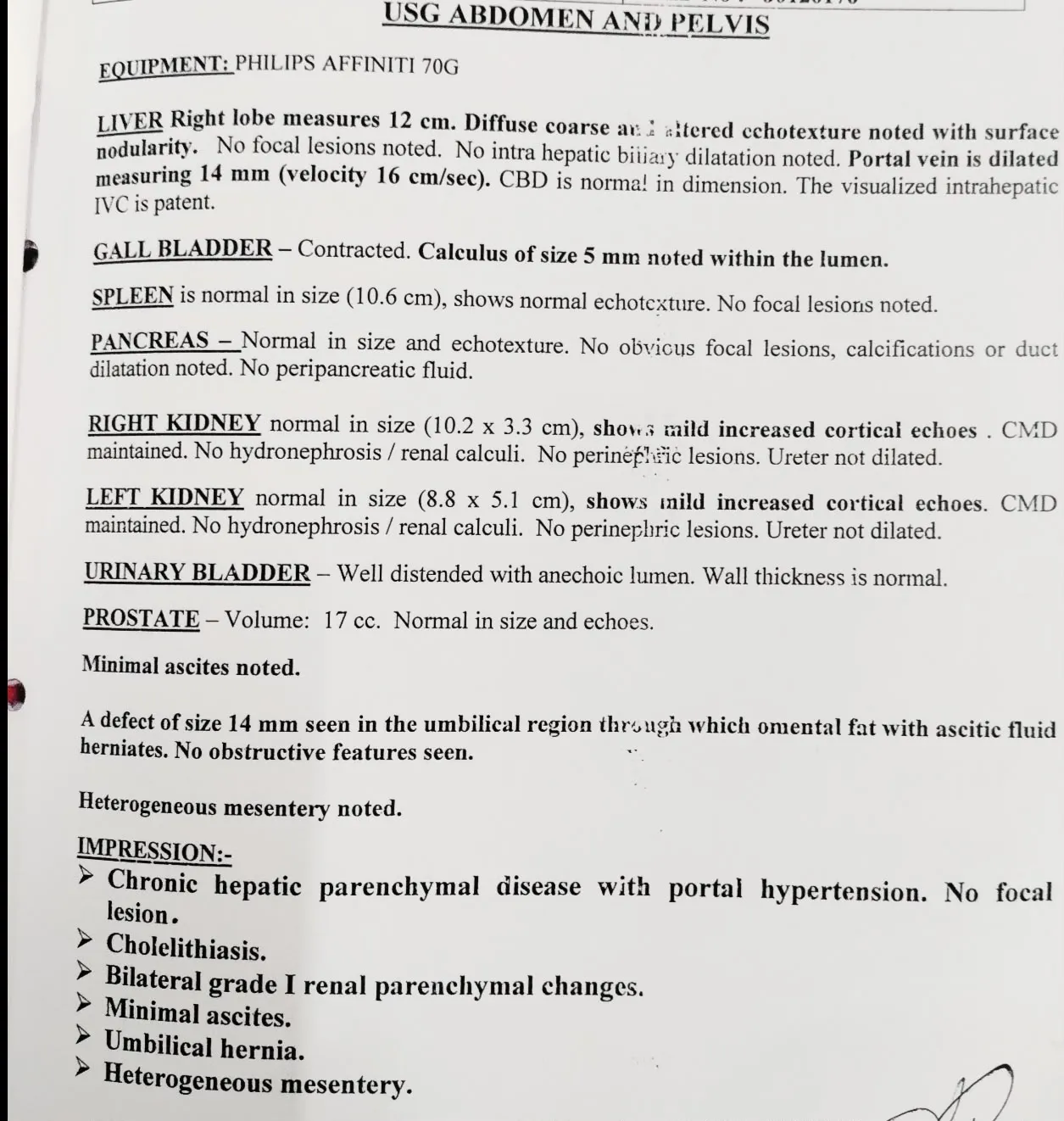
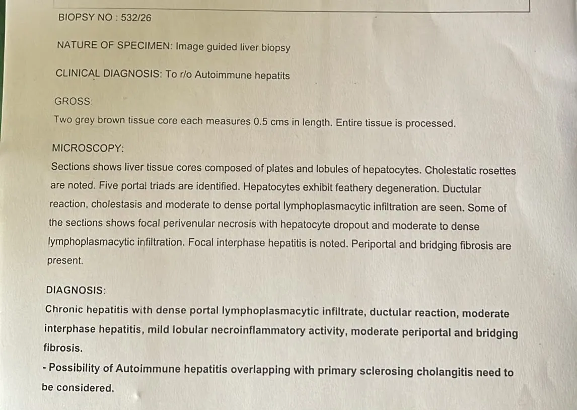
Ultrasonography of the abdomen and pelvis demonstrated chronic hepatic parenchymal disease with portal hypertension, minimal ascites, cholelithiasis, bilateral Grade I renal parenchymal changes, heterogeneous mesentery, and an umbilical hernia, with no focal hepatic lesion. Due to persistent hyperbilirubinemia and inadequate response to corticosteroid therapy, an image-guided liver biopsy was performed after correction of coagulopathy to establish the underlying diagnosis.
During hospitalization, the patient was initially managed with intravenous Cefoperazone–Sulbactam, oral Prednisolone (Wysolone 40 mg), liver supportive medications, correction of electrolyte abnormalities, and monitoring of coagulation parameters. Despite treatment, bilirubin levels continued to rise, and the patient subsequently developed fever with worsening leukopenia. Considering the possibility of sepsis while on corticosteroid therapy, the antibiotic regimen was escalated to intravenous Meropenem. Investigations for opportunistic viral infections revealed a positive Epstein–Barr virus (EBV) PCR, while CMV PCR remained negative. The patient received a single dose of Filgrastim for severe leukopenia, following which the white blood cell count improved. In view of poor response to corticosteroids, the dose of Prednisolone was reduced from 40 mg to 30 mg, and Mycophenolate Mofetil (MMF) 500 mg twice daily was initiated, with a plan to restart immunosuppressive therapy after infectious disease consultation and review of the liver biopsy report. The patient requested discharge after clinical stabilization and was advised to follow up in the outpatient department with the liver biopsy report for further management.
DISCUSSION
In the present case, a 52-year-old man developed acute-on-chronic liver failure (ACLF) secondary to autoimmune hepatitis–primary sclerosing cholangitis (AIH–PSC) overlap syndrome, presenting with fatigue, generalized pruritus, jaundice, hyperbilirubinemia, and elevated liver enzymes. The diagnosis was confirmed by laboratory investigations, imaging, and liver biopsy. This presentation is consistent with the findings of Floreani et al., who described AIH–PSC overlap syndrome as a rare autoimmune liver disease with overlapping hepatocellular and cholestatic features.
Similar to the study by Boberg et al., our patient showed biochemical and histopathological features of both AIH and PSC. However, unlike previously reported cases that responded well to conventional immunosuppressive therapy, our patient presented with ACLF and multiple comorbidities, including type 2 diabetes mellitus, chronic kidney disease, and previous deep vein thrombosis, making management more challenging.
In accordance with current clinical guidelines, corticosteroid therapy and supportive treatment were initiated promptly. However, persistent hyperbilirubinemia and EBV PCR positivity with severe leukopenia required modification of immunosuppressive therapy, escalation of antibiotics, and administration of filgrastim. The patient subsequently showed clinical stabilization and was discharged with advice for close follow-up.
This case highlights that early recognition of AIH–PSC overlap syndrome, timely liver biopsy, and prompt multidisciplinary management are essential for improving outcomes. It also emphasizes the importance of monitoring for infectious complications and treatment response in patients presenting with ACLF associated with autoimmune liver disease.
COMPARISON WITH OTHER CASES:
Autoimmune hepatitis–primary sclerosing cholangitis (AIH–PSC) overlap syndrome is an uncommon autoimmune liver disorder that shares clinical, biochemical, immunological, and histopathological features of both diseases. Although a number of cases have been documented in the literature, each patient's clinical presentation, related comorbidities, and illness severity differ significantly. Understanding the unique traits and management difficulties related to this uncommon illness is made easier by contrasting the current case with previously published studies. In the case study published by Floreani et al., a 48-year-old woman complained of pruritus, exhaustion, and jaundice. Elevated bilirubin, transaminases, alkaline phosphatase, and immunoglobulin G levels with positive antinuclear antibodies were found in laboratory examinations. Primary sclerosing cholangitis was identified by magnetic resonance cholangiopancreatography (MRCP), and autoimmune hepatitis with fibrosis was confirmed by a liver biopsy. Clinical improvement was the outcome of the patient's good response to therapy with prednisolone and ursodeoxycholic acid.
In a similar vein, Boberg et al. described a 42-year-old male who was diagnosed with AIH–PSC overlap syndrome based on biochemical evidence of cholestasis, positive autoimmune markers, endoscopic retrograde cholangiopancreatography (ERCP) bile duct strictures, and histological confirmation. Prednisolone, azathioprine, and ursodeoxycholic acid were used in the patient's treatment, which resulted in long-term disease control and liver function stabilization.
The 52-year-old male patient in this case had several coexisting medical conditions, such as type 2 diabetes mellitus, hypertension, chronic kidney disease, a prior cerebrovascular accident, and persistent deep vein thrombosis, which made clinical management significantly more difficult when compared to these previously published reports. The patient experienced acute-on-chronic liver failure, a severe clinical manifestation that has seldom been documented in AIH–PSC overlap syndrome, in addition to jaundice, anorexia, and weight loss. A mixed hepatocellular and cholestatic pattern with hyperbilirubinemia, elevated transaminases, alkaline phosphatase, gamma-glutamyl transferase, prolonged prothrombin time and international normalized ratio, increased inflammatory markers, and elevated immunoglobulin G levels was found in laboratory investigations. The diagnosis of autoimmune hepatitis with primary sclerosing cholangitis overlap syndrome was confirmed by liver biopsy, positive antinuclear antibody testing, and ultrasonographic features suggestive of biliary cirrhosis.
The current patient needed a multidisciplinary therapeutic approach due to the coexistence of acute-on-chronic liver failure, chronic kidney disease, and persistent venous thrombosis, in contrast to the majority of previously reported cases, which mainly required immunosuppressive therapy and ursodeoxycholic acid. Corticosteroids, N-acetylcysteine, ursodeoxycholic acid, broad-spectrum antibiotics, anticoagulants, diuretics, electrolyte replacement, and supportive care were all part of the management. The patient had significant hepatic dysfunction and several high-risk comorbidities, but their symptoms gradually improved, and they were released in stable condition.
The current case is unique due to the presence of acute-on-chronic liver failure with chronic kidney disease and persistent deep vein thrombosis, which highlights the significance of early diagnosis, thorough clinical evaluation, individualized treatment, and close multidisciplinary follow-up to achieve favorable patient outcomes. Overall, comparison with previously published literature shows that the diagnostic criteria and immunological profile of AIH–PSC overlap syndrome remain similar across reported cases.
CONCLUSION
Acute-on-chronic liver failure secondary to autoimmune hepatitis–primary sclerosing cholangitis overlap syndrome is a rare but potentially life-threatening clinical entity associated with significant morbidity and mortality if not recognized early. This case emphasizes the importance of prompt diagnosis through careful clinical evaluation, laboratory investigations, autoimmune serology, cholangiographic imaging, and histopathological assessment. Early initiation of immunosuppressive therapy, supportive management, and close multidisciplinary follow-up play a crucial role in stabilizing the patient and preventing further disease progression.
This case highlights the need for a high index of suspicion for AIH–PSC overlap syndrome in patients presenting with acute decompensation of chronic liver disease and mixed hepatocellular-cholestatic biochemical abnormalities. Timely recognition and appropriate management can improve clinical outcomes, delay disease progression, and facilitate early identification of patients who may require liver transplantation in advanced stages.
REFERENCES



Akhila D. C., Gayathri Devi B. N., Dr. Chintha Chandran, Dr. Neehar M. Shanavas, Dr. Shaiju S. Dharan, Acute on Chronic Liver Failure – Overlap Syndrome ( Autoimmune Hepatitis with Primary Sclerosing Cholangitis) , Int. J. of Pharm. Sci., 2026, Vol 4, Issue 10, 2451-2459. https://doi.org/10.5281/zenodo.23410490
 10.5281/zenodo.23410490