

We use cookies to ensure our website works properly and to personalise your experience. Cookies policy
1,2,3,4Department of Pharmacology- Late Laxmibai Phadtare College of Pharmacy, At Post. Walchandnagar, Tal. Indapur Dist. Pune-413114.
5Principal- Late Laxmibai Phadtare College of Pharmacy, At Post. Walchandnagar, Tal. Indapur Dist. Pune-413114.
Gastric ulcers are a common gastrointestinal disorder characterized by damage to the gastric mucosa due to an imbalance between protective factors and aggressive factors, such as gastric acid, pepsin, oxidative stress, and inflammatory mediators. Medicinal plants have gained considerable attention as potential sources of gastroprotective agents because of their diverse phytochemical constituents and pharmacological properties. Syzygium cumini and Bryophyllum pinnatum are medicinal plants traditionally used for various therapeutic purposes and possess phytochemicals associated with antioxidant, anti-inflammatory, and other pharmacological activities. Syzygium cumini contains bioactive compounds, including flavonoids, tannins, phenolic acids, and anthocyanins, whereas Bryophyllum pinnatum contains flavonoids, phenolic compounds, bufadienolides, and other secondary metabolites. These constituents may contribute to gastric mucosal protection by reducing oxidative damage, modulating inflammatory responses, and supporting the maintenance of mucosal integrity. This comprehensive review aims to summarize the phytochemical profiles, traditional uses, and available pharmacological evidence related to the gastroprotective potential of both plants. It also explores the possibility of combining their extracts to investigate potential complementary or synergistic effects in experimental gastric ulcer models. However, direct evidence supporting their combined gastroprotective efficacy remains limited and requires systematic investigation.
1.1. Overview of Gastrointestinal Disorders:
Gastrointestinal (GI) disorders represent a diverse group of diseases affecting the digestive tract, including the esophagus, stomach, small intestine, large intestine, and associated organs. These disorders include gastroesophageal reflux disease, gastritis, peptic ulcer disease, inflammatory bowel disease, functional dyspepsia, and gastrointestinal infections. Among these conditions, peptic ulcer disease (PUD) remains an important clinical concern because of its association with abdominal pain, gastrointestinal bleeding, perforation, and other complications. The development of gastrointestinal disorders is influenced by several factors, including microbial infections, dietary habits, chronic medication use, oxidative stress, inflammatory responses, and disruption of mucosal defense mechanisms [1,2]
The gastric mucosa is continuously exposed to potentially damaging factors, including hydrochloric acid, pepsin, alcohol, and certain medications. Under normal physiological conditions, the stomach maintains a protective barrier through the secretion of mucus and bicarbonate, adequate mucosal blood flow, prostaglandin synthesis, and rapid epithelial regeneration. Disruption of these protective mechanisms can result in gastric mucosal injury and subsequent ulceration [1,3].
Peptic ulcer disease is primarily associated with Helicobacter pylori infection and the use of nonsteroidal anti-inflammatory drugs (NSAIDs). These factors impair gastric mucosal integrity through different mechanisms, including inflammation, reduced prostaglandin synthesis, oxidative stress, and disruption of epithelial defence. Consequently, the identification of effective gastroprotective agents remains an important area of pharmacological research. [1&2]
1.2. Gastric Ulcer: Pathophysiology and Clinical Significance:
A gastric ulcer is a localized defect in the gastric mucosa that extends through the muscularis mucosae. It develops when aggressive factors overcome the protective mechanisms of the gastric mucosa. Although gastric acid and pepsin contribute to ulcer formation, the underlying pathology involves a complex interaction between mucosal defense impairment, inflammation, oxidative damage, and disturbances in tissue repair [1,3].
The gastric mucosal barrier consists of epithelial cells, tight junctions, mucus, bicarbonate, and an adequate microcirculation. Prostaglandins, particularly prostaglandin E₂ (PGE₂), play a significant role in maintaining this barrier by stimulating mucus and bicarbonate secretion, regulating mucosal blood flow, and supporting epithelial restitution. When these mechanisms are compromised, gastric acid and pepsin can penetrate the mucosal tissue, leading to inflammation, erosion, and ulcer formation [1,3].
H. pylori infection contributes to gastric ulceration through persistent inflammation and alterations in gastric physiology. Similarly, NSAIDs inhibit cyclooxygenase enzymes, resulting in decreased prostaglandin synthesis and weakening of the gastric mucosal defense system. Additional factors, such as excessive alcohol consumption, smoking, physiological stress, and oxidative injury, may further aggravate gastric mucosal damage [1,2].
Gastric ulcers are clinically significant because they may cause recurrent symptoms, delayed healing, gastrointestinal hemorrhage, and perforation. Therefore, the development of therapeutic agents capable of reducing gastric injury while enhancing mucosal protection and tissue repair is of considerable importance [1].
1.3. Etiological Factors and Risk Factors of Gastric Ulcer:
The development of gastric ulceration is multifactorial. The principal etiological factors and associated risk factors include:
a) Helicobacter pylori infection: Produces chronic gastric inflammation and disrupts mucosal defence mechanisms.
b) NSAID administration: Reduces prostaglandin synthesis through cyclooxygenase inhibition, thereby impairing mucus secretion, bicarbonate production, and mucosal blood flow.
c) Excessive alcohol consumption: Damages the gastric epithelial barrier and promotes oxidative stress.
d) Oxidative stress: Excessive production of reactive oxygen species (ROS) causes lipid peroxidation, cellular damage, and impaired mucosal integrity.
e) Inflammatory responses: Increased production of inflammatory mediators, including tumor necrosis factor-alpha (TNF-α) and interleukin-1 beta (IL-1β), may contribute to tissue injury.
f) Smoking: May impair mucosal blood flow and delay ulcer healing.
g) Physiological stress: Severe physiological stress, particularly in critically ill patients, may contribute to stress-related mucosal injury.
h) Impaired mucosal repair: Reduced epithelial regeneration and inadequate tissue healing may prolong ulceration [1–3].
These factors demonstrate that gastric ulceration is not solely a consequence of excessive acid secretion. Instead, it involves the imbalance between aggressive factors and the defensive mechanisms of the gastric mucosa.
1.4. Limitations of Conventional Antiulcer Therapy:
Conventional pharmacological treatment of gastric ulcers primarily involves proton pump inhibitors (PPIs), histamine H₂-receptor antagonists, antacids, mucosal protective agents, and antimicrobial therapy when H. pylori infection is present.
Proton pump inhibitors, including omeprazole, pantoprazole, and lansoprazole, suppress gastric acid secretion by inhibiting the gastric H⁺/K⁺-ATPase enzyme. Histamine H₂-receptor antagonists reduce acid secretion by blocking histamine-mediated stimulation of gastric parietal cells. Mucosal protective agents, such as sucralfate, support the protection of damaged gastric tissue [1,2].
Although these medications are effective, certain limitations remain:
1. Long-term PPI use has been associated with several potential adverse effects and requires appropriate clinical assessment.
2. Acid suppression alone may not completely address oxidative stress and inflammatory injury.
3. NSAID-associated ulcers may recur when the causative medication is continued.
4. Antibiotic resistance can complicate the eradication of H. pylori.
5. Some patients experience delayed ulcer healing or recurrent symptoms despite conventional treatment [1,2].
These limitations have encouraged research into alternative and complementary therapeutic approaches, particularly medicinal plants containing multiple bioactive constituents with potential antioxidant, anti-inflammatory, and mucosal protective properties.
1.5. Role of Medicinal Plants in Gastroprotection:
Medicinal plants have been widely investigated for their potential role in preventing and treating gastrointestinal disorders. Their therapeutic properties are commonly attributed to secondary metabolites, including flavonoids, phenolic acids, tannins, terpenoids, alkaloids, and other phytochemicals.
Several plant-derived compounds have demonstrated potential gastroprotective effects through antioxidant activity, modulation of inflammatory mediators, enhancement of mucosal defense, and promotion of tissue repair. However, the efficacy of plant extracts depends on factors such as botanical identity, plant part, extraction solvent, phytochemical composition, dosage, and experimental model [3,4].
Among medicinal plants investigated for gastroprotective activity, Syzygium cumini and Bryophyllum pinnatum have attracted scientific interest because of their reported pharmacological properties.
Experimental research has demonstrated that aqueous extracts of S. cumini may reduce indomethacin-induced gastric injury in experimental animals. The reported effects have been associated with antioxidant and anti-inflammatory activity, suggesting a possible role in protecting gastric mucosal integrity [4].
Similarly, B. pinnatum has demonstrated gastroprotective and gastric ulcer-healing properties in experimental studies. Research has reported protective effects of aqueous extracts and isolated mucilage against ethanol-induced gastric injury. Further investigations have demonstrated ulcer-healing activity of leaf extracts in chronic experimental ulcer models, along with changes in oxidative stress and inflammatory markers [5,6].
These findings provide a scientific basis for further investigating the gastroprotective properties of both plants. Nevertheless, the available evidence must be critically evaluated, particularly regarding the reproducibility of experimental findings, identification of active constituents, and potential therapeutic benefits of combined extracts.
1.6. Rationale for Selecting Syzygium cumini and Bryophyllum pinnatum:
Syzygium cumini (L.) Skeels, commonly known as jamun or Indian blackberry, belongs to the family Myrtaceae. Different parts of this plant have been traditionally used for various medicinal purposes. Its phytochemical constituents include phenolic compounds, flavonoids, tannins, and anthocyanins, which have been investigated for antioxidant and anti-inflammatory activities. The gastroprotective potential of S. cumini has been investigated in experimental models of indomethacin-induced gastric ulceration. Chanudom and Tangpong reported that an aqueous extract of S. cumini exhibited antioxidant, anti-inflammatory, and antiulcer effects in an experimental model of acute gastric injury [4]. Bryophyllum pinnatum (Lam.) Oken, also known as Kalanchoe pinnata, belongs to the family Crassulaceae. The plant has a history of traditional use in several regions and contains phytochemicals such as flavonoids, phenolic compounds, and bufadienolides. Previous investigations have demonstrated gastroprotective activity of B. pinnatum extracts in experimental models of gastric ulceration. Pal and Nag Chaudhuri reported antiulcer activity of a methanolic leaf extract in several experimental models, including aspirin-, indomethacin-, ethanol-, and stress-induced gastric lesions [5]. More recent studies have investigated its gastric ulcer-healing activity and the gastroprotective properties of isolated flavonoids [6].
The selection of these two plants is based on their reported pharmacological properties and the possibility that their phytochemical constituents may exert complementary effects on gastric mucosal defense, oxidative stress, inflammation, and tissue repair.
However, the potential benefits of combining S. cumini and B. pinnatum should be considered a research hypothesis until supported by direct experimental evidence. Their individual gastroprotective activities do not independently establish synergistic or additive effects when used together.
1.7. Scope and Objectives of the Review
The present review aims to critically evaluate the phytochemical composition, pharmacological evidence, and gastroprotective potential of Syzygium cumini and Bryophyllum pinnatum, with particular emphasis on their possible application in combined herbal therapy.
The specific objectives of this review are:
1. To summarize the botanical, pharmacognostic, and ethnomedicinal characteristics of S. cumini and B. pinnatum.
2. To critically examine the phytochemical constituents of both plants and their potential contribution to gastroprotective activity.
3. To evaluate the available experimental evidence regarding their antiulcer, antioxidant, anti-inflammatory, and gastric mucosal protective effects.
4. To discuss the proposed mechanisms involved in gastric mucosal protection and ulcer healing.
5. To investigate the scientific rationale and potential prospects for combined therapy using both plant extracts.
6. To identify existing research gaps related to phytochemical standardization, toxicity, dose selection, and combination efficacy.
7. To highlight future research directions for the development of standardized and scientifically validated herbal gastroprotective formulations.
2. FUNDAMENTALS OF GASTROPROTECTION:
Gastroprotection refers to the physiological and pharmacological mechanisms that preserve the integrity of the gastric mucosa against potentially damaging factors, including hydrochloric acid, pepsin, nonsteroidal anti-inflammatory drugs (NSAIDs), alcohol, and reactive oxygen species (ROS). The gastric mucosa possesses several protective mechanisms that maintain its structural and functional integrity under normal physiological conditions. These mechanisms include mucus-bicarbonate secretion, epithelial barrier integrity, adequate mucosal blood flow, prostaglandin synthesis, and rapid epithelial restitution [3,7].
Gastric ulceration occurs when aggressive factors overcome the protective mechanisms of the gastric mucosa. This imbalance may result from excessive acid exposure, impaired mucosal defense, oxidative stress, inflammation, or delayed tissue repair. Therefore, gastroprotective agents may exert their effects through different mechanisms, including the reduction of gastric acid secretion, enhancement of mucosal defense, inhibition of inflammatory responses, and promotion of ulcer healing [1,3].
Medicinal plants containing multiple bioactive constituents have been investigated for their potential gastroprotective properties. Phytochemicals such as flavonoids, tannins, phenolic acids, and terpenoids may contribute to gastric mucosal protection through antioxidant, anti-inflammatory, and cytoprotective mechanisms. These properties provide a pharmacological basis for investigating the gastroprotective potential of Syzygium cumini and Bryophyllum pinnatum [4–6].
2.1. Gastric Mucosal Defence Mechanisms:
The gastric mucosa is continuously exposed to an acidic environment, with gastric pH generally ranging from approximately 1 to 3 under fasting conditions. Despite this acidic environment, the stomach protects itself through a complex system of physical, chemical, and biological defence mechanisms. These mechanisms maintain mucosal integrity and prevent the development of gastric injury [3,7].
The major components of gastric mucosal defence are described below.
2.1.1. Mucus-Bicarbonate Barrier:
The gastric mucus layer forms a protective physical barrier between the gastric epithelium and the acidic gastric contents. It is primarily composed of mucin glycoproteins, particularly MUC5AC, which contribute to the formation of a viscoelastic gel layer over the gastric surface.
Bicarbonate ions secreted by surface epithelial cells become trapped within the mucus layer and neutralize hydrogen ions diffusing from the gastric lumen. This creates a relatively neutral microenvironment at the epithelial surface, protecting gastric cells from acid-mediated injury [7,8] Disruption of the mucus-bicarbonate barrier increases the susceptibility of the gastric mucosa to acid, pepsin, and other damaging agents.
2.1.2. Gastric Epithelial Barrier:
The gastric epithelial barrier consists of surface epithelial cells connected by tight junctions. These junctions regulate the movement of ions and molecules between adjacent cells and prevent the back-diffusion of hydrogen ions into the gastric mucosa.
The integrity of this epithelial barrier is essential for maintaining gastric homeostasis. Damage to epithelial cells or disruption of tight junction proteins may facilitate hydrogen ion penetration, leading to intracellular acidification, inflammation, and mucosal injury [3,8]
2.1.3. Prostaglandins and Mucosal Protection:
Endogenous prostaglandins, particularly prostaglandin E₂ (PGE₂) and prostacyclin (PGI₂), play a central role in maintaining gastric mucosal defence.
Prostaglandins contribute to gastroprotection through the following mechanisms:
NSAIDs inhibit cyclooxygenase (COX) enzymes and reduce prostaglandin synthesis. This reduction weakens gastric mucosal defence and increases the risk of ulcer formation. [7,9]
2.1.4. Gastric Mucosal Blood Flow:
Adequate gastric mucosal blood flow is essential for maintaining epithelial integrity and supporting tissue repair. The gastric microcirculation supplies oxygen and nutrients to epithelial cells and facilitates the removal of hydrogen ions and potentially harmful metabolites.
Reduced mucosal blood flow may result in tissue hypoxia, impaired cellular metabolism, and increased susceptibility to gastric injury. Prostaglandins and nitric oxide (NO) contribute to the regulation of gastric mucosal microcirculation and help maintain the physiological defence system [3,9]
2.1.5. Epithelial Restitution and Regeneration:
Epithelial restitution is an early repair mechanism that involves the migration of viable epithelial cells across an injured surface to restore mucosal continuity. This process occurs rapidly and does not initially require cell proliferation.
Subsequent epithelial proliferation and differentiation replace damaged cells and restore the normal gastric mucosal architecture. Growth factors, including epidermal growth factor (EGF) and transforming growth factor-alpha (TGF-α), contribute to these repair processes [10]
The maintenance of these defence mechanisms is essential for preventing gastric ulceration and supporting the healing of existing mucosal lesions.
2.2. Pathophysiology of Gastric Ulcer Formation:
Gastric ulcer formation is a multifactorial pathological process involving an imbalance between aggressive factors and protective mechanisms of the gastric mucosa. The major aggressive factors include hydrochloric acid, pepsin, H. pylori infection, NSAIDs, alcohol, and oxidative stress. In contrast, the protective mechanisms include mucus-bicarbonate secretion, prostaglandin synthesis, epithelial barrier integrity, mucosal blood flow, and tissue repair [1,3,7]
Under normal physiological conditions, the gastric mucosa maintains a dynamic balance between injury and repair. However, when mucosal defence mechanisms are impaired, aggressive factors penetrate the epithelial barrier and initiate cellular injury.
The principal stages of gastric ulcer formation include:
1. Initiation of mucosal injury: Exposure to ulcerogenic agents, such as NSAIDs, ethanol, or H. pylori, initiates gastric mucosal damage.
2. Disruption of the mucosal barrier: Damage to mucus, bicarbonate secretion, and epithelial tight junctions increases mucosal permeability.
3. Oxidative stress: Excessive production of ROS contributes to lipid peroxidation, protein oxidation, and cellular dysfunction.
4. Inflammatory response: Recruitment of inflammatory cells and release of cytokines aggravate mucosal injury.
5. Ulcer formation: Persistent tissue damage results in erosion and ulceration of the gastric mucosa.
6. Healing or progression: Depending on the balance between injury and repair, the ulcer may heal or progress to chronic ulceration and complications [3,7,10]
The pathophysiological mechanisms of gastric ulceration provide a scientific basis for evaluating compounds that can simultaneously protect the gastric mucosa, reduce oxidative stress, and promote tissue repair.
2.3. Mechanisms of Gastric Mucosal Injury
Gastric mucosal injury may occur through several interrelated mechanisms. The relative contribution of each mechanism depends on the causative agent and the physiological condition of the gastric mucosa.
2.3.1. Acid-Pepsin-Mediated Injury
Hydrochloric acid is secreted by gastric parietal cells and contributes to digestion and antimicrobial defense. Pepsin, a proteolytic enzyme activated under acidic conditions, facilitates the breakdown of dietary proteins.
Although acid and pepsin are essential for normal digestive function, excessive exposure of the gastric mucosa to these substances may aggravate epithelial damage when protective mechanisms are impaired. Hydrogen ion back-diffusion can result in intracellular acidification, cellular dysfunction, and tissue injury [1,7].
2.3.2. NSAID-Induced Gastric Injury
NSAIDs are widely used for their analgesic, antipyretic, and anti-inflammatory properties. However, their administration is associated with gastric mucosal injury. The principal mechanism involves inhibition of COX-1 and/or COX-2 enzymes, leading to decreased prostaglandin synthesis. This reduction impairs mucus secretion, bicarbonate production, mucosal blood flow, and epithelial repair.
In addition, certain NSAIDs may directly affect gastric epithelial cells through topical irritant effects and mitochondrial dysfunction. These mechanisms collectively contribute to gastric erosion and ulcer formation [7,9].
2.3.3. Ethanol-Induced Gastric Injury:
Ethanol can induce acute gastric mucosal injury through direct epithelial damage, disruption of the mucus barrier, increased vascular permeability, and oxidative stress. Ethanol exposure may also cause microvascular disturbances and inflammatory responses, resulting in hemorrhagic lesions and gastric mucosal necrosis. Experimental ethanol-induced ulcer models are frequently used to investigate the cytoprotective and antioxidant properties of potential gastroprotective agents [11].
2.3.4. Helicobacter pylori-Associated Injury:
H. pylori is a Gram-negative bacterium that colonizes the gastric mucosa and contributes to chronic gastritis and peptic ulcer disease. The bacterium produces urease, which facilitates survival in the acidic gastric environment. Its virulence factors, including cytotoxin-associated gene A (CagA) and vacuolating cytotoxin A (VacA), contribute to epithelial injury and inflammatory responses.
Persistent infection may result in increased inflammatory cytokine production, disruption of epithelial integrity, and alterations in gastric acid secretion [1,2].
2.3.5. Oxidative and Inflammatory Injury:
Oxidative stress develops when the production of ROS exceeds the capacity of endogenous antioxidant defence systems. Excessive ROS production can damage cellular lipids, proteins, and DNA. Lipid peroxidation disrupts membrane integrity, while oxidative modification of proteins may impair cellular function.
Inflammatory mediators, including TNF-α, IL-1β, and IL-6, may further aggravate gastric mucosal injury. These processes are particularly relevant to the investigation of medicinal plants containing antioxidant and anti-inflammatory phytochemicals [3,11].
2.4. Oxidative Stress and Inflammatory Responses in Gastric Ulceration:
Oxidative stress and inflammation are important contributors to gastric mucosal injury and ulcer progression. These processes are interconnected and may amplify one another during the development of gastric ulceration.
2.4.1. Role of Reactive Oxygen Species:
Reactive oxygen species include superoxide anion radicals, hydroxyl radicals, and hydrogen peroxide. These molecules are generated during normal cellular metabolism but may accumulate excessively under pathological conditions.
In gastric ulceration, ROS may originate from inflammatory cells, damaged mitochondria, and enzymatic sources. Excessive ROS production can initiate lipid peroxidation, disrupt cellular membranes, and impair epithelial integrity [11].
2.4.2. Lipid Peroxidation:
Lipid peroxidation involves the oxidative degradation of membrane lipids. It may result in the formation of malondialdehyde (MDA) and other reactive products.
Increased MDA levels are commonly used as an indicator of oxidative damage in experimental gastric ulcer studies. Elevated lipid peroxidation may compromise epithelial cell membranes and contribute to gastric mucosal injury [11].
2.4.3. Endogenous Antioxidant Defence:
The gastric mucosa possesses several endogenous antioxidant defense mechanisms that protect cells from oxidative damage.
Important antioxidant enzymes include:
Reduced antioxidant enzyme activity and depletion of GSH may increase susceptibility to gastric mucosal injury [11].
2.4.4. Inflammatory Mediators:
Inflammation is an important component of gastric ulcer pathogenesis. Following mucosal injury, inflammatory cells release cytokines and other mediators that influence tissue damage and repair. TNF-α, IL-1β, and IL-6 may contribute to inflammatory responses, while nuclear factor kappa B (NF-κB) regulates the expression of several inflammation-associated genes.
Excessive activation of inflammatory pathways may aggravate gastric injury. Conversely, controlled inflammatory responses are also necessary for tissue repair and healing [3,11].
2.4.5. Relevance to Herbal Gastroprotection:
Plant-derived compounds may contribute to gastroprotection by reducing oxidative stress, modulating inflammatory pathways, and supporting endogenous antioxidant defence. Flavonoids and phenolic compounds have been investigated for their ability to scavenge free radicals and influence inflammatory signalling pathways. These properties provide a rationale for investigating the potential gastroprotective effects of S. cumini and B. pinnatum.
However, antioxidant activity demonstrated through an in-vitro assay does not independently establish gastroprotective efficacy in vivo. Therefore, biochemical findings should be interpreted alongside gastric lesion scores, histopathological observations, and other pharmacological outcomes.
2.5. Mechanisms of Gastroprotective Agents:
Gastroprotective agents act through different pharmacological mechanisms to prevent gastric mucosal injury, reduce ulcer severity, and promote healing. Depending on their mechanism of action, these agents may be classified as antisecretory agents, mucosal protective agents, antioxidant agents, anti-inflammatory agents, and agents that enhance gastric tissue repair [1,3,7].
2.5.1. Inhibition of Gastric Acid Secretion:
Antisecretory agents reduce gastric acid secretion and minimize acid-mediated injury. Proton pump inhibitors (PPIs), such as omeprazole and pantoprazole, inhibit the gastric H⁺/K⁺-ATPase enzyme in parietal cells, thereby reducing gastric acid secretion. Histamine H₂-receptor antagonists, such as famotidine, inhibit histamine-mediated stimulation of acid secretion. These agents reduce the acidic environment that contributes to ulcer formation and facilitate mucosal healing [1,7].
2.5.2. Enhancement of Gastric Mucosal Defence:
Mucosal protective agents strengthen the gastric defence system by enhancing mucus and bicarbonate secretion, improving epithelial integrity, and supporting mucosal blood flow. Sucralfate forms a protective layer over damaged mucosal tissue, whereas misoprostol, a prostaglandin E₁ analogue, enhances mucus and bicarbonate secretion and reduces gastric acid secretion. These mechanisms help protect the gastric mucosa against chemical and pharmacological injury [7,9].
2.5.3. Antioxidant Activity
Antioxidant agents protect gastric mucosal cells by reducing excessive ROS production and limiting oxidative damage.
Potential mechanisms include:
Flavonoids and phenolic compounds present in medicinal plants have been investigated for these activities. However, the specific antioxidant mechanisms of individual plant extracts require experimental validation [11].
2.5.4. Anti-inflammatory Activity:
Anti-inflammatory agents may reduce gastric mucosal injury by modulating the production of inflammatory cytokines and regulating inflammation-associated signaling pathways.
Potential mechanisms include inhibition of excessive NF-κB activation, reduction of TNF-α and IL-1β production, and modulation of inflammatory cell recruitment. The anti-inflammatory properties of medicinal plant extracts may contribute to their gastroprotective effects, particularly in experimental models involving ethanol, NSAIDs, or other ulcerogenic agents [3,11].
2.5.5. Promotion of Gastric Ulcer Healing:
Gastric ulcer healing involves a coordinated sequence of epithelial restitution, cell proliferation, angiogenesis, extracellular matrix remodelling, and restoration of mucosal architecture.
Growth factors, including EGF and TGF-α, contribute to epithelial repair. Adequate mucosal blood flow and controlled inflammatory responses are also important for successful healing [10]. Potential gastroprotective agents may support these processes by enhancing epithelial regeneration, maintaining mucosal integrity, and reducing excessive oxidative and inflammatory damage.
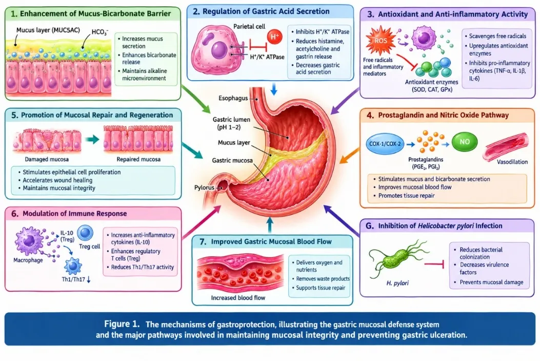
Figure 01: The mechanisms of gastroprotection, illustrating the gastric mucosal defence system and the major pathways involved in maintaining mucosal integrity and preventing gastric ulceration.
2.6. Experimental Models for Evaluating Gastroprotective Activity
Experimental models are essential for investigating the pharmacological activity of potential gastroprotective agents. These models provide information regarding the ability of test substances to prevent gastric mucosal injury, reduce ulcer severity, and promote gastric ulcer healing. Experimental evaluation generally includes the induction of gastric lesions, administration of the test substance, assessment of gastric injury, and biochemical or histopathological analysis. Different experimental models are selected according to the mechanism of gastric injury and the therapeutic objective of the investigation [12,13].
2.6.1. Ethanol-Induced Gastric Ulcer Model:
The ethanol-induced gastric ulcer model is widely used for evaluating the gastroprotective activity of medicinal plants and pharmacological agents. Ethanol produces acute gastric mucosal injury through direct epithelial damage, disruption of the mucus barrier, oxidative stress, and microvascular disturbances.
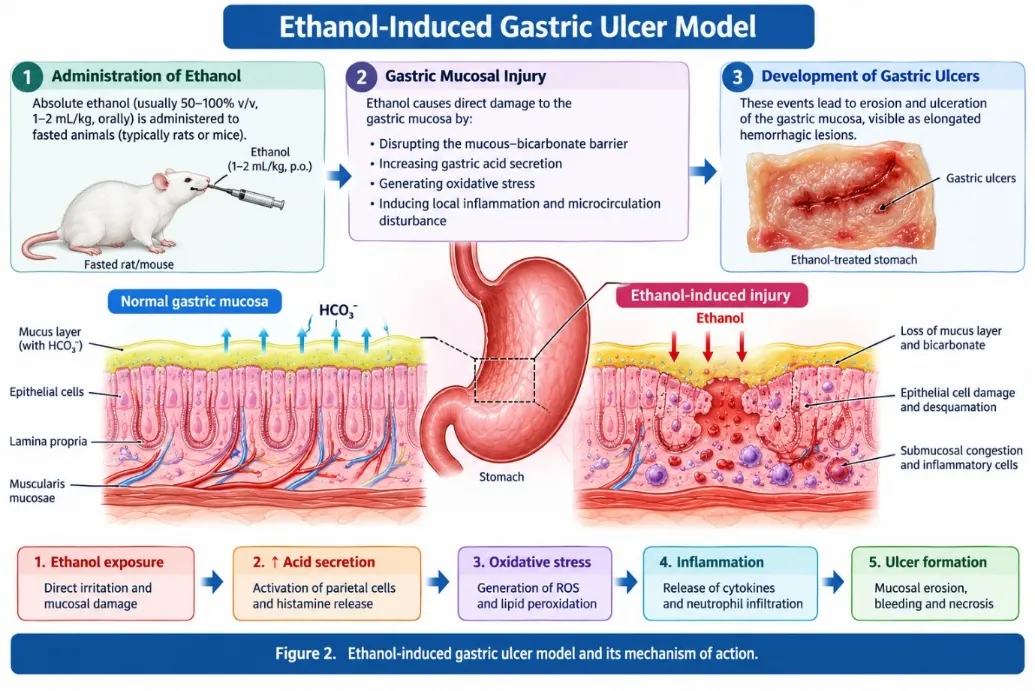
Figure 02: Ethanol-Induced Gastric Ulcer Model
The severity of gastric injury is commonly evaluated through macroscopic examination, ulcer scoring, biochemical assessment, and histopathological analysis. This model is particularly useful for investigating cytoprotective and antioxidant effects [11,12].
2.6.2. NSAID-Induced Gastric Ulcer Model:
NSAID-induced gastric ulcer models are used to investigate gastric injury associated with the inhibition of prostaglandin synthesis.
Indomethacin and aspirin are commonly used ulcerogenic agents in experimental studies. Their administration reduces prostaglandin-mediated mucosal protection and may result in gastric lesions. The gastroprotective activity of test substances can be evaluated by assessing ulcer severity, gastric acidity, mucus content, oxidative stress markers, and histopathological changes.
These models are relevant for investigating agents that enhance mucosal defense or reduce NSAID-associated gastric injury [7,9,13].
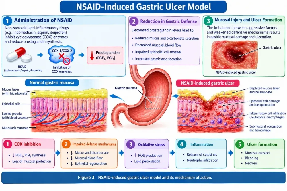
Figure 03: NSAID-Induced Gastric Ulcer Model
2.6.3. Pylorus-Ligation-Induced Gastric Ulcer Model:
The pylorus-ligation model involves surgical ligation of the pyloric region of the stomach, resulting in the accumulation of gastric secretions.
The accumulated gastric juice contributes to gastric injury through acid-pepsin-mediated mechanisms.
Commonly evaluated parameters include:
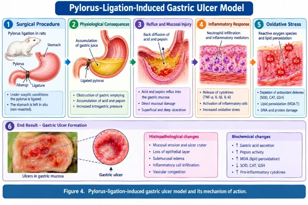
Figure 04: Pylorus-Ligation-Induced Gastric Ulcer Model
This model is particularly useful for evaluating the effects of test substances on gastric acid secretion and ulcer formation [12,13].
2.6.4. Stress-Induced Gastric Ulcer Model:
Stress-induced gastric ulcer models are used to investigate gastric mucosal injury associated with physiological stress. Experimental stress may produce gastric lesions through alterations in mucosal blood flow, autonomic activity, oxidative stress, and inflammatory responses.
Depending on the study design, stress may be induced through restraint, water immersion, or other validated experimental procedures. The severity of gastric lesions and associated biochemical changes are assessed to determine the gastroprotective activity of test substances [12,13].
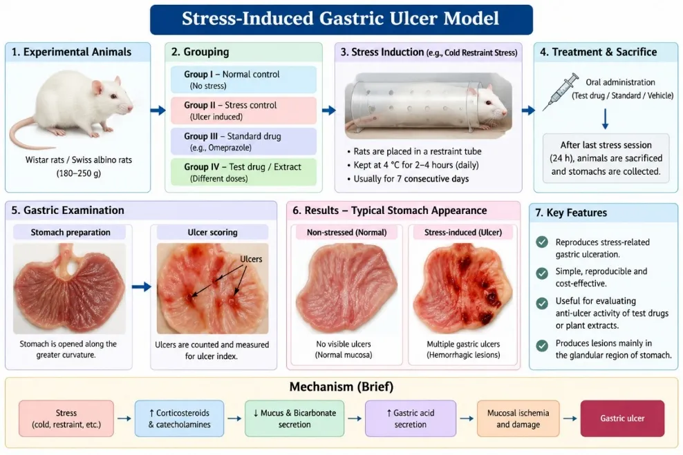
Figure 05: Stress-Induced Gastric Ulcer Model.
2.6.5. Evaluation Parameters in Experimental Gastroprotection:
Table 01: Experimental Parameters Used for Evaluating Gastroprotective Activity
| Evaluation parameter | Scientific significance |
|---|---|
| Ulcer index | Quantifies the severity of gastric lesions |
| Percentage ulcer inhibition | Determines the reduction in ulcer formation compared with the ulcer control group |
| Gastric juice volume | Evaluates gastric secretory activity |
| Gastric pH | Determines the acidity of gastric contents |
| Free acidity | Measures the concentration of free hydrochloric acid |
| Total acidity | Estimates the total titratable acidity of gastric juice |
| Gastric mucus content | Evaluates the mucosal protective barrier |
| Malondialdehyde (MDA) | Indicates lipid peroxidation and oxidative damage |
| Superoxide dismutase (SOD) | Evaluates antioxidant defense |
| Catalase (CAT) | Assesses hydrogen peroxide detoxification |
| Reduced glutathione (GSH) | Indicates intracellular antioxidant status |
| Histopathological examination | Evaluates mucosal damage, inflammation, and tissue repair |
The percentage inhibition of ulcer formation may be calculated using the following equation:
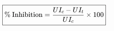
Where:
A higher percentage of ulcer inhibition indicates a greater reduction in ulcer severity relative to the ulcer control group.
3. BOTANICAL AND PHARMACOGNOSTIC PROFILE OF SELECTED MEDICINAL PLANTS:
This section provides a brief overview of the botanical classification, geographical distribution, botanical characteristics, and traditional medicinal uses of Syzygium cumini and Bryophyllum pinnatum. Both plants are widely recognized in traditional medicine and contain phytochemicals that have been investigated for their pharmacological properties [14,15].
3.1. Botanical Profile of Syzygium cumini:
Syzygium cumini (L.) Skeels, commonly known as jamun, Indian blackberry, or black plum, is an evergreen tree belonging to the family Myrtaceae. The plant is native to the Indian subcontinent and is widely distributed throughout tropical and subtropical regions. Different parts of the plant, including leaves, bark, fruits, and seeds, have been traditionally used for medicinal purposes [14].
3.1.1. Taxonomy and Botanical Classification:
Table 02: Taxonomy of Syzygium Cumini (L.)
| Classification | Description |
|---|---|
| Kingdom | Plantae |
| Family | Myrtaceae |
| Genus | Syzygium |
| Species | Syzygium cumini (L.) Skeels |
| Common name | Jamun, Indian blackberry |
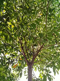
Figure 06: Syzygium cumini (L.) the Accepted Botanical Name Is Syzygium cumini (L.) Skeels, Belonging to the Family Myrtaceae [14,15].
3.1.2. Botanical Description and Geographical Distribution
Syzygium cumini is a large evergreen tree with a dense crown, greyish-brown bark, simple leathery leaves, and small white flowers. Its fruits are fleshy berries that become dark purple to nearly black when ripe. The plant is native to tropical and subtropical Asia and is widely cultivated in India and other tropical regions [14,15].
3.1.3. Traditional and Ethnomedicinal Uses:
Different parts of S. cumini, including leaves, bark, seeds, and fruits, have been traditionally used for the management of diabetes, dysentery, and gastrointestinal complaints. The plant is also reported to possess antioxidant, anti-inflammatory, and antimicrobial properties. These traditional uses have encouraged investigations into its potential pharmacological applications [15,16].
3.2. Botanical Profile of Bryophyllum pinnatum.
Bryophyllum pinnatum (Lam.) Oken, commonly known as the miracle leaf plant, air plant, or Patharchatta, is a succulent perennial herb belonging to the family Crassulaceae. It is characterized by its fleshy leaves, which can produce adventitious plantlets along their margins. The plant is widely distributed in tropical and subtropical regions and is used in traditional medicine for various ailments [17].
3.2.1. Taxonomy and Botanical Classification:
Table 03: Taxonomy of Bryophyllum Pinnatum
| Classification | Description |
|---|---|
| Kingdom | Plantae |
| Family | Crassulaceae |
| Genus | Bryophyllum |
| Species | Bryophyllum pinnatum (Lam.) Oken |
| Common name | Miracle leaf, Patharchatta |
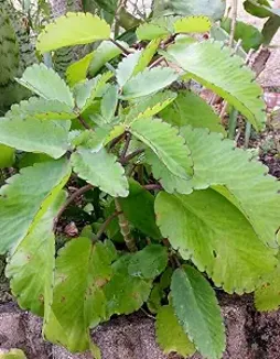
Figure 07: Kalanchoe pinnata. the Plant Is Also Known by the Synonym Kalanchoe pinnata (Lam.) Pers. and Is Recognized for Its Characteristic Vegetative Propagation Through Leaf-Margin Plantlets [17,18].
3.2.2. Botanical Description and Geographical Distribution
Bryophyllum pinnatum is a succulent herb generally reaching 40–150 cm in height. It possesses fleshy, pinnately compound leaves with crenate margins, tubular flowers, and reddish-purple corollas. The plant is native to Madagascar and is widely naturalized in tropical and subtropical regions, including India [17,18].
3.2.3. Traditional and Ethnomedicinal Uses
The leaves of B. pinnatum are traditionally used for the management of wounds, inflammation, urinary disorders, fever, and gastrointestinal complaints. Its medicinal properties are attributed to various phytochemicals, including flavonoids, phenolic compounds, bufadienolides, and polysaccharides. Reported pharmacological activities include antioxidant, anti-inflammatory, antimicrobial, and antiulcer effects [18].
4. PHYTOCHEMICAL CONSTITUENTS AND PHARMACOLOGICAL PROPERTIES:
The phytochemical composition of Syzygium cumini and Bryophyllum pinnatum varies according to the plant part, geographical origin, extraction solvent, and processing conditions. Both plants contain bioactive constituents associated with antioxidant, anti-inflammatory, and gastroprotective effects. The major reported phytochemicals and pharmacological properties of both plants are summarized in the following comparative table. [16,18–20]
Table 04: Phytochemical Constituents and Pharmacological Properties
| Plant | Major phytochemical constituents | Reported pharmacological properties | Potential relevance to gastroprotection |
|---|---|---|---|
| Syzygium cumini L. | Leaves: quercetin, myricetin, myricitrin, flavonol glycosides, tannins, and terpenoids. Seeds: gallic acid, ellagic acid, quercetin, hydrolysable tannins, and phytosterols. Fruits: anthocyanins, flavonoids, phenolic acids, and other polyphenols. [15,16,19] | Antioxidant, anti-inflammatory, antidiabetic, antimicrobial, antidiarrheal, and reported anti-ulcer activity. Seed and tannin-rich extracts have shown gastroprotective effects in experimental ulcer models. [16,19,20] | Phenolic compounds and tannins may reduce oxidative injury and support mucosal protection. Reported anti-ulcer effects suggest potential to limit gastric mucosal damage, although the effect depends on extract type and experimental model. [16,19,20] |
| Bryophyllum pinnatum (Lam.) Oken | Flavonoids such as quercetin, kaempferol, apigenin, and rutin; phenolic compounds; bufadienolides; terpenoids; tannins; organic acids; and other secondary metabolites. [18,21] | Antioxidant, anti-inflammatory, antimicrobial, wound-healing, and reported gastroprotective/anti-ulcer activity. Experimental studies have also reported gastric ulcer healing and protection from mucosal injury. [6,18,21] | Flavonoids and phenolic constituents may contribute to antioxidant and anti-inflammatory effects, while reported gastric protection and ulcer-healing activity support further evaluation of the plant in experimental gastroprotection. [6,18,21] |
5. MECHANISTIC INSIGHTS INTO GASTROPROTECTION:
5.1. Modulation of Gastric Acid Secretion:
Gastric acid secretion is regulated by histamine, gastrin, and acetylcholine through activation of parietal cells. Excessive acid secretion may contribute to gastric mucosal injury. Gastroprotective agents may reduce gastric acidity by inhibiting acid secretion or maintaining the balance between aggressive and defensive factors. Experimental findings with B. pinnatum suggest that its aqueous extract may reduce gastric volume and free and total acidity. [22,23]
5.2. Enhancement of Gastric Mucosal Defense:
The gastric mucosal barrier protects the stomach against acid, pepsin, and other damaging agents. It consists of epithelial cells, mucus, bicarbonate, and mucosal blood flow. Plant-derived bioactive constituents may strengthen this barrier by preserving epithelial integrity and reducing mucosal damage. Studies on B. pinnatum have demonstrated protective effects against experimentally induced gastric lesions. [24,25]
5.3. Regulation of Oxidative Stress and Antioxidant Enzymes:
Oxidative stress plays an important role in gastric ulcer development through the excessive generation of reactive oxygen species (ROS). Antioxidant mechanisms involving superoxide dismutase (SOD), catalase (CAT), glutathione (GSH), and reduced lipid peroxidation may help preserve gastric tissue integrity. S. cumini and B. pinnatum contain antioxidant phytoconstituents that may contribute to gastroprotection. [26,27]
5.4. Modulation of Inflammatory Mediators:
Inflammatory mediators such as tumor necrosis factor-alpha (TNF-α), interleukin-1 beta (IL-1β), and nuclear factor-kappa B (NF-κB) contribute to gastric mucosal inflammation. Suppression of excessive inflammatory responses may reduce tissue damage and support ulcer healing. Experimental investigations of B. pinnatum have demonstrated reductions in inflammatory markers in gastric ulcer models. [28,29]
5.5. Stimulation of Mucus and Prostaglandin Production:
Gastric mucus and bicarbonate form an essential protective layer against gastric acid and pepsin. Prostaglandins, particularly PGE₂, contribute to mucus secretion, bicarbonate production, and mucosal blood flow. Enhancement of these defensive mechanisms may improve gastric mucosal resistance. The protective effects of plant extracts may involve preservation of mucus content and mucosal integrity. [30,31]
5.6. Promotion of Gastric Tissue Repair and Healing:
Gastric ulcer healing involves epithelial regeneration, angiogenesis, cellular migration, and restoration of the mucosal architecture. Antioxidant and anti-inflammatory constituents may facilitate these processes by reducing tissue injury and supporting cellular repair. Studies involving B. pinnatum leaf extract have reported improvements in gastric ulcer healing in experimental chronic ulcer models. [32,33]
5.7. Proposed Molecular Mechanisms of Gastroprotection
The proposed molecular mechanisms of gastroprotection include antioxidant activity, modulation of inflammatory signaling, preservation of mucosal defense, and regulation of gastric acid secretion. Flavonoids and phenolic compounds may contribute to these effects through interactions with oxidative and inflammatory pathways. However, the precise molecular targets of the combined S. cumini and B. pinnatum extracts require further experimental validation. [34,35]
6. PROSPECTS FOR COMBINED THERAPY USING SYZYGIUM CUMINI AND BRYOPHYLLUM PINNATUM:
6.1. Rationale for Combined Herbal Therapy:
The combination of S. cumini and B. pinnatum may provide a broader gastroprotective effect by targeting multiple mechanisms involved in gastric ulcer development. Their potential antioxidant, anti-inflammatory, and mucosal protective activities provide a scientific rationale for investigating combined therapy. [36]
6.2. Complementary Phytochemical Profiles
S. cumini contains flavonoids, phenolic acids, tannins, and other bioactive constituents, whereas B. pinnatum contains flavonoids, phenolic compounds, and bufadienolides. These different phytochemical profiles may provide complementary biological activities. However, the composition of each extract depends on the plant part, extraction solvent, and processing conditions. [37]
6.3. Potential Additive and Synergistic Gastroprotective Effects:
The combined extracts may produce additive or synergistic effects by influencing different gastroprotective pathways. For example, one extract may primarily contribute antioxidant activity, while the other may enhance mucosal protection or reduce inflammation. Nevertheless, synergy must be demonstrated experimentally using appropriate combination studies rather than assumed from individual plant activities. [38]
6.4. Possible Interactions Between Bioactive Constituents
Interactions between flavonoids, phenolic compounds, tannins, and other phytoconstituents may influence antioxidant activity, absorption, metabolism, and biological effectiveness. Such interactions may enhance or reduce the overall pharmacological activity of combined extracts. Therefore, phytochemical profiling and interaction studies are essential for evaluating the safety and efficacy of the proposed combination. [37,38]
6.5. Evidence Supporting Combined Gastroprotective Therapy:
Preclinical studies have reported gastroprotective and ulcer-healing activities of B. pinnatum, while S. cumini has demonstrated anti-inflammatory and anti-ulcer effects in experimental models. These findings support the investigation of the two plants as a potential combination. However, direct experimental evidence establishing the efficacy of their combined extracts remains necessary. [22,24&28]
6.6. Challenges in Developing Combined Plant Extract Formulations:
The development of combined herbal formulations may be affected by variations in phytochemical composition, extraction yield, stability, solubility, and bioavailability. Differences in plant source and extraction methods may also influence reproducibility. Standardization of both extracts is therefore essential for developing a consistent and effective formulation. [37,39]
6.7. Future Experimental Approaches for Evaluating Combination Efficacy:
Future studies should evaluate individual extracts and their combinations using appropriate gastric ulcer models. Different combination ratios should be investigated to determine the optimal dose and potential synergistic effects. Ulcer index, percentage protection, gastric acidity, mucus content, oxidative stress markers, inflammatory mediators, and histopathological changes may be assessed. [38,40]
7. COMPARATIVE EVALUATION OF EXISTING EVIDENCE:
7.1. Comparison of Individual Gastroprotective Activities:
Existing experimental studies indicate that B. pinnatum possesses gastroprotective and ulcer-healing properties. S. cumini has also demonstrated anti-inflammatory and antioxidant effects in gastric ulcer models. However, differences in experimental design and extract composition limit direct comparison between the two plants. [22,24,28]
7.2. Comparison of Phytochemical Constituents and Mechanisms
Both plants contain phytoconstituents associated with antioxidant and anti-inflammatory activities. S. cumini is characterized by phenolic compounds and flavonoids, whereas B. pinnatum contains several flavonoid glycosides. Their different chemical profiles may contribute to complementary gastroprotective mechanisms. [26,27,37]
7.3. Comparison of Experimental Models and Study Outcomes:
Experimental investigations have employed ethanol-induced, indomethacin-induced, and acetic acid-induced gastric ulcer models. These models represent different mechanisms of gastric injury, including oxidative damage, impaired mucosal defense, and chronic ulceration. Differences in animal species, dose, treatment duration, and outcome measures should be considered when interpreting results. [22,24,28]
7.4. Strengths and Limitations of Available Evidence
The available evidence includes experimental evaluation of gastric lesions, biochemical markers, and histopathological changes. These findings provide preliminary support for the gastroprotective potential of the plants. However, limitations include variation in extract preparation, limited mechanistic validation, and insufficient clinical evidence. [24,28,39]
7.5. Evidence Gaps in Combined Therapy
Although individual gastroprotective activities have been reported, the combined efficacy of S. cumini and B. pinnatum requires systematic investigation. Important research gaps include optimal combination ratios, dose-dependent effects, phytochemical interactions, toxicity, and reproducibility of the formulation. [38,40]
8. SAFETY, TOXICITY, AND TRANSLATIONAL CONSIDERATIONS:
8.1. Acute and Subacute Toxicity Studies
Toxicity studies are essential to establish the safety of herbal extracts before pharmacological evaluation. Acute toxicity studies assess immediate adverse effects following single-dose administration, whereas subacute toxicity studies investigate the effects of repeated exposure. The combined extracts should be evaluated for behavioral, biochemical, and histopathological changes. [39,40]
8.2. Dose Selection and Therapeutic Window:
Appropriate dose selection is necessary to determine the effective and safe concentration of the combined extracts. Dose-response studies should be conducted using individual and combined treatments. The therapeutic window should be established by comparing pharmacological efficacy with potential toxic effects. [38,40]
8.3. Potential Adverse Effects and Herb–Drug Interactions:
Herbal extracts may interact with conventional medicines through pharmacodynamic or pharmacokinetic mechanisms. Such interactions may influence drug absorption, metabolism, or therapeutic effects. Therefore, the combined extracts should be investigated for potential interactions with commonly used anti-ulcer agents and other medications. [39,40]
8.4. Standardization and Quality Control of Plant Extracts:
Standardization is necessary to ensure the identity, purity, quality, and reproducibility of herbal extracts. Parameters such as botanical authentication, extraction yield, physicochemical properties, phytochemical profiles, and marker compound content should be evaluated. Chromatographic techniques such as TLC, HPTLC, and HPLC may support quality control. [37,39]
8.5. Challenges in Translating Preclinical Findings into Clinical Applications
The translation of preclinical findings into clinical applications requires confirmation of safety, efficacy, dosage, and bioavailability in humans. Variability in plant composition, differences in metabolism, and the lack of standardized formulations may limit clinical translation. Further pharmacological and clinical investigations are therefore required. [39,40]
9. RESEARCH GAPS AND FUTURE PERSPECTIVES:
9.1. Limitations of Existing Preclinical Research
Existing research is primarily based on experimental animal models, and differences in extraction procedures, dose selection, and experimental protocols limit the reproducibility of findings. Further standardized investigations are required to establish the gastroprotective potential of the individual and combined extracts. [24,28]
9.2. Need for Phytochemical Standardization:
Phytochemical standardization is essential to maintain consistency between batches of plant extracts. Future studies should identify suitable chemical markers and establish validated analytical methods for quantifying important bioactive constituents of both plants. [37,39]
9.3. Identification of Active Gastroprotective Constituents:
The identification of active constituents may help clarify the pharmacological mechanisms responsible for gastroprotection. Bioassay-guided fractionation, chromatographic analysis, and structural characterization may be used to identify compounds associated with the observed biological activities. [27,37]
9.4. Investigation of Molecular Targets and Signalling Pathways:
Future research should investigate molecular pathways associated with oxidative stress, inflammation, mucosal defence, and tissue repair. Important targets may include NF-κB, Nrf2, COX-2, antioxidant enzymes, and inflammatory cytokines. These investigations may provide a better understanding of the mechanisms underlying gastroprotective activity. [28,34]
9.5. Need for Combination Studies and Synergy Assessment:
Direct combination studies are required to establish whether S. cumini and B. pinnatum produce additive, synergistic, or antagonistic effects. Appropriate experimental designs, including dose-response analysis and combination-index approaches, may help determine the optimal extract ratio. [38,40]
9.6. Formulation Development and Bioavailability Enhancement:
Formulation development may improve the stability, solubility, and bioavailability of the combined extracts. Conventional herbal formulations, capsules, suspensions, and suitable gastroretentive systems may be investigated. Formulation optimization should be supported by physicochemical and pharmacological evaluation. [39,40]
9.7. Prospects for Clinical Evaluation
Following adequate preclinical evaluation, standardized combined extracts may be considered for further clinical investigation. Clinical studies should assess safety, tolerability, dose, efficacy, and potential interactions with conventional anti-ulcer medicines. The development of evidence-based herbal gastroprotective formulations may provide an additional research direction for managing gastric mucosal injury. [39,40]
CONCLUSION:
The present review highlights the gastroprotective potential of Syzygium cumini and Bryophyllum pinnatum, which contain various bioactive phytoconstituents with antioxidant, anti-inflammatory, and mucosal protective properties. These plants may contribute to gastric mucosal defense by regulating oxidative stress, inflammatory responses, gastric acid secretion, and tissue repair mechanisms. Their complementary phytochemical profiles provide a scientific rationale for investigating their combined therapeutic potential in gastric ulcer management. However, the efficacy and safety of the combined extracts remain to be established through systematic experimental investigations. Future research should focus on phytochemical standardization, dose optimization, toxicity assessment, molecular mechanism elucidation, and evaluation of possible synergistic effects. Comprehensive preclinical and clinical studies are essential to validate the therapeutic potential of this herbal combination and support the development of a standardized, safe, and effective gastroprotective formulation.
REFERENCES

Sakshi Pise, Saloni Talekar, Shabnam Mulani, Harshada Wadile, Dr. Pravin Utekar, Gastroprotective Potential of Syzygium Cumini and Bryophyllum Pinnatum: A Comprehensive Review of Phytochemistry, Pharmacological Evidence, and Prospects for Combined Therapy., Int. J. of Pharm. Sci., 2026, Vol 4, Issue 11, 1062-1081. https://doi.org/10.5281/zenodo.23972950
 10.5281/zenodo.23972950