

We use cookies to ensure our website works properly and to personalise your experience. Cookies policy
1,2MBBS Third Year, Department of Pharmacology, Fergana Medical Institute of Public Health, Uzbekistan.
3Assistant Professor Department Of Pharmacology, Fergana Medical Institute of Public Health, Uzbekistan.
T lymphocytes must change their metabolic wiring to survive, expand, differentiate and persist, and the quality of that wiring increasingly appears to be a determinant of immune outcome rather than a passive by-product of activation. Naive cells rely on efficient mitochondrial oxidation, whereas activated effector cells switch to a program dominated by aerobic glycolysis, amino acid uptake and biosynthesis, and memory and regulatory subsets return to a more oxidative, lipid-supported state. Mitochondria sit at the centre of this circuit. They supply energy and biosynthetic intermediates, generate signalling reactive oxygen species, and provide substrates, such as acetyl-CoA, alpha-ketoglutarate and S-adenosylmethionine, that are used by chromatin-modifying enzymes. When mitochondrial mass, dynamics or respiratory capacity deteriorate under chronic antigen exposure, nutrient competition, hypoxia or inflammation, T cells lose fitness and enter dysfunctional states, including exhaustion and senescence. In parallel, epigenetic remodelling fixes these states and limits the durability of therapeutic reinvigoration. This review integrates current evidence on T-cell metabolic programs, mitochondrial function and failure, the metabolic-epigenetic interface, and the signalling nodes (mTOR, AMPK, HIF-1, Myc and PD-1) that coordinate them. We then discuss how these mechanisms shape autoimmune disease and tumour immunity, and we critically appraise therapeutic strategies, including metabolic inhibitors, mitochondrial enhancers, nutrient supplementation and engineering of cellular therapies. Finally, we outline outstanding challenges, including metabolic heterogeneity, systemic toxicity, translational gaps and the need for single-cell metabolic profiling.
The adaptive immune response is unusual among physiological processes because it demands an abrupt and very large change in cellular behaviour. A single antigen-specific T cell can divide many times within days, produce cytokines and cytotoxic molecules in large quantities, migrate through tissues of very different nutrient composition, and then, after the threat has resolved, contract into a small, long-lived memory pool. Each of these steps has an energetic and biosynthetic cost, and over the past two decades it has become clear that the cell's metabolic program is not simply a consequence of its functional state but one of the instructions that creates it [1,2]. This realisation gave rise to the field of immunometabolism, which examines how nutrient availability, metabolic enzymes and metabolites regulate immune cell fate and function [3,4].
Mitochondria have emerged as a particularly important organising centre within this field. Beyond oxidative phosphorylation (OXPHOS), these organelles control calcium buffering, redox balance, apoptosis, nucleotide and amino acid synthesis, and the generation of metabolites that act as cofactors for epigenetic enzymes [5]. T cells with impaired mitochondria generally fail to proliferate appropriately, show defective cytokine production and, under chronic stimulation, adopt the transcriptional and functional phenotype of exhaustion, a state in which cells retain antigen specificity but lose effector capacity [6]. More recent work shows that the exhausted state is also reinforced at the chromatin level, which explains why checkpoint blockade often produces only partial or temporary functional recovery [7,8].
These observations have clinical relevance in two opposing contexts. In autoimmune and chronic inflammatory diseases, T cells display metabolic features that favour excessive activation, survival or pathogenic differentiation, and correcting these features can suppress disease in experimental models [9,10]. In cancer, the opposite problem predominates: tumour-infiltrating T cells face glucose deprivation, hypoxia, acidity and lipid overload that suppress their mitochondrial function and drive dysfunction [11,12]. A growing therapeutic literature now attempts to exploit these vulnerabilities, either by dampening inappropriate metabolism in autoimmunity or by restoring metabolic fitness in anti-tumour cells [13,14].
The aim of this review is to provide an integrated account of T-cell immunometabolism that connects three themes that are often treated separately: metabolic reprogramming during activation and differentiation, mitochondrial dysfunction as a cause of T-cell failure, and epigenetic control as both an output and a regulator of metabolism. We then consider how these principles are manifested in autoimmune disease and cancer, review the therapeutic strategies under development, and discuss limitations that must be addressed before clinical translation can be achieved.
2. Overview of T-Cell Metabolism
2.1 Quiescent naive T cells
Naive T cells circulate in a resting state with low biosynthetic demands. They take up modest amounts of glucose, amino acids and fatty acids and generate most of their ATP by oxidising these substrates in the mitochondria through the tricarboxylic acid (TCA) cycle and the electron transport chain [15,16]. Quiescence is not metabolic inactivity, however. It is an actively maintained state that depends on tonic cytokine signals such as interleukin-7 (IL-7) and low-level T-cell receptor (TCR) contact with self-peptide/MHC, which together sustain glucose transporter expression, mitochondrial integrity and survival [16,17]. Efficient oxidative metabolism in this state keeps the cell energetically stable while preserving the capacity to respond rapidly to antigen.
2.2 Nutrient uptake as a gatekeeper of activation
The first major change after antigen recognition is a rise in nutrient import. Co-stimulation through CD28 activates the phosphoinositide 3-kinase (PI3K)-Akt pathway and increases surface expression of the glucose transporter GLUT1, thereby linking the second activating signal to glucose uptake [18,19]. Studies using T-cell-specific deletion of GLUT1 showed that glucose import is essential for effector CD4+ T-cell expansion and function, while having comparatively little effect on regulatory T-cell (Treg) development [20]. Amino acid transporters are induced with similar speed. The large neutral amino acid transporter system and the glutamine transporter ASCT2 are upregulated following TCR stimulation, supplying leucine and glutamine that are needed both for protein synthesis and for activation of mechanistic target of rapamycin complex 1 (mTORC1) [21,22].
2.3 Metabolic signatures of T-cell subsets
Different T-cell lineages adopt characteristic, though not exclusive, metabolic programs. Effector CD4+ subsets such as T helper 1 (Th1) and Th17 cells show high rates of glycolysis, and their differentiation can be shifted by manipulating glycolytic flux [23,24]. A central mediator is hypoxia-inducible factor 1 alpha (HIF-1α), which promotes Th17 differentiation and suppresses Foxp3 stability [24,25]. Tregs, in contrast, rely more heavily on oxidative metabolism, including fatty acid oxidation, and show lower glycolytic dependence in resting conditions [26,27]. Nevertheless, activated Tregs also upregulate glycolysis and mTORC1 activity in order to proliferate and exert suppression, which indicates that these programs are context dependent rather than fixed [28,29]. In addition, mitochondrial complex III activity is required for the suppressive function of Tregs, demonstrating that oxidative capacity is not only a survival feature but a functional requirement [30].
Memory CD8+ T cells are distinguished by a larger mitochondrial mass, greater spare respiratory capacity and a dependence on fatty acid oxidation (FAO) [31,32]. Importantly, the fatty acids they oxidise are partly generated through cell-intrinsic lipolysis, which maintains a mitochondrial fuel supply even when external lipids are limited [33]. This oxidative reserve helps memory cells survive, but it is not their only metabolic feature: they can also rapidly re-engage glycolysis upon re-stimulation, enabling immediate production of effector molecules such as interferon gamma (IFN-γ) [34].
2.4 Amino acids and one-carbon metabolism
Glucose and lipids are not the only determinants of T-cell function. Glutamine supports the TCA cycle, nucleotide synthesis and glutathione production, and its transport and catabolism are coordinated with MAPK/ERK signalling during activation [35,36]. Glutaminase-dependent metabolism differentially regulates Th1 and Th17 differentiation [37]. Serine provides one-carbon units for purine synthesis and is required for the expansion of effector T cells [38], while arginine availability influences survival and anti-tumour activity, partly by altering mitochondrial and metabolic gene expression [39]. Together these findings suggest that T-cell activation depends on a coordinated network of nutrient inputs and that depletion of any one may produce a distinct functional deficit.
3. Metabolic Reprogramming During T-Cell Activation
3.1 The glycolytic switch
Stimulation of a naive T cell triggers a transition that resembles the Warburg effect described in cancer cells, in which glucose is converted to lactate even when oxygen is available. This aerobic glycolysis is less efficient than complete oxidation in terms of ATP per glucose molecule, but it permits rapid carbon flux through biosynthetic pathways, including the pentose phosphate pathway, serine synthesis and the generation of NADPH, and it helps maintain redox balance in rapidly dividing cells [15,40]. The transcription factor Myc is a master regulator of this program. Following TCR engagement, Myc is rapidly induced and drives the expression of genes for glycolysis and glutaminolysis, and cells lacking Myc fail to undergo appropriate growth and proliferation [41].
The consequences of aerobic glycolysis extend beyond energy supply. Glyceraldehyde-3-phosphate dehydrogenase (GAPDH), when not engaged in glycolysis, can bind the 3′ untranslated region of IFN-γ mRNA and suppress its translation; high glycolytic flux engages GAPDH in its enzymatic role and thereby releases the transcript for translation [42]. In this way glycolysis is directly coupled to effector cytokine output. Early after stimulation, TCR signalling itself can induce glycolysis through mechanisms that precede large changes in gene expression, indicating that metabolic changes accompany, rather than merely follow, the earliest steps of activation [43]. Conversely, restricting glucose reduces IFN-γ production and mTOR-dependent signalling in effector CD8+ T cells [44].
3.2 Mitochondrial priming and the bioenergetic transition
Although glycolysis dominates the metabolic phenotype of activated T cells, mitochondrial function increases rather than decreases. CD28 co-stimulation primes mitochondria by promoting carnitine palmitoyltransferase 1a (CPT1a)-dependent fatty acid oxidation and remodelling of cristae, which enables the cell to switch quickly to OXPHOS when needed [45]. Mitochondrial biogenesis and proteome remodelling in the first 24 hours after stimulation support one-carbon metabolism, particularly serine-dependent pathways that supply nucleotides for division [46]. These findings overturn the earlier view that activated T cells simply switch from mitochondrial to glycolytic metabolism; instead they construct a larger and more specialised mitochondrial network in parallel with high glycolytic flux.
3.3 Metabolites as signalling and epigenetic effectors
Activated T cells also use metabolic intermediates as signals. Phosphoenolpyruvate (PEP), a glycolytic intermediate, sustains calcium–NFAT signalling by inhibiting sarco/endoplasmic reticulum calcium ATPase (SERCA) activity, and its depletion in glucose-poor tumours weakens TCR signalling [47]. Mitochondrial reactive oxygen species (ROS), generated at complex III, act as second messengers that are necessary for NFAT activation and IL-2 production [48]. Furthermore, glycolytic flux supports acetyl-CoA availability for histone acetylation at the Ifng locus, which connects metabolic state to chromatin accessibility and promotes Th1 differentiation [49]. These examples demonstrate that metabolic pathways in T cells function as information-processing systems and not merely as energy supplies.
3.4 Biosynthetic demands of proliferation
A proliferating T cell must double its mass, genome, lipid membranes and protein content within hours. Nucleotide synthesis draws on glutamine, serine, glycine and aspartate; lipid synthesis relies on citrate exported from mitochondria; and protein synthesis depends on amino acid sufficiency and mTORC1 activity [46,50]. Since each of these demands is met by different nutrients, the metabolic environment can constrain the response in a selective manner. For example, restricting one-carbon metabolism impairs proliferation without necessarily altering early activation markers, whereas glucose restriction impairs cytokine output more than division [38,44]. This partial independence of proliferation and effector function is relevant for therapeutic design because it suggests that inhibiting individual nodes can bias rather than abolish T-cell responses.
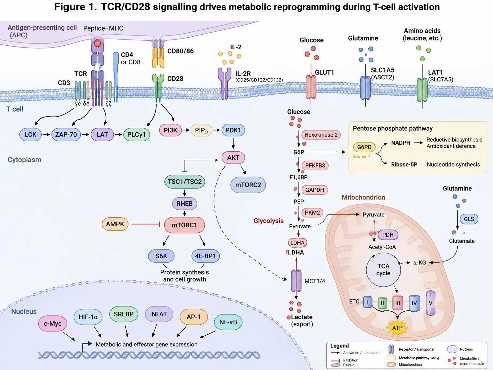
Figure 1. TCR/CD28 signalling drives metabolic reprogramming during T-cell activation. Antigen recognition (peptide–MHC/TCR–CD3) and co-stimulation (CD80/86–CD28) activate LCK–ZAP-70–LAT–PLCγ1 and PI3K–PDK1–AKT signalling, relieving TSC1/2 inhibition of RHEB and activating mTORC1 (opposed by AMPK). Upregulated GLUT1, ASCT2 and LAT1 fuel glycolysis (HK2, PFKFB3, GAPDH, PKM2, LDHA), the pentose phosphate pathway (G6PD), glutaminolysis (GLS) and mitochondrial TCA/ETC activity, while c-Myc, HIF-1α, SREBP, NFAT, AP-1 and NF-κB drive metabolic and effector gene expression.
4. Mitochondrial Function in T Cells
4.1 Mitochondria as biosynthetic and signalling hubs
The mitochondrion in a T cell does much more than produce ATP. The TCA cycle provides intermediates for amino acid, lipid and nucleotide synthesis, while the electron transport chain regenerates NAD+, which is required for aspartate synthesis and for sustained proliferation [5,46]. Complex IV (cytochrome c oxidase) activity acts as a checkpoint for activation and differentiation, as T cells with defective complex IV assembly show impaired activation, altered cytokine production and distinct fate decisions [51]. Mitochondrial respiration also supports lysosomal function and helps control inflammatory T-cell responses, linking organelle quality control to effector regulation [52].
4.2 Mitochondrial dynamics and cristae architecture
The shape of the mitochondrial network is a determinant of metabolic program and cell fate. Effector T cells display fragmented mitochondria with loose cristae that favour aerobic glycolysis, whereas memory T cells have fused networks with tight cristae that favour efficient electron transport chain assembly and OXPHOS [53]. Genetic or pharmacological manipulation of fusion and fission proteins, such as OPA1 and DRP1, can therefore bias T cells towards memory-like or effector-like states [53]. Thus mitochondrial morphology is not a passive feature of metabolic state but an upstream regulator of it.
4.3 Metabolite output and cell fate
Mitochondrial metabolism can influence T-cell differentiation independently of overall ATP production. In one model, the functional capacity of effector cells could be uncoupled from differentiation by manipulating specific aspects of mitochondrial metabolism, suggesting that distinct modes of mitochondrial activity control different aspects of T-cell programming [54]. Mitochondria-derived metabolites such as 2-hydroxyglutarate, which accumulates in activated CD8+ T cells, can alter histone and DNA methylation and regulate T-cell fate and anti-tumour capacity [55]. These findings introduce the idea that mitochondria act as an interface between metabolic state and gene regulation, a concept that we explore further in Section 6.
4.4 Mitochondrial quality, membrane potential and ageing
Mitochondrial quality control determines how well a T cell sustains function over time. Mitochondrial membrane potential has been used as a marker of cellular fitness: CD8+ T cells with low membrane potential exhibit stem-like properties and superior persistence and anti-tumour activity after transfer, even when glycolytic capacity is retained [56]. At the organismal level, T cells with defective mitochondria, caused by deletion of the mitochondrial transcription factor TFAM, can drive inflammatory ageing, tissue senescence and premature mortality in mice, which underlines the systemic effect that T-cell mitochondrial dysfunction may have [57]. These data suggest that mitochondrial integrity is important for the long-term health of both individual T cells and the host.
5. Mitochondrial Dysfunction and T-Cell Exhaustion
5.1 Exhaustion as a metabolic as well as a transcriptional state
Exhaustion arises when T cells are exposed to persistent antigen and inflammatory signals, as occurs in chronic viral infection and in tumours. It is characterised by sustained expression of inhibitory receptors, reduced cytokine production and impaired proliferative capacity [6]. Although it was initially defined by surface markers and transcriptional signatures, a series of studies has shown that metabolic failure is an early and probably causal feature. In a mouse model of chronic viral infection, antigen-specific CD8+ T cells showed reduced glucose uptake, decreased mitochondrial mass and altered mitochondrial polarisation at a time when functional decline was still partial, and these changes were linked to PD-1 signalling and downregulation of the coactivator PGC-1α [58]. This pattern, termed bioenergetic insufficiency, could be partially reversed by forced expression of PGC-1α, which improved mitochondrial function and effector capacity [58].
PD-1 itself appears to be a metabolic regulator. Engagement of PD-1 suppresses glycolysis and the amino acid-dependent metabolic changes that follow CD28 signalling, while increasing fatty acid oxidation and lipolysis, thereby shifting the cell towards a lower-output metabolic state [59]. Because CD28 is a principal target of PD-1 signalling, the receptor effectively cuts off the co-stimulatory signal that primes both glycolysis and mitochondria. In the tumour setting, repression of PGC-1α-driven mitochondrial biogenesis by persistent Akt signalling was shown to produce intratumoural T cells with low mitochondrial mass and reduced respiratory capacity, and enforcing PGC-1α expression partly restored function [12].
5.2 Defects in mitochondrial dynamics and self-renewal
More recent studies have refined the picture by showing that dysfunctional mitochondria in tumour-infiltrating lymphocytes (TILs) are not only low in number but also structurally abnormal. CD8+ TILs show disturbed mitochondrial dynamics, with altered fusion and fission, accumulation of damaged mitochondria and a transcriptional program that reinforces exhaustion; improving mitochondrial dynamics in these cells enhanced anti-tumour function [60]. Under conditions of persistent antigen, impaired OXPHOS has also been shown to limit the self-renewal of T cells and to cause accumulation of reductive stress and metabolic by-products that disrupt the cell's capacity to maintain stem-like precursors [61]. In human renal cell carcinoma, CD8+ T cells infiltrating the tumour exhibit mitochondrial dysregulation together with glycolytic insufficiency, and their dysfunction correlates with a depleted metabolic profile [62].
It is important to note that exhausted cells are not uniform. Virus-specific CD8+ T cells in the same host can show distinct metabolic dependencies depending on whether they remain functional or have become exhausted, with the exhausted fraction displaying depolarised mitochondria and reduced capacity to respond to metabolic stress [63]. Heterogeneity within the exhausted pool, including progenitor-like and terminally differentiated subsets, is therefore likely to be mirrored by metabolic heterogeneity, a point that has implications for interpreting bulk metabolic measurements.
5.3 Environmental drivers of mitochondrial failure
Several features of the tumour microenvironment (TME) converge on the T-cell mitochondrion. Hypoxia increases HIF-1 activity and, when combined with chronic stimulation, can promote effector genes in the short term while limiting oxidative capacity [64,65]. Lipid-rich environments also contribute. In melanoma, enhancing fatty acid catabolism in CD8+ T cells with a PPAR-alpha agonist improved their capacity to function in a metabolically challenging tumour and increased the efficacy of immunotherapy [66], whereas cholesterol accumulation in the TME induces expression of endoplasmic reticulum stress mediators and immune checkpoints and promotes exhaustion [67]. In pancreatic tumours, long-chain fatty acids accumulate and impair mitochondrial function in infiltrating CD8+ T cells by overwhelming their oxidation capacity [68]. These results emphasise that lipid availability may help or harm depending on the amount, the type and the cell's capacity to oxidise it.
An additional and unexpected mechanism of mitochondrial loss was recently reported: tumour cells can transfer mitochondria carrying mutated DNA to T cells through nanotube-like structures, and this transfer is associated with senescence-like features in the recipient lymphocytes [69]. Whether this process is common across tumour types remains to be established, but it illustrates the degree to which T-cell mitochondria are vulnerable to the surrounding cells.
Table 1: Metabolic and Mitochondrial Features of Major T-cell States.
| T-cell state | Dominant metabolic features | Mitochondrial characteristics | Key references |
|---|---|---|---|
| Naive | Low-rate OXPHOS; low biosynthesis; IL-7-dependent survival | Small, functional network; quiescent | [15,16] |
| Effector | Aerobic glycolysis; glutaminolysis; mTORC1 and Myc activity | Fragmented mitochondria, loose cristae; ROS signalling | [41,48,53] |
| Memory | FAO supported by lipolysis; high spare respiratory capacity; rapid glycolytic recall | Fused network, tight cristae, high mass | [31-34,53] |
| Regulatory | Oxidative metabolism; lactate tolerance; context-dependent glycolysis | Complex III required for suppression | [26-30] |
| Exhausted | Reduced glycolysis and OXPHOS; PD-1-linked metabolic restriction | Low mass, depolarisation, abnormal dynamics | [12,58-62] |
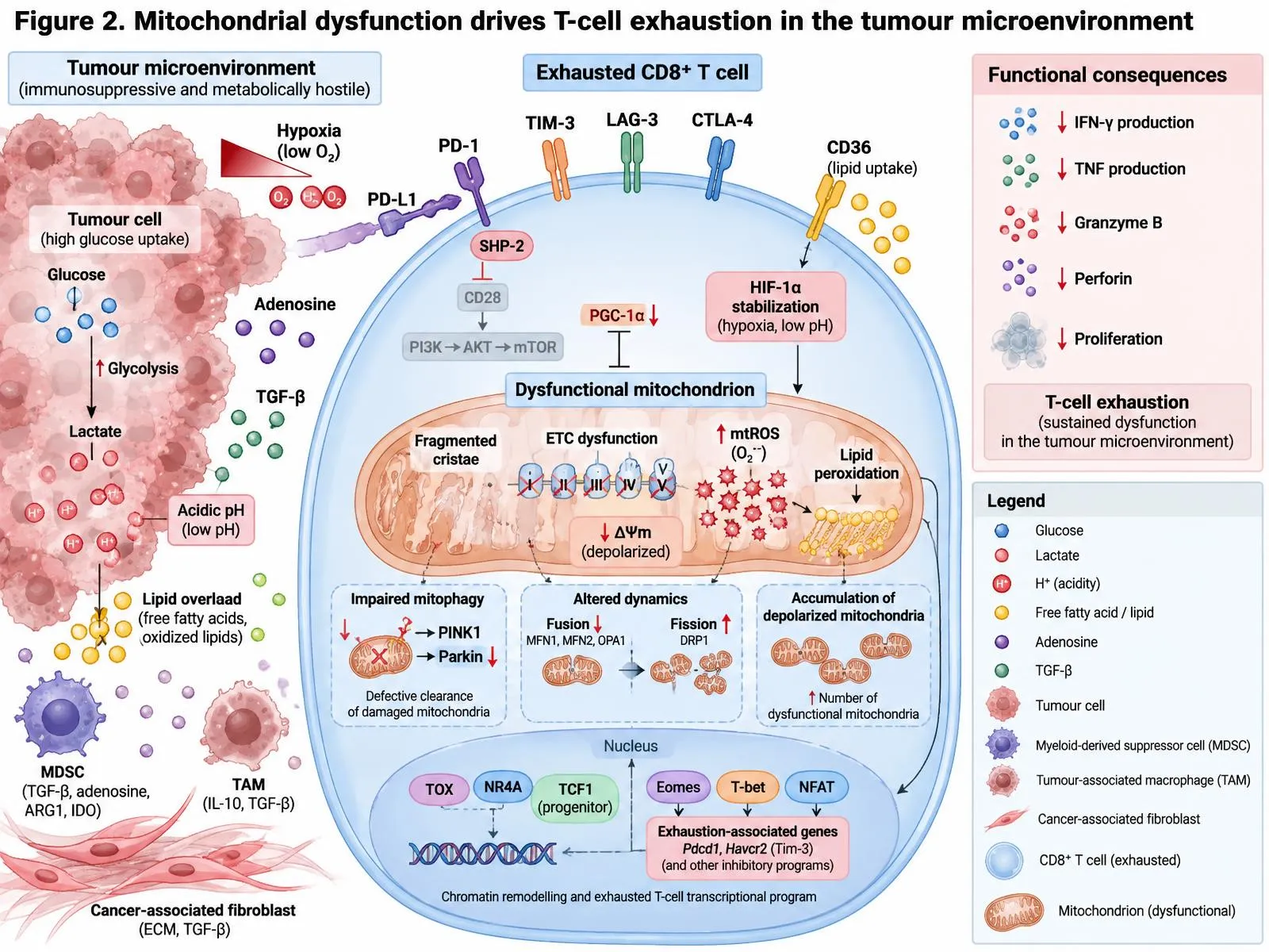
Figure 2. Mitochondrial dysfunction drives T-cell exhaustion in the tumour microenvironment. Hypoxia, glucose competition, lactate/H+ acidity, lipid overload, adenosine and TGF-β, together with PD-L1–PD-1/SHP-2 inhibition of CD28–PI3K–AKT–mTOR signalling and loss of PGC-1α, produce fragmented cristae, ETC failure, reduced ΔΨm, mtROS, lipid peroxidation, impaired PINK1/Parkin mitophagy and imbalanced fusion (MFN1/2, OPA1) versus fission (DRP1). TOX, NR4A, Eomes, T-bet and NFAT consolidate the exhaustion program, reducing IFN-γ, TNF, granzyme B, perforin and proliferation.
6. Epigenetic Regulation of T-Cell Metabolism
6.1 Metabolites as substrates and cofactors of chromatin enzymes
Chromatin-modifying enzymes depend on metabolites that are generated by the pathways described above, and their activity therefore reflects the cell's metabolic state [70]. Histone acetyltransferases use acetyl-CoA, which in many cells is generated by ATP-citrate lyase from citrate exported from mitochondria [71]. Histone and DNA methyltransferases use S-adenosylmethionine produced through one-carbon metabolism, while the Jumonji family of histone demethylases and TET enzymes of DNA demethylation require alpha-ketoglutarate and are inhibited by succinate, fumarate and 2-hydroxyglutarate [70,72]. More recently, a wider range of histone acylations, including crotonylation, succinylation and lactylation, has been linked to metabolic state [72]. These relationships indicate that mitochondrial and glycolytic fluxes directly influence the chromatin landscape and that metabolic reprogramming can be translated into stable transcriptional programs [73].
6.2 Evidence from T-cell activation and differentiation
Several T-cell examples illustrate this coupling. Aerobic glycolysis maintains lactate dehydrogenase A activity, which in Th1 cells keeps acetyl-CoA high and thereby promotes histone acetylation at the Ifng locus [49]. Memory CD8+ T cells maintain permissive chromatin at effector genes, marked by histone modifications that allow rapid transcription on re-challenge [74], and they require acetate-derived acetyl-CoA for optimal recall responses, particularly when glucose is limited [75,76]. Alpha-ketoglutarate-dependent demethylases are in turn influenced by 2-hydroxyglutarate, which promotes a memory-like phenotype with improved persistence and anti-tumour activity [55]. These findings suggest that the metabolites produced in the course of activation do not simply fuel the response but record it in chromatin.
6.3 Epigenetic fixation of exhaustion
The epigenetic landscape of exhausted T cells differs substantially from that of effector and memory cells and is remarkably stable. Genome-wide chromatin accessibility profiling showed that exhausted CD8+ T cells have a distinct open-chromatin signature, and that this signature is only minimally altered after PD-1 blockade, which explains why reinvigoration is frequently incomplete or short-lived [77,78]. Tumour-specific T cells pass through chromatin states, with an early plastic state that can be reprogrammed and a later fixed state that cannot, which provides a conceptual framework for the timing of interventions [79]. De novo DNA methylation programs also limit the rejuvenation capacity of PD-1-blocked cells, and pharmacological inhibition of DNA methyltransferases can improve responses [80]. At the PDCD1 locus, chronic infection leads to demethylation that maintains PD-1 expression even after antigen decline [81].
Transcription factors link this epigenetic imprinting with metabolism. TOX is induced by chronic stimulation and programs the exhausted state transcriptionally and epigenetically [82,83]. In parallel, polycomb repressive complex 2 (PRC2) restricts multipotency and drives terminal differentiation of effector CD8+ T cells [84]. Metabolic restriction can also act through epigenetic routes: in tumour models, glucose limitation reduces expression of the methyltransferase EZH2 through microRNA-dependent mechanisms and thereby weakens polyfunctionality [85].
6.4 A bidirectional relationship
The interaction between metabolism and the epigenome is bidirectional. Metabolic inputs shape the chromatin state; chromatin state in turn determines which metabolic genes can be expressed, including those controlling mitochondrial biogenesis and glycolysis. Exhausted cells that have lost mitochondrial fitness may therefore have restricted access to the metabolites required to remodel chromatin, creating a self-reinforcing loop [7,54]. This concept suggests that effective rescue may require simultaneous restoration of metabolic fitness and chromatin plasticity, rather than either approach alone, an idea that is discussed in the therapeutic section below.
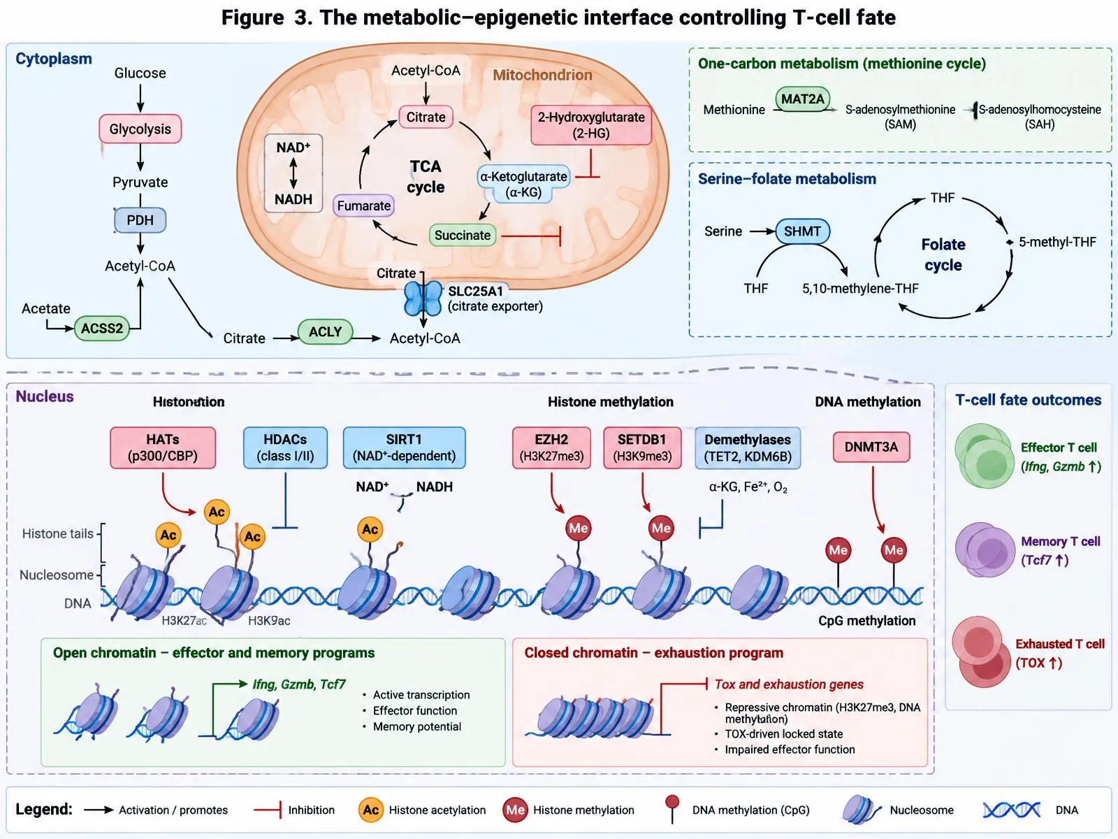
Figure 3: The Metabolic–Epigenetic Interface Controlling T-cell Fate. Acetyl-CoA (via PDH, SLC25a1–ACLY And ACSS2), Α-Ketoglutarate, Succinate, Fumarate, 2-Hydroxyglutarate, NAD+ and S-adenosylmethionine (MAT2a; Serine–Folate Cycle via SHMT) Act as Substrates or Cofactors for HATs (P300/CBP), HDACs, SIRT1, EZH2, SETDB1, DNMT3a and Α-Kg/Fe2+-Dependent Demethylases (TET2, KDM6B), Determining Open Chromatin at Effector/Memory Loci (Ifng, Gzmb, Tcf7) or a TOX-driven, Repressed Exhaustion State.
7. Metabolic Signaling Pathways in T Cells
7.1 mTOR
The serine/threonine kinase mTOR integrates growth factor, nutrient and energy signals and is probably the best studied regulator linking T-cell signalling to metabolism [86]. It operates in two complexes. mTORC1 responds to amino acids, particularly leucine and glutamine, and promotes glycolytic and lipid synthesis programs through HIF-1α and SREBP, whereas mTORC2 controls Akt activation and FOXO phosphorylation [87]. In the absence of mTOR, naive CD4+ T cells fail to differentiate into Th1, Th2 or Th17 lineages and instead acquire a Foxp3+ regulatory phenotype, indicating that mTOR activity is necessary for effector commitment [88]. The two complexes make different contributions: mTORC1 is required for Th1 and Th17 differentiation, while mTORC2 is needed for Th2 differentiation [89].
In CD8+ T cells, strong mTORC1 signalling favours terminal effector differentiation, while its partial inhibition promotes memory formation. Treatment with rapamycin during the expansion phase of the response enhanced the quality and number of memory CD8+ T cells, which suggests that mTOR acts as a rheostat between short-lived effector and long-lived memory fates [90,91]. In Tregs, mTORC1 signalling is required for their proliferation, lipid synthesis and suppressive capacity, even though high mTOR activity in conventional T cells tends to oppose regulatory differentiation [29]. The pathway is thus not simply pro-inflammatory, and its effect depends on the cell type and the magnitude and timing of signalling [92].
7.2 AMPK
AMP-activated protein kinase (AMPK) is an energy sensor activated by a high AMP/ATP ratio that restores energy homeostasis by promoting catabolic pathways and suppressing anabolic ones, including mTORC1 [93]. In T cells, AMPK is not strictly required for activation under replete conditions, but it becomes important when nutrients are limiting. T cells lacking AMPKα1 showed greater mTORC1 activity and glycolysis, together with elevated stress and impaired survival and effector function during infection in vivo, which indicates that AMPK restrains excessive anabolic activity and maintains metabolic adaptation [94]. Because tumour and chronically inflamed tissues are often nutrient poor, AMPK may be a particularly important buffer for T cells in these sites, although the optimal degree of activation for therapy is not clear.
7.3 HIF-1 and Myc
HIF-1α is stabilised by hypoxia but also by mTORC1 signalling and TCR stimulation under normal oxygen conditions. It promotes the expression of glycolytic enzymes and glucose transporters and, as noted above, favours Th17 over Treg differentiation [24,25]. In cytotoxic T cells, HIF activity enhances effector molecule expression and migration-related genes, and sustained HIF signalling can enhance responses to persistent antigen [64]. However, a HIF-1α and VEGF-A axis in CD8+ T cells also modulated tumour progression, showing that the pathway has complex effects in the TME [65]. Myc, as discussed in Section 3, works in parallel with HIF-1 to establish the glycolytic and glutaminolytic program of activated cells [41]. Both factors respond to signalling from the TCR and cytokines such as IL-2, and both can be limited by nutrient restriction, which explains in part why activated T cells are particularly sensitive to glucose and glutamine deprivation.
7.4 Co-stimulation, checkpoints and cross-talk
CD28 acts through PI3K and Akt to increase glucose uptake and mitochondrial priming [18,19,45], whereas PD-1 engagement opposes this signalling and shifts the cell to a low-output, fatty acid-oxidising state [59]. Mitochondrial ROS, produced downstream of TCR engagement, feed back to sustain NFAT activity [48], while metabolic stress activates AMPK to limit mTORC1 [93,94]. These pathways are interconnected: mTORC1 supports Myc and HIF-1α; HIF-1α and Myc increase glycolytic enzymes; AMPK and PD-1 counteract these effects. The balance between these inputs may be considered as a metabolic rheostat that decides whether a cell adopts a short-lived effector, long-lived memory, regulatory or exhausted fate [95,96]. This integrative model also illustrates why single-pathway inhibitors produce nuanced, context-dependent outcomes.
8. Immunometabolic Alterations in Autoimmune Diseases
8.1 General principles
In autoimmune disease, T cells receive repeated or inappropriately strong activating signals and exist in a tissue environment that is frequently inflammatory and hypoxic. As a result, they may adopt metabolic programs that favour sustained survival and pathogenic differentiation. Common features reported across diseases include increased glycolytic activity, altered mitochondrial function with excess oxidative stress, heightened mTOR signalling and an imbalance of Th17 and Treg cells [9,10]. Because these metabolic features are linked to disease-relevant functions, they have been proposed as therapeutic targets, although the same pathways are also needed by protective immune cells, which limits the therapeutic window.
8.2 Rheumatoid arthritis
Rheumatoid arthritis (RA) provides one of the clearest examples of a metabolic phenotype that departs from the expected pattern. Naive CD4+ T cells from patients with RA were found to have reduced expression of the glycolytic enzyme 6-phosphofructo-2-kinase/fructose-2,6-bisphosphatase 3 (PFKFB3) and lower glycolytic flux, together with reduced ATP and lactate production [97]. Instead of using glucose for energy, these cells diverted it towards the pentose phosphate pathway, producing excess NADPH and reducing intracellular ROS, which pushed them into a hyperproliferative state that favoured Th1 and Th17 differentiation and tissue invasion [97]. In this sense RA T cells are metabolically reprogrammed in a way that reduces redox signalling and promotes inflammation, a finding that cautions against assuming that autoimmunity simply reflects higher glycolysis. Subsequent work has characterised the early and late stages of the disease and described how metabolic features change with chronicity and ageing [9].
8.3 Systemic lupus erythematosus
Systemic lupus erythematosus (SLE) is characterised by mitochondrial abnormalities in T cells. Lupus T cells show mitochondrial hyperpolarisation and ATP depletion, which predispose them to necrosis rather than apoptosis and thereby increase exposure of nuclear antigens [98]. Oxidative stress, depletion of glutathione and activation of mTOR have been described as central features of the disease and as targets for treatment [99]. In patients, N-acetylcysteine, a precursor of glutathione, reduced disease activity by blocking mTOR in T cells [100], and rapamycin lowered disease activity and normalised T-cell calcium handling [101]. A later single-arm phase 1/2 trial of sirolimus in patients resistant to or intolerant of standard therapy reported clinical benefit and expansion of Treg cells, though the open-label design limits the strength of the conclusions [102]. In mouse models, combined inhibition of glycolysis and mitochondrial metabolism, using 2-deoxyglucose and metformin, normalised CD4+ T-cell metabolism and reversed disease [10]. Taken together, SLE shows how mitochondrial dysfunction and redox imbalance can be both a cause and a target.
8.4 Multiple sclerosis and other conditions
In experimental autoimmune encephalomyelitis, a model of multiple sclerosis (MS), inhibition of fatty acid metabolism was reported to ameliorate disease activity [103], and Th17 cells depend on glycolysis, which is HIF-1α-dependent, for pathogenic differentiation [24,25]. Dimethyl fumarate, an approved therapy for MS, was shown to inhibit the glycolytic enzyme GAPDH through succination, thereby reducing aerobic glycolysis in activated immune cells, which provides a metabolic explanation for part of its therapeutic effect [104]. Beyond specific diseases, mitochondrial dysfunction in T cells has been associated with premature immune ageing and chronic inflammation, as shown in mice with T-cell-specific loss of mitochondrial transcription factor TFAM [57]. These findings suggest that metabolic features that contribute to autoimmunity may overlap with those that promote inflammaging.
8.5 Interpretation and caveats
The evidence summarised above has several limitations. Many mechanistic findings arise from mouse models or from circulating T cells, which may not reflect cells in inflamed tissues. In addition, patient therapy and disease activity influence metabolic measurements, making causal inference difficult. The apparently contradictory metabolic findings across diseases (reduced glycolysis in RA but increased glycolysis in other settings) suggest that disease-specific and subset-specific analyses are needed [9,97]. Finally, because metabolic pathways are shared with protective immunity, broad inhibitors may increase infection risk, an issue we discuss in the section on challenges.
9. T-Cell Metabolic Dysfunction in Cancer
9.1 Metabolic competition
Tumours are metabolically demanding tissues, and the cells that compose them compete with infiltrating T cells for limited nutrients. Experiments in mouse sarcoma models showed that tumour cells can consume glucose at a rate that restricts T-cell access, reducing glycolytic capacity, mTOR activity and IFN-γ production in infiltrating cells, and that checkpoint blockade can partially relieve the restriction by reducing tumour glycolysis [11]. Glucose restriction also lowers levels of the glycolytic intermediate phosphoenolpyruvate, thereby weakening calcium–NFAT signalling, and increasing PEP production in T cells through enzyme overexpression improved anti-tumour responses [47]. Consequently, the tumour acts not only as an immunosuppressive environment through checkpoint ligands, but also as a metabolic competitor that deprives T cells of the fuel needed for effector function.
9.2 Lactate and acidity
The consequence of tumour glycolysis is accumulation of lactate and protons in the extracellular space. Lactic acid inhibits proliferation and cytokine production of human T cells and impairs cytotoxic activity in vitro [105]. In melanoma models, lactate dehydrogenase A (LDHA)-associated lactic acid production by the tumour blunted the activity of T and natural killer cells and weakened immune surveillance, while LDHA silencing improved immune control [106]. Tregs, by contrast, appear better adapted to this environment: Foxp3 reprograms their metabolism so that they tolerate low glucose and high lactate and can even use lactate to support their function [27]. This asymmetry could contribute to a relative advantage of suppressive cells within tumours.
9.3 Amino acids, oxygen and lipids
Depletion of amino acids is another significant constraint. L-arginine promotes T-cell survival and anti-tumour activity by shifting metabolism from glycolysis towards oxidative phosphorylation, and its depletion in the TME may contribute to dysfunction [39]. Glutamine is consumed avidly by many tumour cells, but pharmacological blockade of glutamine metabolism produced divergent effects, suppressing tumour cell metabolism while inducing oxidative and long-lived phenotypes in effector T cells, which enhanced anti-tumour immunity in preclinical models [107]. This finding is an important reminder that T cells and tumour cells adapt differently to the same nutrient deprivation and that selective effects can be exploited.
Hypoxia and lipid abnormalities, described in Section 5, further impair mitochondrial function [12,64-68]. Chronic Akt-driven suppression of mitochondrial biogenesis contributes to the intratumoural metabolic insufficiency of T cells [12], and epigenetic restriction through reduced EZH2 and increased TOX activity maintains the dysfunctional state [82,83,85]. In summary, TILs face a convergence of glucose and amino acid deprivation, hypoxia, acidity, lipid stress and chronic stimulation, and these constraints together act on mitochondrial and epigenetic mechanisms to produce a stable dysfunctional phenotype [108].
9.4 Implications for immunotherapy
Because checkpoint blockade acts on T cells that have already been altered metabolically, its efficacy depends in part on whether the cells retain sufficient mitochondrial and chromatin plasticity to respond [7,58,77]. Metabolic features of the tumour and of the T cells may therefore serve as biomarkers of response. In addition, strategies that increase T-cell stemness and metabolic fitness, for example by inhibiting LDH in combination with IL-21 to promote stem-like CD8+ T cells, have been suggested to improve anti-tumour immunity [109]. These findings provide the conceptual basis for the therapeutic approaches considered in the next section.
10. Therapeutic Strategies Targeting T-Cell Metabolism
10.1 Rebalancing glycolysis and oxidative metabolism
Although glycolysis is required for effector function, excessive reliance on it may reduce longevity. In a landmark study, inhibition of glycolysis with 2-deoxyglucose during T-cell expansion ex vivo generated CD8+ T cells with a memory-like phenotype and markedly improved anti-tumour activity after adoptive transfer [110]. Selecting or generating cells with a mitochondrial profile characteristic of stem-like cells, such as those with low membrane potential, yielded better persistence in cellular therapy [56]. These approaches suggest that adoptive cell products may be improved by manipulating their metabolism during manufacture, a step that is practical because the cells are handled outside the patient.
10.2 Enhancing mitochondrial function and combining with checkpoint blockade
Several agents have been tested for their capacity to improve mitochondrial fitness in vivo. Metformin, which inhibits mitochondrial complex I and activates AMPK, protected CD8+ TILs from apoptosis and enhanced anti-tumour effects in mice [111]. A separate study showed that metformin reduced tumour oxygen consumption and hypoxia and thereby increased the efficacy of PD-1 blockade [112]. Agents that activate mitochondrial biogenesis, including bezafibrate, which acts through PPAR and PGC-1α, synergised with PD-1 blockade to improve T-cell-dependent anti-tumour activity [113]. In the same direction, PPAR-alpha stimulation improved fatty acid catabolism in TILs and increased the efficacy of immunotherapy in melanoma [66]. These data are promising, but they are largely preclinical, and metformin in particular has pleiotropic effects on tumour cells and the host that complicate interpretation.
10.3 Nutrient supplementation and metabolic checkpoint modulation
Nutritional strategies include supplying arginine to improve T-cell survival and anti-tumour activity [39] and modulating glutamine availability to favour T cells over tumour cells [107]. Increasing glycolytic intermediates, for example PEP, can strengthen calcium signalling in glucose-poor tumours [47]. Provision of acetate supports effector function in low-glucose environments [76], although whether it could be used safely in patients is uncertain. A general challenge is to deliver nutrients selectively to the TME without feeding the tumour.
10.4 Engineering cellular therapies
Chimeric antigen receptor (CAR) T cells are a particularly suitable platform for metabolic engineering. The choice of costimulatory domain shapes metabolism: CD28-based CARs favour glycolysis and effector-like differentiation, while 4-1BB-based CARs promote mitochondrial biogenesis, oxidative metabolism and memory-like features [114]. In line with this, 4-1BB costimulation reduces exhaustion caused by tonic CAR signalling [115]. Another approach is to protect CAR T cells from oxidative stress. Co-expression of catalase reduced intracellular ROS and preserved anti-tumour function of CAR T cells under oxidative conditions and protected neighbouring cells [116]. Further strategies include overexpression of PGC-1α, manipulation of mitochondrial dynamics and epigenetic editing, but these remain at an early stage [53,58].
10.5 Targeting metabolism in autoimmunity
In autoimmunity the goal is the opposite: to dampen pathogenic activation without causing broad immunosuppression. Inhibition of mTOR with sirolimus, redox modulation with N-acetylcysteine and inhibition of glycolysis or mitochondrial complex I have shown activity in lupus models or patients [10,100-102]. Dimethyl fumarate acts through GAPDH inhibition in MS [104]. Low-dose mTOR inhibition has also been reported to improve immune function in elderly volunteers, as measured by enhanced antibody responses to influenza vaccine, illustrating that the effects of mTOR modulation depend on dose and context [117]. Combining these metabolic interventions with immunomodulators may allow lower doses of each, although this has not been rigorously tested.
10.6 Combining metabolic and epigenetic approaches
Because exhaustion is stabilised epigenetically, interventions that only improve metabolism may fail to reverse established dysfunction. Combining DNA methyltransferase inhibition with PD-1 blockade improved rejuvenation in preclinical models [80], and manipulation of mitochondrial metabolism can alter the epigenetic state through metabolites such as 2-hydroxyglutarate [55]. Thus a rational strategy might include early metabolic support to prevent entry into the fixed epigenetic state, followed by combined metabolic and epigenetic modulation for established dysfunction [7,79].
Table 2: Selected Therapeutic Strategies Targeting T-cell Metabolism.
| Strategy | Mechanism | Setting | Evidence level | Key references |
|---|---|---|---|---|
| 2-Deoxyglucose during ex vivo expansion | Restricts glycolysis; promotes memory-like cells | Adoptive T-cell therapy | Preclinical | [110] |
| Metformin | Complex I inhibition, AMPK activation, reduced tumour hypoxia | Cancer; lupus models | Preclinical; some clinical associations | [10,111,112] |
| Bezafibrate / PPAR agonists | Enhances FAO and mitochondrial biogenesis | Cancer with PD-1 blockade | Preclinical | [66,113] |
| Arginine or glutamine modulation | Supports oxidative, long-lived T cells | Cancer | Preclinical | [39,107] |
| 4-1BB CAR design; catalase co-expression | Memory-like metabolism; reduced oxidative stress | CAR T cells | Preclinical | [114-116] |
| Sirolimus; N-acetylcysteine | mTOR inhibition; redox restoration | Systemic lupus erythematosus | Early clinical | [100-102] |
| Dimethyl fumarate | GAPDH inhibition; reduced aerobic glycolysis | Multiple sclerosis | Approved therapy | [104] |
11. Challenges and Future Perspectives
11.1 Heterogeneity and measurement
A central limitation of current knowledge is that most metabolic measurements are made on bulk populations or on cells activated in vitro in nutrient-rich media that do not resemble tissue conditions. Cells in vivo experience much lower and more variable concentrations of glucose, amino acids and oxygen, and the metabolic phenotype of T cells cultured in standard media may differ from that of the same cells in a tumour or inflamed joint [3,4]. In addition, the heterogeneity of T-cell subsets means that bulk measurements of oxygen consumption or extracellular acidification may average out important differences between progenitor-like and terminally differentiated cells [63]. In tumours, nutrient partitioning among cell types is itself cell-programmed, with myeloid cells taking up a large share of glucose and tumour cells preferring glutamine, so that conclusions based on total tissue measurements can be misleading [118]. Single-cell metabolic profiling, spatial metabolomics and in vivo isotope tracing are therefore important methodological priorities.
11.2 Specificity and safety
Because metabolic pathways are shared by almost all proliferating cells, systemic inhibition risks toxicity to non-immune tissues and to protective T cells, and can increase the risk of infection or impair vaccine responses. Examples include the dose-dependent effects of mTOR inhibitors, which can be immunosuppressive or immunostimulatory depending on regimen [117]. Metabolic drugs such as metformin also act on tumour cells, myeloid cells and host metabolism, so effects on T cells are not easily separated [111,112]. Strategies that target metabolism within the T cell itself, for example by engineering of adoptive cell products, may therefore offer better selectivity than systemic drugs [114,116]. In the context of autoimmunity, long-term safety and the risk of impairing anti-infective immunity will need careful evaluation [102].
11.3 Timing and epigenetic durability
Because exhaustion becomes epigenetically fixed over time, the timing of intervention is likely to be crucial, yet the window for reprogramming is not defined in patients [77,79]. Combination strategies that couple metabolic correction with epigenetic modulation or with checkpoint blockade are conceptually appealing, but the sequencing, dosing and safety of such combinations remain to be defined [7,80]. Biomarkers that can identify cells still capable of metabolic recovery, such as mitochondrial mass or membrane potential, would help patient selection [56,58].
11.4 Translational gaps
Most evidence for therapeutic efficacy comes from mouse models, where tumours grow rapidly and T cells are often transgenic with uniform specificity and affinity, which does not reproduce the diversity of human responses. Few metabolic interventions have been tested in controlled clinical trials of immune outcomes, and the open-label design of the available lupus data illustrates the need for larger randomised studies [102]. Moreover, the metabolic status of human T cells varies with age, diet, microbiome, comorbidity and prior therapy, all of which could modify responses and are rarely controlled in experiments [57,119].
11.5 Future directions
Several directions seem particularly promising. First, integration of single-cell transcriptomic, epigenomic and metabolomic data may reveal metabolic states that predict therapeutic response. Second, synthetic biology approaches, such as metabolic gene circuits or receptors that sense local nutrient levels, may allow cells to adapt to hostile environments. Third, the interplay between T-cell mitochondria and neighbouring cells, including mitochondrial transfer, warrants further exploration [69]. Fourth, the effect of diet and the microbiome on T-cell metabolism could open non-pharmacological options. Finally, adaptive trial designs that include metabolic pharmacodynamic endpoints could accelerate translation.
CONCLUSION
T-cell fate and function are controlled by metabolic programs that are tightly coupled to mitochondrial state and chromatin regulation. Naive, effector, memory, regulatory and exhausted cells differ not only in their transcriptional profiles but also in how they obtain and use nutrients, how their mitochondria are organised, and which metabolites are available to chromatin-modifying enzymes. In chronic infection and cancer, sustained stimulation, nutrient competition, hypoxia and lipid stress erode mitochondrial function and push cells into an exhausted state that becomes epigenetically stable, whereas in autoimmune disease, aberrant metabolic and redox programs favour pathogenic survival and differentiation.
These insights have enabled metabolic and mitochondrial interventions, from modulation of glycolysis in cell manufacturing to enhancement of oxidative capacity and engineering of CAR T cells, as well as metabolic and redox modulation in autoimmunity. However, most data remain preclinical, systemic interventions lack selectivity, and the optimal timing and combination with epigenetic and checkpoint therapies remain uncertain. Progress will depend on better in vivo and single-cell measurement of T-cell metabolism, careful clinical testing of selected interventions and recognition that metabolic strategies need to be tailored to the disease, the T-cell subset and the stage of dysfunction.
REFERENCES

Ranajeet Patil, Malleshappa Birajadar, Shivprasad Dhage, Immunometabolic Reprogramming of T Cells: Mitochondrial Dysfunction, Epigenetic Control, and Therapeutic Opportunities, Int. J. of Pharm. Sci., 2026, Vol 4, Issue 11, 1015-1035. https://doi.org/10.5281/zenodo.23972695
 10.5281/zenodo.23972695